_____________________________________________________________________________________________________
*Corresponding author: E-mail: [email protected];
www.sciencedomain.org
Possible Effects of Moringa oleifera versus Ginger
(Zingiber officinalis) on Experimental Colitis in Mice
Ahlam Elmasry
1*, Mohamed-Hesham Daba
1and Amro A. El-Karef
21
Department of Clinical Pharmacology, Faculty of Medicine, Mansoura University, Egypt. 2
Department of Pathology, Faculty of Medicine, Mansoura University, Egypt.
Authors’ contributions
This work was carried out in collaboration between all authors. Author MHD designed the study, review the protocol, managed the experimental process and review manuscript. Author AE managed the literature searches, wrote the protocol, wrote the first draft of the manuscript, managed the experimental process and analyses of the study and performed the spectroscopy analysis. Author AAEK managed hisopathological and immunohistochemistery analysis and wrote its related part in the results. All authors read and approved the final manuscript.
Article Information
DOI: 10.9734/BJMMR/2016/26312 Editor(s): (1) Divya Kesanakurti, Department of Cancer Biology and Pharmacology, University of Illinois
College of Medicine, USA. Reviewers: (1) Anonymous, Ain Shams University, Cairo, Egypt. (2)Hossam El-Din M. Omar, Assiut University, Egypt. Complete Peer review History:http://sciencedomain.org/review-history/14855
Received 11th April 2016 Accepted 20th May 2016 Published 31st May 2016
ABSTRACT
Aims: We evaluated effects of extracts of both Moringa oleifera (MOR) leaves and ginger (GIN)
root on dextran sodium sulphate (DSS) induced colitis mice.
Study Design: experimental study
Place and Duration of Study: Clinical Pharmacology dep., Mansoura Faculty of Medicine. To
weeks study.
Methodology: Forty BALB/c mice were used throughout this study. Mice were divided into 5
groups (n=8). Group (1) received plain filtered water. Group (2) received DSS. Group (3) received DSS and MOR. Group (4) received DSS and GIN. Group (5) received DSS plus MOR & GIN. All mice were sacrificed after 14 days of colitis induction and colon was removed. Length of the colon was detected and examined microscopically and immunohistochemistery for detection of NFKβ. Biochemical assessment of TNFα was done in serum, while, MDA and GSH were done in colonic tissue homogenate.
Results: Both MOR and/ or GIN showed significant reduction in DAI, microscopic lesion score,
NFKβ expression as well as significant improvement in TNFα, MDA, and GSH levels as compared to group (2).
Conclusion: The ethanolic extract of leaves of MOR and/ or extract of GIN root showed significant
improvement of experimentally induced colitis, which may be attributed to its anti-inflammatory and antioxidant properties. Combination therapy of GIN and MOR showed non additive benefit than GIN alone. These natural plants could be used as additive to drug therapy of IBD.
Keywords: IBD; Moringa; ginger; TNFα; NFκB.
1. INTRODUCTION
Inflammatory Bowel disease (IBD) is a group of chronic inflammation of the colon and small digestive tract, comprising of Cohn's (CD) and Ulcerative Colitis (UC), it shows acute pain, vomiting, and diarrhea followed by remission [1]. Multiple etiological factors claimed to be involved in the pathogenesis of IBD; external environment, genetics, immune system and intestinal flora [2]. Many medical treatments are available like biological drugs or immunosuppressants. Although they are powerful, but they are associated with significant side effects, in particular infection and increased risk of malignancy, and elevated costs which require optimal medical treatment adjustment [3].
Dextran Sodium Sulfate (DSS) is an ideal mouse model of colitis as it has the ability to interrupt intestinal barrier function and stimulating local and systemic inflammation, thus it produces a clinical and histological picture mimic that of inflammatory bowel disease with ulcerative colitis [4].
Ginger (Zingiber officinalis, GIN) is widely used in cooking and drinks, and herbal remedy. It affects both non-specific proliferation of T lymphocyte and cell-mediated immune response, and thus may show favorable effects in chronic inflammation and autoimmune disease [5,6]. It produce biological effects through inhibition of NFKβ [7]. It shows antioxidant, and antiemetic effects [8], as well as antibacterial against S. aureus and E. faecalis [9]. The ginger (GIN) contains active ingredient called zingerone and 6-gingerol [10,11].
Leaves of moringa oleifera (MOR) contains - Glycoside niazirin, niazirinin, 4-[4’-O-acetyl- α -L-rhamnosyloxy) benzyl] isothiocyanate, niaziminin A and B 14-15, pterygospermin, 4-(α -L-rhamnopyranosyloxy) benzyl glucosinolate and carotenoids (including β-carotene or pro-vitamin A) [12].
Hyperactivated immune cells in intestine produce high levels of cytokines like (TNFα, IL-6, INFδ) resulted in chronic damage of mucosa. These mediators have the ability to activate NFKβ which is a key regulator in inflammatory process responsible for chronic IBD. NFKβ blockade is a target for many new therapeutic approach [13].
Isolated compounds of MOR inhibited the production of TNFα and IL-2 [14] and NFKβ [15]. Also, MOR showed antioxidant properties in vivo and in vitro [16]. It also shows antibacterial activities against Shigella, Escherichia coli, Pseudomonas aeruginosa, Staphylococcus aureus, and Klebsiella pneumoniae [17,18,19]. The antibacterial activities of the plant extracts were comparable to those of antibiotics, ciprofloxacin, cotrimoxazole and chloramphenicol [20].
Based on these criteria of MOR and GIN, we have tried them in a model of ulcerative colitis as the pathogenesis of ulcerative colitis includes inflammation, immune modulation, oxidative stress and bacterial infection and both herbs can ameliorate this pathogenesis. Many researchers tried MOR and GIN in different model of IBD, but no one up to our knowledge, tried them together in DSS colitis model. In this study we are trying to find an explanation to their beneficial effects in IBD, if any especially we have used a different model of IBD, which more simulating human pathology, and to find out if their use in combination is more beneficial than their sole use.
2. MATERIALS AND METHODS
2.1 Animals Used
2.2 Drugs and Chemicals Used
• Moringa oleifera (the plant extraction is prepared in pharmacognosy department, faculty of Pharmacy, Mansoura University).
• Ginger (the plant extraction is prepared in pharmacognosy department, faculty of Pharmacy, Mansoura University).
• Dextran sodium sulfate (purchased from TDB consultancy, Sweden; MW 40,000).
2.3 Induction of Colitis [22]
Acute colitis was induced by administration of 5% DSS dissolved in sterile filtered water for 7 days which is changed every day then return to plain filtered water drinking at day 8 [22].
2.4 Grouping of Animals and
Experimental Design
Mice were divided into the following groups (8 in each group): Calculation of sample size by power analysis method using G*power software. β: is the probability of committing a Type II error (0.05). P: Power of a study is probability of finding an effect, it is 80%- 90% [23].
• Group 1: control healthy group received plain filtered water.
• Group 2: control non treated group receiving 5% DSS for 7 days.
• Group 3: DSS-induced colitis mice
treated with MOR orally via
a gastric tube (200 mg/kg/d) starting from day 5 to day 14 [11].
• Group 4: DSS-induced colitis mice treated
with GIN orally via a gastric tube (100 mg/kg/d) starting from day 5 to day 14
[10].
• Group 5: DSS-induced colitis mice treated with MOR and GIN orally via a gastric tube in the same pervious concentrations starting from day 5 to day 14.
N.B All groups take the same food with special composition according to Knapka [24] to exclude possible diet effect on our experimental model.
2.5 Assessment
The Disease Activity Index (DAI) of the DSS-induced colitis was assessed, including the body weight, an evaluation of stool consistency and
presence of blood in the stools. After the end of each group treated protocol, animals were sacrificed using an over dose of thiopental (10 mg/kg intraperitoneal). 1-2 min later, blood samples were collected from the heart using syringe [25]. The abdomen was dissected and distal colon is removed. Then the following measurements have been done.
2.5.1 Measurements of
− Serum TNFα according to Brenner et al. [26].
− MDA and GSH according to Draper and Hadley [27] and Reddy et al. [28] respectively in supernatant extracted from colonic tissue homogenate.
2.5.2 Histopathology assessment
2.5.2.1 Histological examinations of the distal colon
Parts from distal colon from each animal were stained with hematoxylin and eosin (H&E) for detection of the severity of the colitis and scored according to the criteria listed in Table (1). Individual scores and the sum of all scores were calculated [29].
2.5.2.2 Immunohistochemical staining of NFKβ
Mouse NFKβ/65 Rabbit Polyclonal Antibody antibodies (Thermo Fisher Scientific, CA, USA) were applied to 5 um cut sections after deparaffiniztion and rehydration. After removal of the unbound primary antibodies by rinsing with PBS, slides were incubated with secondary antibody. The analysis of antibody binding was performed with a diaminobenzidine (DAB), and counterstained by hematoxylin. Expression of NFKB was detected in the cytoplasm of inflammatory cells at the areas of inflammation and fibrosis (according to manufacturer instructions). Expression in different animal groups was compared as regards to the intensity (compared to endothelium as an internal control) and distribution of positive areas.
2.6 Statistical Analysis
Table 1. Histopathological scoring
Grade Extent of inflammation Infiltrating neutrophils + lympho-histocytes Extent of crypt damage Crypt abscess Sub mucosal edema Loss of goblet cells Reactive epithelia hyperplasia
0 None None None None None None None
1 Mucosa Focal Basal one third Focal Focal Focal Focal 2 Mucosa +
submucosa
Multifocal Basal two third Multifocal Multifocal Multifocal Multifocal
3 Mucosa +
submucosa+muscle layer
Diffuse Entire crypt damage
Diffuse Diffuse Diffuse
4 Transmural Entire crypt
damage+ ulceration
3. RESULTS
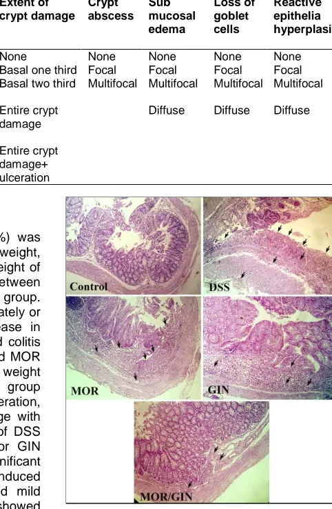
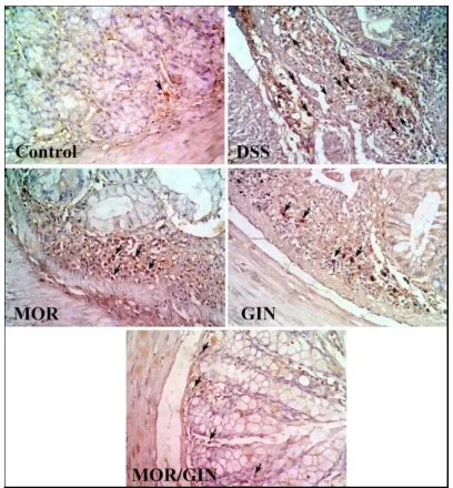
Induction of colitis in mice by DDS (5%) was obvious with increased DAI (loss of weight, diarrhea and blood in stool). The body weight of the mice showed significant reduction between DSS induced colitis group and control group. Mice treated either by GIN or MOR separately or in combination showed significant increase in body weight compared with DSS induced colitis group. Mice treated by combined GIN and MOR showed significant increase in body weight compared with MOR only treated group (Table 2). The colon showed mucosal ulceration, inflammatory cell infiltration, crypt damage with loss of goblet cells (Fig. 1). Treatment of DSS induced colitis mice with either MOR or GIN alone or in combination resulted in significant decrease DAI in relation to control DSS induced colitis mice. Single treatments produced mild improvement while combined treatment showed marked improvement (Fig. 1). NFKβ was highly expressed in inflammatory cells infiltrating colon of DSS group, while groups treated with either MOR and/ or GIN showed mild to moderate reduction of its expression (Fig. 2). Sum of histological scoring showed significant improvement in MOR or/ and GIN compared by DSS induced colitis group. GIN treated group alone or in combination with MOR showed significant improvement compared by MOR treated group alone.
DSS induced colitis mice showed significant decrease in colon length compared with control normal group. Treatment DSS colitis mice with GIN produced significant increase in colon length compared with DSS colitis mice. Combination therapy with GIN and MOR showed significant increase in colon length compared with MOR only treated group (Table 3).
Fig. 1. NOR: Normal control mice. DSS: mice received DSS, showed marked transmural
changes of ulcerative colitis; mucosal ulceration, inflammatory cell infiltration, crypt
damage with loss of goblet cells with sever degree of macro- and micro-vesicular steatosis. Treatment with GIN or MOR produced mild improvement of pathological changes. Combination of MOR/GIN produced
marked improvement comparable to normal control group (H&E)
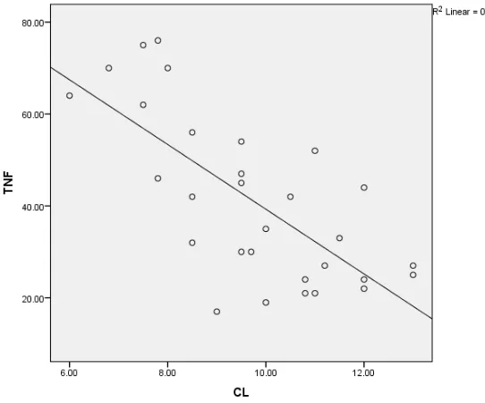
group, while combined therapy group showed significant decrease of TNFα compared with MOR group and no significant changes compared to GIN treated group (Table 3). There is a negative correlation between TNFα and colon length in different groups (Fig. 3).
Fig. 2. Immunohistochemical staining with NFKβ showed markedly expressed in the cytoplasm of inflammatory cells infiltrate of
DSS-treated group. Treatment with GIN or MOR produced mild to moderate reduction of
NFKβ expression compared to combination of MOR/GIN-treated group (x400)
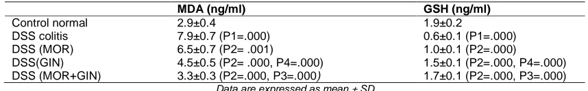
Oxidative stress markers are significantly changed (increased MDA, decreased GSH) in DSS induced colitis group compared with control normal group. Treatment of DSS induced colitis mice either with GIN or MOR alone or combination resulted in significant change in oxidative stress markers from control DSS induced colitis mice. GIN alone treated group showed significant changes in these parameters compared with MOR treated group. However, combination therapy with GIN and MOR produced significant changes in oxidative stress compared with MOR group and no significant changes compared with GIN treated group (Table 4).
4. DISCUSSION
In this study, administration of DSS to mice for 7 days produced an elevation of the disease activity index (DAI) and histological damage
score. This is consistent with the study of Elkatay et al. [30]. These effects could be explained by the fact that DSS could promote inflammation by variable biological pathways including direct cytotoxic effects [29] and apoptotic damage of colonic epithelial cells [31]. DSS induced colitis is a reproducible model of IBD in mice. It resembles IBD in human morphologically and symptomatic-cally [32]. MOR and/or GIN have the ability to improve disease activity and histological damage score. These results are in accordance with previous studies which showed that GIN improved histological examination in acetic acid induced colitis in rats [33,34]. In addition, GIN extracts ameliorate colonic atrophy in DSS induced colitis in rats [11]. On the other hand, MOR leave extract improved DAI, macroscopic and microscopic lesion in acetic acid induced ulcer in rats [35]. While, MOR seed improves all signs of inflammation, and severity of colitis in acetic acid induced colitis in rats [36].
Abnormality of cytokine generation and signaling mechanisms by intestinal epithelial cells, macrophages and lymphocytes have been supposed to be included in the pathogenesis of IBD, and the transcription factor NFKβ is one of the major regulatory components in this complex steps [37]. Pro-inflammatory cytokines are integrated together to produce mucosal tissue
damage in IBD. For example, TNFα and IL-1 involved in production of matrix
metalloproteinase which cause severe damage of the extracellular matrix and mucosal degradation while, NFKβ -induced cytokines are included in further enhancement and differentiation of lamina propria immune cells, leading to the prolongation of mucosal inflammation and differentiation of T-helper (Th)1 cells, the most important cells involved in the pathogenesis of IBD [38,39]. In IBD patients, the increased expression of NFKβ in mucosal macrophages is related to an expanded capacity of these cells to synthesis and release TNFα. The expression and actuation of NFKβ is strongly induced in the inflamed gut of IBD patients in amounts correlated significantly with the seriousness of intestinal inflammation [13].
Oz et al. [41] found that inflammatory cytokine levels like TNFα was significantly increased in DSS-induced colitis in mice. This is also is in consistence with Elkatary et al. [30] who showed considerable increase in TNFα in DSS colitis mice and Ajayi et al. [11] who showed significant expression of NFKβ in colon of DSS mice. GIN and/ or MOR showed significant decrease of TNFα and decrease the expression of NFKβ in
colon. Similar findings were reported by El-Abhar et al. [33], who showed significant
decrease in TNFα in mice treated with GIN.
Arablou et al. [42] showed that GIN decrease the levels of TNFα in patients with type 2 DM. GIN different components decrease T cell proliferation and cytokine production like IL-2 [43]. GIN extracts ameliorate the expression of NFKβ in trinitrobenzene sulphonic acid-induced colitis in mice [10,11]. In addition, MOR decreased TNFα and NFKβ in lipopolysaccharide (LPS)-stimulated macrophage cells [44] and smoking induced macrophages [45]. The negative correlation between TNFα and colon length supports the role of TNF α in IBD.
Table 2. Effects of MOR and/ or GIN on body weight and histopathological scoring in different experimental groups
Weight (gm) Histopathological scoring
Control normal 20.0±1.4 ---
DSS colitis 13.0±0.9 (P1=.000) 14.8±1.5
DSS (MOR) 15.2±1.2 (P2= .03) 9.8±1.5 (P1=.000)
DSS(GIN) 16±1.1(P2= .02) 7.5±1.0 (P2=.000, P4=.01)
DSS (MOR+GIN) 18.2±1.3 (P2=.000, P3=.03) 5.5±1.0 (P2=.000, P3=.000) Data are expressed as mean ± SD
P1: Significant changes from control normal, P2: Significant changes from DSS control group, P3: Significant changes of combined MOR & GIN from MOR treated group, P4: Significant changes of GIN from MOR treated group
Table 3. Effects of MOR and/ or GIN on colon length and TNFα in different experimental groups
Colon length (cm) TNFα (Pg/ml)
Control normal 11.1±1.5 21.6±3.6
DSS colitis 7.3±0.7 (P1=.000) 69.5±5.6 (P1=.000)
DSS (MOR) 9.1±1.1 (P2= .07) 49.2±5.6 (P2=.000)
DSS(GIN) 10±1.2 (P2= .03) 38.3±6.9 (P2=.000, P4= .01)
DSS (MOR+GIN) 11.1±1.1 (P2=.000, P3=.04) 26.7±4.3 (P2=.000, P3=.000) Data expressed as mean ± SD
P1: Significant changes from control normal, P2: Significant changes from DSS control group, P3: Significant changes of combined MOR & GIN from MOR treated group, P4: Significant changes of GIN from MOR treated group
Table 4. Effects of MOR and/ or GIN on oxidative stress markers (MDA & GSH)
MDA (ng/ml) GSH (ng/ml)
Control normal 2.9±0.4 1.9±0.2
DSS colitis 7.9±0.7 (P1=.000) 0.6±0.1 (P1=.000)
DSS (MOR) 6.5±0.7 (P2= .001) 1.0±0.1 (P2=.000)
DSS(GIN) 4.5±0.5 (P2= .000, P4=.000) 1.5±0.1 (P2=.000, P4=.000) DSS (MOR+GIN) 3.3±0.3 (P2=.000, P3=.000) 1.7±0.1 (P2=.000, P3=.000)
Data are expressed as mean ± SD
P1: Significant changes from control normal, P2: Significant changes from DSS control group.
P3: Significant changes of combined MOR & GIN from MOR treated group, P4: Significant changes of GIN from MOR treated group
In the present study, inflammation in the colon led to the disturbance of oxidative balance as indicated from a significant increase in the MDA and decrease of GSH levels in the colonic tissue in comparison to the control mice. Similar findings have been previously reported by (Elkatary et al. [30] and Pandurangan et al. [46]. The production and release of ROS species especially MDA by immune cells are claimed to have a crucial effect in the pathophysiology of colitis [47]. Elevated levels of MDA in stress condition is responsible for lipid membrane destruction and tissue injury in UC [48].
In addition, there was reduction of GSH level in the colonic tissue compared to the control group in the present study. GSH is one of the protective mechanisms of the body to prevent accumulation of ROS and damage of cells or pathological changes [49]. Consumption of GSH is one of the important factors responsible for colonic damage occurring both in human IBD and in animal models. This consumption could be a result of increased release of free radicals and could represent a particular disease due to an affection colitis ability of GSH producing enzyme [49,50].
In the present study, MOR and GIN separately or in combination had the ability to improve the distorted (GSH& MDA) balance. This is in coherence with El-Abhar et al. [31] who reported that GIN restored GSH levels and decreased MDA in acetic acid induced colitis in rats and Nwozo et al. [51] who prescribed similar changes in ethanol induced hepatic injury in rats. MOR showed antioxidant properties invitro and invivo (rat treated with CCL4) [16].
Ginger could be used safely in humans in doses up to (2-4 gm daily) [52,53]. It is could be used safely in pregnant woman to treat nausea and vomiting [53]. It showed antioxidant effects, as well as antibacterial effects against E. coli [9] which may be included in the pathogenesis of IBD [54]. Ginger affects non-specific proliferation of T lymphocyte in addition to cell-mediated
immune response [5,6]. These effects encourage their use in clinical trials on patients with IBD. Despite the promising clinical uses of ginger it should be used carefully with other herbs or medications which affect clotting factors in the body like garlic, ginkobiloba, heparin, cumarin and asprin [55,56]. It increased the platelet activity of nifidipine (antihypertensive drug), so careful monitoring is very important when ginger is used in doses less than 4 gm and contraindicated if it is used in doses higher than 4 gm daily [57]. It should be used with caution in patient with arrhythmia or CNS depression as it may cause arrhythmia or CNS depression [58].
5. CONCLUSION
MOR may produce its beneficial effects through decrease TNFα and NFKβ levels, so interrupt the inflammatory pathway in IBD and also improve oxidative stress balance. Further investigations are needed to assess the effect of MOR both in animals and clinical trials. GIN could be used as a food supplement to treat patients with IBD as GIN safety is approved by FDA (see appendix 1). Combination of both herbs has no additive effects as GIN more effective than MOR in this study.
CONSENT
It is not applicable.
ETHICAL APPROVAL
All authors hereby declare that all experiments have been examined and approved by the appropriate our local ethics committee with No. R 49, Faculty of Medicine, Mansoura University.
ACKNOWLEDGEMENTS
of GIN and MOR extract. We also very thankful to Dr/ Ehsan Mohammad, Clinical Pathology Dep., Faculty of Medicine, Mansoura University, for her help in assessment of TNF α in serum of mice.
COMPETING INTERESTS
Authors have declared that no competing interests exist.
REFERENCES
1. Keohane J1, O'Mahony C, O'Mahony L, O'Mahony S, Quigley EM, Shanahan F. Irritable bowel syndrome-type symptoms in patients with inflammatory bowel disease: a real association or reflection of occult inflammation. Am J Gastroenterol. 2010; 105(8):1788,1789-94.
2. Scaldaferri F, Fiocchi C. Inflammatory bowel disease: Progress and current concepts of etiopathogenesis. J Dig Dis. 2007;8(4):171-8.
3. Annaházi A, Molnár T. Optimal Endpoint of Therapy in IBD: An update on factors determining a successful drug withdrawal. Gastroenterol Res Pract. 2015;2015: 832395
4. Aharoni R, Kayhan B, Brenner O, Domev H, Labunskay G, Arnon R. Immunomodulatory therapeutic effect of glatiramer acetate on several murine models of inflammatory bowel disease. J Pharmacol Exp Ther. 2006;318:68-78. 5. Thomson M, Al-Qattan KK, Al-Sawan SM,
Alnaqeeb MA, Khan I, Ali M. The use of ginger (Zingiber officinale Rosc.) as a potential anti-inflammatory and antithrombotic agent. Prostaglandins, Leukotrienes and Essential Fatty Acids. 2002;67(6):475-478.
6. Zhou Hl, Deng YM, Xie QM. The modulatory effects of the volatile oil of ginger on the cellular immune response in vitro and in vivo in mice. J Ethnopharmacol. 2006;105:301-305.
7. Rahmani AH, Shabrmi FM, Aly SM. Active ingredients of ginger as potential candidates in the prevention and treatment of diseases via modulation ofbiological activities. Int J Physiol Pathophysiol Pharmacol. 2014;6(2):125-36.
8. Ensiyeh J, Sakineh MA. Comparing ginger and vitamin B6 for the treatment of nausea and vomiting in pregnancy: A randomised
controlled trial. Midwifery. 2009;25(6):649-653.
9. Peixoto JR, Silva GC, Costa RA, de Sousa Fontenelle JR, Vieira GH, Filho AA, et al. In vitro antibacterial effect of aqueous and ethanolic Moringa leaf extracts. Asian Pac J Trop Med. 2011;4(3):201-4.
10. Hsiang CY, Lo HY, Huang HC, Li CC, Wu SL, Ho TY. Ginger extract and zingerone ameliorated trinitrobenzene sulphonic acid-induced colitis in mice via modulation of nuclear factor-κB activity and interleukin-1β signalling pathway. Food Chem. 2013; 136(1):170-7.
11. Ajayi BO, Adedara IA, Farombi EO. Pharmacological activity of 6-gingerol in dextran sulphate sodium induced ulcerative colitis in BALB/c mice. Phytother Res. 2015;29(4):566-72.
12. Enwa FO, Omojate CG, Adonu CC. A Review on the phytochemical profile and the antibacterial susceptibility pattern of some clinical isolates to the ethanolic leaves extract of Moringa oleifera Lam (Moringaceae). IJAR. 2013;1(5):226-238. 13. Atreya I, Atreya R, Neurath MF. NF-κB in
inflammatory bowel disease (Review). J Intern Med. 2008;263:591-596.
14. Mahajan SG, Mali RG, Mehta AA. Effect of
Moringa oleifera Lam. Seed extract on
toluene di-isocyanate-induced Immune-mediated inflammatory responses in rats. J Immunotoxicol. 2007;4:85-96.
15. Berkovich L, Earon G, Ron I, Rimmon A, Vexler A, Lev-Ari S. Moringa oleifera aqueous leaf extract down-regulates nuclear factor-kappaB andincreases cytotoxic effect of chemotherapy in pancreatic cancer cells. BMC Complement Altern Med. 2013;13:212.
16. Verma AR, Vijayakumar M, Mathela CS, Rao CV. In vitro and in vivo antioxidant properties of different fractions of Moringa oleifera leaves. Food Chem Toxicol. 2009;47:196–201.
17. Jabeen R, Shahid M, Jamil A, Ashraf M. Microscopic evaluation of the antimicrobial activity of seed extracts of Moringa
oleifera. Pak. J. Bot. 2008;40:1349.
18. Nepolean P, Anitha J, Emilin RR. Isolation, analysis and identification of phytochemicals of antimicrobial activity of
Moringa oleifera Lam. Curr. Biotica. 2009;
3:33-39.
steam distillate of Moringa oleifera Lam. J. Pharm. Sci. Res. 2010;2:34-37.
20. Doughari JH, Pukuma MS, De N. Antibacterial effects of Balanites aegyptiaca Del and Moringa oleifera Lam.
on Salmonella typhi. Afri. J. Biotech. 2007; 6(19):2212-2215.
21. Perše M, Cerar A. Dextran sodium sulphate colitis mouse model: Traps and tricks. J Biomed Biotechnol. 2012; 2012:718617.
DOI: 10.1155/2012/718617
22. Tsuchiya T, Fukuda S, Hamada H, Nakamura A, Kohama Y, Ishikawa H, et al. Role of gamma delta T cells in the inflammatory response of experimental colitis mice. J Immunol. 2003;171(10): 5507-13.
23. Charan J, Kantharia ND. How to calculate sample size in animal studies? Journal of Pharmacology and Pharmacotherapeutics. 2013;4(4):303.
24. Knapka JJ. Nutrition. In the mouse in biomedical research. Foster HL, Small JD, Fox JG, eds. New York: Academic Press. 1983;3:51–67.
25. University of Minnesota. Euthanasia Guidelines; 2013.
Available:http://www.ahc.umn.edu/rar/euth anasia.html
26. Brenner DA, O'Hara M, Angel P, Chojkier M, Karin M. Prolonged activation of JUN and collagenase genes by tumour necrosis factor-alpha. Nature. 1989;337:661-663.
27. Draper W, Hadley M. Indirect
determination of oxygen free radicals. Methods Enzymol. 1990;186:421- 431. 28. Reddy YN, Murthy SV, Krishna DR,
Prabhakar MC. Role of free radicals and antioxidants in tuberculosis patients. Indian J Tuberc. 2004;51:213-218.
29. Cooper HS, Murthy SN, Shah RS, Sedergran DJ. Clinicopathologic study of dextran sulfate sodium experimental murine colitis. Lab. Invest. 1993;69:238-249.
30. Elkatary R, Abdelrahman K, Hassanin A, Elmasry A, Karef A. Comparative study between effect of simvastatin (5 mg/Kg) and simvastatin (50 mg/Kg) in an early treatment of experimentally induced colitis in mice. BJMMR. 2015;8(11):937-947. 31. Renes IB, Verburg M, Van Nispen DJ,
Taminiau JA, Buller HA, Dekker J, and Einerhand AW. Epithelial proliferation, cell death, and gene expression in experimental colitis: Alterations in carbonic
anhydrase I, mucin MUC2, and trefoil factor 3 expression. J. Int J Colorectal Dis. 2002;17:317-326.
32. Randhawa PK, Singh K, Singh N, Jaggi AS. A review on chemical-induced inflammatory bowel disease models in rodents. Korean J Physiol Pharmacol. 2014;18(4):279-288.
33. El-Abhar HS, Hammad LN, Gawad HS. Modulating effect of ginger extract on rats with ulcerative colitis. J Ethnopharmacol. 2008;118(3):367-72.
34. Rashidian A, Mehrzadi S, Ghannadi AR, Mahzooni P, Sadr S, Minaiyan M. Protective effect of ginger volatile oil against acetic acid induced colitis in rats: A light microscopic evaluation. J Integr Med. 2014;12(2):115-20.
35. Das S, Kanodia L, Mukherjee A, Hakim A. Effect of ethanolic extract of leaves of
Paederia foetida Linn. on acetic acid
induced colitis in albino rats. Indian J Pharmacol. 2013;45(5):453-7.
36. Minaiyan M, Asghari G, Taheri D, Saeidi M, Nasr-Esfahani S. Anti-inflammatory effect of Moringa oleifera Lam. seeds on acetic acid-induced acute colitis in rats. Avicenna J Phytomed. 2014;4(2):127-36. 37. Li Q, Verma IM. NF-kappaB regulation in
the immune system. Nat Rev Immunol. 2002;2:725-34.
38. Pallone F, Monteleone G. Mechanisms of tissue damage in inflammatory bowel disease. Curr Opin Gastroenterol. 2001; 17:307–12.
39. Holtmann MH, Neurath MF. Differential TNF-signaling in chronic inflammatory disorders. Curr Mol Med. 2004;4:439–44. 40. Nishiyama Y, Kataoka T, Yamato K,
Taguchi T, Yamaoka K. Suppression of dextran sulfate sodium-induced colitis in mice by radon inhalation. Mediators of Inflammation. 2012;2012:239617.
41. Oz H, Zhong J, de Villiers W. Pattern recognition scavenger receptors, SR-A and CD36, have an additive role in the development of colitis in mice. Dig Dis Sci. 2009;54(12):2561–2567.
42. Arablou T, Aryaeian N, Valizadeh M, Sharifi F, Hosseini A, Djalali M. The effect of ginger consumption on glycemic status, lipid profile and some inflammatory markers in patients with type 2 diabetes mellitus. Int J Food Sci Nutr. 2014; 65(4):515-20
lymphocyte proliferation and cytokine synthesis by [6]-Gingerol, [8]-Gingerol, and [10]-Gingerol. Phytother Res. 2015; 29(11):1707-13.
44. Lee HJ, Jeong YJ, Lee TS, Park YY, Chae WG, Chung IK, et al. Moringa fruit inhibits LPS-induced NO/iNOS expression through suppressing the NF-κ B activation in RAW264.7 cells. Am J Chin Med. 2013; 41(5):1109-23.
45. Kooltheat N, Sranujit RP, Chumark P, Potup P, Laytragoon-Lewin N, Usuwanthim K. An ethyl acetate fraction of Moringa
oleifera Lam. Inhibits human macrophage
cytokine production induced by cigarette smoke. Nutrients. 2014;6(2):697-710. 46. Pandurangan AK, Ismail S, Saadatdoust Z,
Esa NM. Allicin Alleviates Dextran Sodium Sulfate (DSS)-induced ulcerative colitis in BALB/c mice. Oxid Med Cell Longev. 2015;2015.Article ID 605208, 13 page. 47. Zhu H, Li YR. Oxidative stress and redox
signaling mechanisms of inflammatory bowel disease: Updated experimental and clinical evidence. Exp Biol Med (Maywood). 2012;237(5):474–80.
48. Lenoir L1, Rossary A, Joubert-Zakeyh J, Vergnaud-Gauduchon J, Farges MC, Fraisse D, Texier O, Lamaison JL, Vasson MP, Felgines C. Lemon verbena infusion consumption attenuates oxidative stress in dextran sulfate sodium-induced colitis in the rat. Digestive Diseases and Sciences. 2011;56(12):3534-45.
49. Clarkson PM, Thompson HS. Antioxidants: what role do they play in physical activity and health? Am J Clin Nutr. 2000; 72(Suppl):637S-646S.
50. Koch O, Pani G, Borrello S, Colavitti R, Cravero A, Farrè S, et al. Oxidative stress
and antioxidant defenses in
ethanolinduced cell injury. Molecular Aspects of Medicine. 2004;25(1-2):191-198.
51. Nwozo SO, Osunmadewa DA, Oyinloye BE. Anti fatty liver effects of oils from
Zingiber officinale and Curcuma longa on
ethanol-induced fatty liver in rats. J Integr Med. 2014;12(1):59-65.
52. Kraft K, Hobbs C. Pocket guide to herbal medicine. Stuttgart: Theieme; 2004;70-71. 53. Bryer E. A literature review of the
effectiveness of ginger in alleviating mild-to-moderate nausea and vomiting of pregnancy. J Midwifery Womens Health. 2005;50(1):e1-3.
54. Rhodes JM. The role of Escherichia coli in inflammatory bowel disease. Gut. 2007; 56(5):610–612.
55. Abebe W. Herbal medication: Potential for adverse interactions with analgesic drugs. J Clin Pharm Ther. 2002;27(6):391-401. 56. Brinker F. Herbal contraindications and
drug interactions: Plus herbal adjuncts with medicines, 4th edition. Eclectic Medical Publications; 2010.
57. Young HY, Liao JC, Chang YS, Luo YL, Lu MC, Peng WH. Synergistic effect of ginger and nifedipine on human platelet aggregation: A study in hypertensive patients and normal volunteers. Am J Chin Med. 2006;34(4):545-51.
11 APPENDIX
FDA Home3 Medical Devices4 Databases5
CFR Code of Federal Regulations Title 21
The information on this page is current as of April 1 2015.
For the most up-to-date version of CFR Title 21, go to the Electronic Code of Federal Regulations (eCFR).6
New Search Help7 | More About 21CFR8 [Code of Federal Regulations]
[Title 21, Volume 3]
[Revised as of April 1, 2015] [CITE: 21CFR182.20]
TITLE 21--FOOD AND DRUGS
CHAPTER I--FOOD AND DRUG ADMINISTRATION DEPARTMENT OF HEALTH AND HUMAN SERVICES
SUBCHAPTER B--FOOD FOR HUMAN CONSUMPTION (CONTINUED) PART 182 -- SUBSTANCES GENERALLY RECOGNIZED AS SAFE
Subpart A--General Provisions
Sec. 182.20 Essential oils, oleoresins (solvent-free), and natural extractives (including distillates).
12
Common name Botanical name of plant source
Alfalfa Medicago sativa L.
Allspice Pimenta officinalis Lindl.
Almond, bitter
(free from prussic acid) Prunus amygdalus Batsch, Prunus armeniaca L., or Prunus persica (L.) Batsch.
Ambrette (seed) Hibiscus moschatus Moench.
Angelica root Angelica archangelica L.
Angelica seed Do.
Angelica stem Do.
Angostura (cusparia
bark) Galipea officinalis Hancock.
Anise Pimpinella anisum L.
Asafetida Ferula assa-foetida L. and related spp. of Ferula.
Balm (lemon balm) Melissa officinalis L.
Balsam of Peru Myroxylon pereirae Klotzsch. Basil Ocimum basilicum L.
Basil Ocimum basilicum L.
Bay leaves Laurus nobilis L.
Bay (myrcia oil) Pimenta racemosa (Mill.) J. W. Moore.
Bergamot (bergamot Citrus aurantium L. subsp. bergamia Wright et Arn. orange)
Bitter almond (free Prunus amygdalus Batsch, Prunus armeniaca L.,
from prussic acid) or Prunus persica (L.) Batsch.
Bois de rose Aniba rosaeodora Ducke.
Cacao Theobroma cacao L.
Camomile (chamomile)
flowers, Hungarian Matricaria chamomilla L.
Camomile (chamomile)
flowers, Roman or Anthemis nobilis L. English
13
Capsicum Capsicum frutescens L. and Capsicum annuum L. Caraway Carum carvi L.
Cardamom seed
(cardamon) Elettaria cardamomum Maton.
Carob bean Ceratonia siliqua L.
Carrot Daucus carota L.
Cascarilla bark Croton eluteria Benn.
Cassia bark, Chinese Cinnamomum cassia Blume.
Cassia bark, Padang or
Batavia Cinnamomum burmanni Blume.
Cassia bark, Saigon Cinnamomum loureirii Nees.
Celery seed Apium graveolens L.
Cherry, wild, bark Prunus serotina Ehrh.
Chervil Anthriscus cerefolium (L.) Hoffm.
Chicory Cichorium intybus L.
Cinnamon bark, Ceylon Cinnamomum zeylanicum Nees.
Cinnamon bark, Chinese Cinnamomum cassia Blume.
Cinnamon bark, Saigon Cinnamomum loureirii Nees.
Cinnamon leaf, Ceylon Cinnamomum zeylanicum Nees.
Cinnamon leaf, Chinese Cinnamomum cassia Blume.
Cinnamon leaf, Saigon Cinnamomum loureirii Nees.
Citronella Cymbopogon nardus Rendle.
Citrus peels Citrus spp.
Clary (clary sage) Salvia sclarea L. Clover Trifolium spp.
Coca (decocainized) Erythroxylum coca Lam. and other spp. of
Erythroxylum.
Coffee Coffea spp.
Cola nut Cola acuminata Schott and Endl., and other spp. of Cola.
Coriander Coriandrum sativum L.
Cumin (cummin) Cuminum cyminum L. Curacao orange peel
14
Cusparia bark Galipea officinalis Hancock.
Dandelion Taraxacum officinale Weber and T. laevigatum
DC.
Dandelion root Do.
Dog grass (quackgrass,
triticum) Agropyron repens (L.) Beauv.
Elder flowers Sambucus canadensis L. and S. nigra I. Estragole (esdragol,
esdragon, tarragon) Artemisia dracunculus L.
Estragon (tarragon) Do.
Fennel, sweet Foeniculum vulgare Mill.
Fenugreek Trigonella foenum-graecum L.
Galanga (galangal) Alpinia officinarum Hance.
Geranium Pelargonium spp.
Geranium, Eas t IndianCymbopogon martini Stapf.
Geranium, ros e Pelargonium graveolens L'Her.
Ginger Zingiber officinale Rosc.
Grapefruit Citrus paradisi Macf.
Guava Psidium spp.
Hickory bark Carya spp.
Horehound (ho arhound)Marrubium vulgare L.
Hops Humulus lupulus L.
Horsemint Monarda punctata L.
Hyssop Hyssopus officinalis L.
Immortelle Helichrysum augustifolium DC.
Jasmine Jasminum officinale L. and other spp. of
Jasminum.
Juniper (berries) Juniperus communis L.
Kola nut Cola acuminata Schott and Endl., and other spp. of Cola.
Laurel berries Laurus nobilis L.
15
Lavender Lavandula officinalis Chaix.
Lavender, spike Lavandula latifolia Vill.
Lavandin Hybrids between Lavandula officinalis Chaix and
Lavandula latifolin Vill.
Lemon Citrus limon (L.) Burm. f.
Lemon balm (see balm)
Lemon grass Cymbopogon citratus DC. and Cymbopogon lexuosus Stapf.
Lemon peel Citrus limon (L.) Burm. f.
Lime Citrus aurantifolia Swingle. Linden flowers Tilia spp.
Locust bean Ceratonia siliqua L,
Lupulin Humulus lupulus L.
Mace Myristica fragrans Houtt.
Mandarin Citrus reticulata Blanco.
Marjoram, sweet Majorana hortensis Moench.
Mate Ilex paraguariensis St. Hil.
Melissa (see balm)
Menthol Mentha spp.
Menthyl acetate Do.
Molasses (extract) Saccarum officinarum L.
Mustard Brassica spp.
Naringin Citrus paradisi Macf.
Neroli, bigarade Citrus aurantium L.
Nutmeg Myristica fragrans Houtt.
Onion Allium cepa L.
Orange, bitter,
flowers Citrus aurantium L.
Orange, bitter, peel Do.
Orange leaf Citrus sinensis (L.) Osbeck.
Orange, sweet Do.
Orange, sweet, flowers Do.
16
Origanum Origanum spp.
Palmarosa Cymbopogon martini Stapf.
Paprika Capsicum annuum L.
Parsley Petroselinum crispum (Mill.)Mansf.
Pepper, black Piper nigrum L.
Pepper, white Do.
Peppermint Mentha piperita L.
Peruvian balsam Myroxylon pereirae Klotzsch.
Petitgrain Citrus aurantium L.
Petitgrain lemon Citrus limon (L.) Burm. f.
Petitgrain mandarin or
tangerine Citrus reticulata Blanco.
Pimenta Pimenta officinalis Lindl.
Pimenta leaf Pimenta officinalis Lindl.
Pipsissewa leaves Chimaphila umbellata Nutt.
Pomegranate Punica granatum L.
Prickly ash bark Xanthoxylum (or Zanthoxylum) Americanum Mill. or Xanthoxylum clava-herculis L.
Rosa alba L., Rosa centifolia L., Rosa
Rose absolute damascena Mill., Rosa gallica L., and vars. of these spp.
Rose (otto of roses,
attar of roses) Do.
Rose buds Do.
Rose flowers Do.
Rose fruit (hips) Do.
Rose geranium Pelargonium graveolens L'Her.
Rose leaves Rosa spp.
Rosemary Rosmarinus officinalis L.
17
Sage Salvia officinalis L.
Sage, Greek Salvia triloba L.
Sage, Spanish Salvia lavandulaefolia Vahl.
St. John's bread Ceratonia siliqua L.
Savory, summer Satureia hortensis L.
Savory, winter Satureia montana L.
Schinus molle Schinus molle L.
Sloe berries
(blackthorn berries) Prunus spinosa L.
Spearmint Mentha spicata L.
Spike lavender Lavandula latifolia Vill.
Tamarind Tamarindus indica L.
Tangerine Citrus reticulata Blanco.
Tarragon Artemisia dracunculus L.
Tea Thea sinensis L.
Thyme Thymus vulgaris L. and Thymus zygis var.
gracilis Boiss.
Thyme, white Do.
Thyme, wild or
creeping Thymus serpyllum L.
Triticum (see dog grass)
Tuberose Polianthes tuberosa L.
Turmeric Curcuma longa L.
Vanilla Vanilla planifolia Andr. or Vanilla tahitensis
J. W. Moore.
Violet flowers Viola odorata L.
Violet leaves Do.
Violet leaves absolute Do.
Wild cherry bark Prunus serotina Ehrh.
Ylang-ylang Cananga odorata Hook. f. and Thoms.
18
[42 FR 14640, Mar. 15, 1977, as amended at 44 FR 3963, Jan. 19, 1979;
47 FR 29953, July 9, 1982; 48 FR 51613, Nov. 10, 1983; 50 FR 21043 and
21044, May 22, 1985]
Links on this page:
1. http://www.addthis.com/bookmark.php?u508=true&v=152&username=fdamain
2. http://www.addthis.com/bookmark.php
3. http://www.fda.gov/default.htm
4. http://www.fda.gov/MedicalDevices/default.htm
5. http://www.fda.gov/MedicalDevices/DeviceRegulationandGuidance/Databases/default.htm
6. http://www.ecfr.gov/cgi-bin/text-idx?
SID=3ee286332416f26a91d9e6d786a604ab&mc=true&tpl=/ecfrbrowse/Title21/21tab_02.tpl
7. /scripts/cdrh/cfdocs/search/default.cfm?FAQ=true
8. http://www.fda.gov/MedicalDevices/DeviceRegulationandGuidance/Databases/ucm135680.htm
Page Last Updated: 08/21/2015
Note: If you need help accessing information in different file formats, see Instructions for Downloading Viewers and Players.
Accessibility Contact FDA Careers FDA Basics FOIA No FEAR Act Site Map Transparency Website Policies
U.S. Food and Drug Administration 10903 New Hampshire Avenue Silver Spring, MD 20993
19 Contact FDA
For Government For Press
Combination Products Advisory Committees Science & Research Regulatory Information Safety Emergency Preparedness International Programs News & Events Training and Continuing Education Inspections/Compliance State & Local Officials Consumers Industry Health Professionals FDA Archive
Links on this page:
1. http://www.addthis.com/bookmark.php?u508=true&v=152&username=fdamain
2. http://www.addthis.com/bookmark.php
3. http://www.fda.gov/default.htm
4. http://www.fda.gov/MedicalDevices/default.htm
5. http://www.fda.gov/MedicalDevices/DeviceRegulationandGuidance/Databases/default.htm
6. http://www.ecfr.gov/cgi-bin/text-idx?
SID=3ee286332416f26a91d9e6d786a604ab&mc=true&tpl=/ecfrbrowse/Title21/21tab_02.tpl 7. /scripts/cdrh/cfdocs/search/default.cfm?FAQ=true
8. http://www.fda.gov/MedicalDevices/DeviceRegulationandGuidance/Databases/ucm135680.htm
_____________________________________________________________________________________________________________________________
© 2016 Elmasry et al.; This is an Open Access article distributed under the terms of the Creative Commons Attribution License (http://creativecommons.org/licenses/by/4.0), which permits unrestricted use, distribution, and reproduction in any medium, provided the original work is properly cited.
Peer-review history: