e82
A
therosclerosis is a complex multifactorial disease, and individual susceptibility to plaque development is influ-enced by many genetic factors. We previously showed that plaque size in mice is dependent on the strain and vascular location in the early stage of atherosclerosis.1–4 At the aorticroot, apolipoprotein E–deficient (Apoe−/−) mice on a DBA/2J
background (DBA/2J-Apoe−/−) develop larger plaques than
those on a C57BL/6J background (C57BL/6J-Apoe−/−),
whereas Apoe−/− mice on a 129S6 background (129S6-Apoe−/−)
are resistant to plaque development compared with DBA/2J-Apoe−/− or C57BL/6J-Apoe−/− mice. In the aortic arch area,
however, 129S6-Apoe−/− and DBA/2J-Apoe−/− mice show
larger lesion size than C57BL/6J-Apoe−/− mice. These
obser-vations clearly indicate that the location specificity of plaque development is genetically controlled.
Our QTL analysis using an intercross between DBA/2J-Apoe−/− and 129S6-Apoe−/− revealed Aath4, an atherosclerosis
QTL for the aortic arch area on the distal part of chromosome 2 (Peak: 137 Mb, confidence interval: 123−148 Mb).4 The
DBA/2J allele of Aath4 confers susceptibility to atheroscle-rosis, whereas the 129S6 allele confers resistance. Aath4 was
not detected in a C57BL/6J-Apoe−/− × 129S6-Apoe−/− cross,2
indicating that Aath4 sequences unique to DBA/2J are respon-sible for the different phenotypes. Many candidate genes are present in the chromosomal region in which Aath4 resides, including several phagocytosis-related genes.
One of the candidates is c-mer proto-oncogene tyrosine kinase (Mertk) located at 128.5 Mb, which encodes a member of the TAM (Tyro3, Axl, and Mer) receptor tyrosine kinase family. MERTK is primarily expressed in monocytes as well as in epithelial and reproductive tissues,5 and it is involved
in phagocytosis of apoptotic cells.6,7 It has 2
immunoglob-ulin-like domains and 2 fibronectin type III domains in the extracellular region, and binds to apoptotic cells via bridg-ing molecules such as GAS6 and protein S.5,8,9 The binding
promotes phosphorylation of the tyrosine kinase domain located within the intracellular region of MERTK; this phos-phorylation leads to activation of downstream signaling and induces structural changes in cytoskeletons, enabling the cell to engulf its target cells.10 Macrophages from mice
with an inactivated Mertk kinase domain (Mertk−/−) are
defi-cient in the clearance of apoptotic cells (efferocytosis).6 The
© 2017 American Heart Association, Inc.
Arterioscler Thromb Vasc Biol is available at http://atvb.ahajournals.org DOI: 10.1161/ATVBAHA.117.309522 Objective—Arch atherosclerosis 4 (Aath4) is a quantitative trait locus for atherosclerotic plaque formation in the inner curve
of the aortic arch previously identified in an F2 cross of Apoe−/− mice on DBA/2J and 129S6 backgrounds. C-mer proto-oncogene tyrosine kinase (Mertk), coding for a ligand-activated transmembrane tyrosine kinase, is a candidate gene within the same chromosomal region. Our objective was to determine whether strain differences in Mertk influence plaque formation. Approach and Results—To dissect the strain effects of Mertk on atherosclerosis, we first established a congenic mouse line
(Aath4aDBA/DBA) in which a 5′ region of Aath4 of DBA/2J, including Mertk, was backcrossed onto a 129S6-Apoe−/− background. The resulting Aath4aDBA/DBA male mice developed significantly larger plaques compared with control mice (Aath4a129/129), proving that the DBA/2J allele of Aath4a is proatherogenic. Thioglycollate-elicited peritoneal macrophages from Aath4aDBA/ DBA mice express less than 50% of Mertk mRNA and cell-surface MERTK protein compared with those from the control mice. Moreover, both large and small peritoneal Aath4aDBA/DBA macrophages showed reduced phagocytosis of apoptotic cells. When Mertk cDNAs from 129S6 and DBA/2J mice were overexpressed in HEK293T (human embryonic kidney 293T) cells, phagocytosis of apoptotic cells was equally enhanced in direct proportion to Mertk levels, indicating that phagocytosis is modulated by the amount of MERTK, but that it is not affected by MERTK amino acid differences between 129S6 and DBA/2J. Conclusions—Reduced transcription of Mertk, rather than differences in MERTK protein structure, determines the reduced
efficiency of apoptotic cell clearance in the Aath4aDBA/DBA mice, which, in turn, contributes to their increased susceptibility to atherosclerosis.
Visual Overview—An online visual overview is available for this article. (Arterioscler Thromb Vasc Biol. 2017;37:e82-e91. DOI: 10.1161/ATVBAHA.117.309522.)
Key Words: aorta ◼ atherosclerosis ◼ macrophages ◼ mice ◼ phagocytosis
Received on: December 12, 2016; final version accepted on: April 24, 2017.
From the Department of Pathology and Laboratory Medicine, University of North Carolina at Chapel Hill.
The online-only Data Supplement is available with this article at http://atvb.ahajournals.org/lookup/suppl/doi:10.1161/ATVBAHA.117.309522/-/DC1.
Correspondence to Nobuyo Maeda, PhD, CB no. 7525, 701 Brinkhous-Bullitt Bldg, Chapel Hill, NC 27599. E-mail [email protected]
Mertk Expression, Restricts Efferocytosis, and Increases
Susceptibility to Atherosclerosis
Yukako Kayashima, Natalia Makhanova, Nobuyo Maeda
Mertk−/− mice are viable, but show retinal degeneration,
because of the failure of the retinal pigment epithelium to engulf the outer segments of photoreceptors.7,11 MERTK also
plays a pivotal role in atherosclerotic plaque development via its effects on efferocytosis: Mertk deficiency in Apoe−/− mice
promotes accumulation of apoptotic cells and expansion of necrotic cores within plaques12; and low-density lipoprotein
receptor-deficient (Ldlr−/−) mice with Mertk−/− bone
mar-row show accumulation of apoptotic cells and accelerated atherosclerosis.13
In this study, we have generated and studied a mouse line, Aath4aDBA/DBA, in which a 5′ region of the Aath4 from DBA/2J
has been transferred onto a 129S6-Apoe−/− background. We
show that these mice have elevated plaque susceptibility and reduced efferocytosis by macrophages. We also demonstrate that decreased transcription of Mertk, not DBA-unique amino acid alterations in MERTK, determines the limited efferocyto-sis that occurs in Aath4aDBA/DBA mice.
Materials and Methods
Materials and Methods are available in the online-only Data Supplement.
Results
Generation of Aath4aDBA/DBA MiceAath4aDBA/DBA congenic mice were constructed by
backcross-ing DBA/2J-Apoe−/− mice to the 129S6-Apoe−/− strain for more
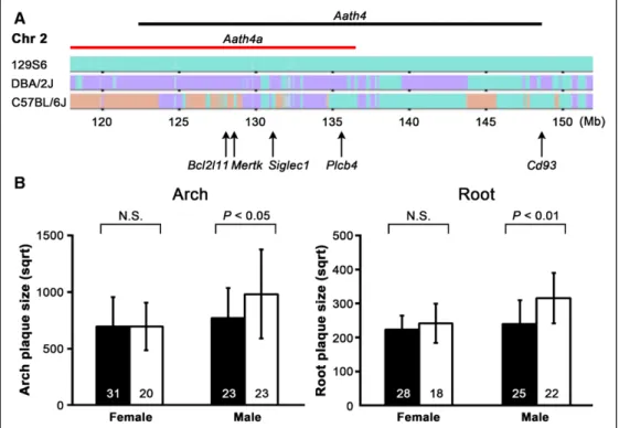
than 7 generations. The backcrossed genomic segments in chro-mosome 2 (117–137 Mb) are shown in Figure 1A. The DBA allele of SNP (single nucleotide polymorphism) rs27446327 was used as a marker for Chr 2: 128 Mb. Heterozygous mice were mated to generate homozygotes. SNPs rs30884601, rs36935260, rs48427643, and rs30243498 were also typed at early generations to ensure that DBA alleles at other athero-sclerosis QTLs (Chr 1: 155 Mb, Chr 1: 165 Mb, Chr 2: 143 Mb, and Chr 10: 86 Mb) were eliminated.
Aath4aDBA/DBA Male Mice Develop Larger
Plaques With Increased Calcium Deposits
As summarized in Table 1, Aath4a129/129 control mice and
Aath4aDBA/DBA mice showed no significant differences in body
weight. In males, plasma cholesterol levels were almost the same between controls and Aath4aDBA/DBA, whereas
triglycer-ides were significantly higher in Aath4aDBA/DBA (P=0.002). In
female Aath4aDBA/DBA mice, plasma total cholesterol was lower
(P=0.010) and HDL-cholesterol was higher (P=0.016) com-pared with Aath4a129/129 controls, but triglycerides were not
significantly different.
At 5 months of age, Aath4aDBA/DBA male mice developed ≈50% larger plaques at the inner curve of the aortic arch com-pared with control mice (P<0.05; Figure 1B, left). Plaque size at Nonstandard Abbreviations and Acronyms
Aath4 arch atherosclerosis 4
LPMs large peritoneal macrophages
SPMs small peritoneal macrophages
Figure 1. Aath4aDBA/DBA males develop larger atherosclerotic plaques. A,Haplotype maps of Chr 2 taken from the Mouse Phylogeny
Viewer (http://msub.csbio.unc.edu/#viewer). 129S6 sequence is colored in green (top), genomic regions where DBA/2J share the same sequences as 129S6 are shown in green, and DBA/2J-specific sequences are highlighted with purple (middle), and C57BL/6J unique regions are colored in peach (bottom). The backcrossed region in the congenic strain Aath4aDBA/DBA (red bar), position of Aath4 (black bar),
and the location of some of the candidate genes (arrows) are indicated. B,Comparison of plaque size at the aortic arch (left) and root (right) between the control Aath4a129/129 (filled bars) and Aath4aDBA/DBA (open bars) mice at 5 mo old. Plaque size is indicated in square root
(sqrt) of area in μm2. Data are shown as the mean±SD. Numbers of mice are indicated in the bars. N.S. indicates not significant.
the aortic root was also significantly larger in Aath4aDBA/DBA males
(P<0.01; Figure 1B, right). In contrast, females did not show sig-nificant differences in the size of either aortic arch or aortic root plaques. Male Aath4aDBA/DBA mice also developed significantly
larger brachiocephalic artery lesions compared with the control (P<0.05), but did not differ in the left common carotid and left subclavian arteries. In females, no significant difference was observed in any of these branches between Aath4aDBA/DBA mice
and controls (Figure I in the online-only Data Supplement). The plaques developing in the aortic roots of Aath4aDBA/DBA
and control mice ranged from a monolayer of foam cells to complex lesions with inflammation and necrotic cores (Figure 2A; Figure II in the online-only Data Supplement). When plaques of similar size and locations were examined histo-logically, the early raised lesions of Aath4aDBA/DBA contained
more scattered basophilic calcium deposits (Figure 2A and 2B) than the Aath4a129/129 control plaques. Multiple calcium
deposits were detectable, often associated with the acellular necrotic cores, in the raised plaques in 28 of 36 Aath4aDBA/DBA
mice compared with 6 of 28 Aath4a129/129 control mice. In
more mature plaques, calcium deposits were detected equally in both Aath4aDBA/DBA mice and Aath4a129/129 controls, and they
were located deeper in the plaques, near the internal lamina (Figure IIE and IIF in the online-only Data Supplement, arrowheads). Enhanced cell death or reduced clearance of dead cells could be the source of these calcium deposits. Although very few, TUNEL (terminal deoxynucleotidyl trans-ferase dUTP nick-end labeling)-positive nuclei were detected within the plaques of both Aath4a129/129 and Aath4aDBA/DBA mice
(Figure III in the online-only Data Supplement).
Because average lesions were significantly larger in size and more advanced in Aath4aDBA/DBA mice than in Aath4a129/129,
we selected 7 raised lesions with similar size from Aath4a129/129
and Aath4aDBA/DBA mice to compare the components of the early
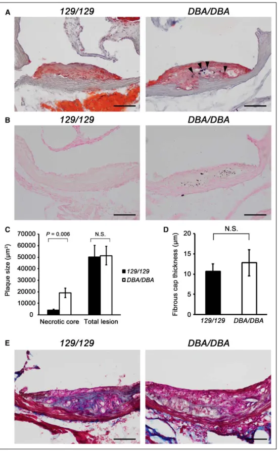
to intermediate stage plaques. Necrotic area was ≈5× larger in
Aath4aDBA/DBA than in control mice (Figure 2C). Fibrous cap
thickness was variable, and the difference was not statistically significant between the 2 groups (Figure 2D). However, col-lagen content in these lesions was less in the Aath4aDBA/DBA
mice, suggesting that resolution of inflammation associated with the plaque development is likely delayed in Aath4aDBA/DBA
(Figure 2E). General inflammation in these mice was low, judged by the low concentration of transforming growth factor-β1 and undetectable interleukin-10 in plasma. No sig-nificant differences in mRNA levels of interleukin-1β and transforming growth factor-β1 in the aorta were observed between control Aath4a129/129 and Aath4aDBA/DBA mice or in
plasma transforming growth factor-β1 levels (Figure IVA and IVB in the online-only Data Supplement), probably because these mice were fed with normal chow.
Together these results indicate that the DBA/2J allele of the 5′ region of Aath4 enhances atherosclerosis in both the aortic arch and the root. Increased cell death in the early stages of plaque development may be associated with this enhancement.
Reduced Mertk Expression in
Aath4aDBA/DBA Macrophages
The Aath4a QTL includes Mertk, which is important for efferocytosis, the phagocytotic removal of apoptotic cells.4
MERTK is localized on cell surface where it is proteolytically cleaved by ADAM metallopeptidase domain 17 (ADAM17) to produce soluble MER (sMER). sMER attenuates MERTK-triggered intracellular signaling by blocking bridging mol-ecules.14 In thioglycollate-elicited peritoneal macrophages
isolated from Aath4aDBA/DBA, Mertk mRNA levels were less
than 50% of that in Aath4a129/129 control macrophages (Figure
3A). In parallel with the mRNA expression, the amount of MERTK protein in cultured macrophages was also reduced in Aath4aDBA/DBA (Figure 3B), as well as cell-surface expression
of MERTK (Figure 3C).
sMER in the conditioned medium of Aath4aDBA/DBA
mac-rophage and plasma sMER in Aath4aDBA/DBA mice were also
reduced compared with those in controls, consistent with the reduction of protein amounts in macrophages (Figure 3B and 3D). MERTK protein was abundantly present within the ath-erosclerotic plaques of the Aath4a129/129 control mice,
border-ing the macrophage marker CD68-positive area, whereas the amount was significantly decreased in Aath4aDBA/DBA plaques
(Figure V in the online-only Data Supplement).
Reduced Phagocytosis in Aath4aDBA/DBA Macrophages MERTK is important for the normal execution of efferocytosis during atherosclerotic plaque development.12,13 We, therefore,
examined phagocytosis of apoptotic Jurkat T cells by perito-neal macrophages of Aath4aDBA/DBA mice and found that the
uptake of apoptotic cells was reduced to ≈40% of Aath4a129/129
controls (Figures 4A through 4C; Figure VI in the online-only Data Supplement).
Ghosn et al15 have reported that there are two types of
peri-toneal macrophages: large periperi-toneal macrophages (LPMs) and small peritoneal macrophages (SPMs).16 LPMs
predom-inate in the steady state peritoneal cavity but are decreased by inflammatory stimuli such as lipopolysaccharide and Table. Body Weights and Plasma Lipids in the Control and
Aath4aDBA/DBA Mice
Aath4a129/129 (n) Aath4aDBA/DBA (n)
Body weight, g
Male 28.59±0.37 (33) 28.08±0.42 (23) Female 21.30±0.38 (27) 21.37±0.37 (20) T-Chol, mg/dL
Male 680.9±23.0 (37) 738.2±29.2 (23) Female 572.3±27.5 (26) 482.2±31.3* (20) HDL-C, mg/dL
Male 79.4±5.0 (33) 77.1±9.5 (9) Female 45.1±3.7 (26) 60.2±4.2* (20) TG, mg/dL
Male 79±9 (33) 125±8† (23) Female 48±4 (25) 58±5 (19)
Data are shown as the mean±SE. HDL-C indicates high-density lipoprotein cholesterol; T-Chol, total cholesterol; and TG, triglyceride.
*P<0.05.
†P<0.01 vs Aath4a129/129.
Figure 2. Aath4aDBA/DBA mice develop plaques with increased calcium deposits, larger necrotic core, and less collagen content. A,
Repre-sentative early but raised plaques at the aortic root of control Aath4a129/129 (129/129) and Aath4aDBA/DBA (DBA/DBA) mice at 5 mo of age. The
plaques with similar sizes stained with SudanIVB and hematoxylin are shown. Aath4aDBA/DBA mouse (right) contains extensive calcium
depos-its (arrowheads). B,Calcium deposits detected by von Kossa stain. Numerous deposits were observed in the early plaques of Aath4aDBA/DBA
mice (right), whereas rarely seen in those of control Aath4a129/129 mice (left). Bar=100 μm. C,Necrotic core size in the plaques of Aath4a129/129
(129/129) and Aath4aDBA/DBA (DBA/DBA) mice at 5 mo old. Advanced lesions that are similar in total size were selected and necrotic area was
measured in the selected lesions (n=7). D, Fibrous cap thickness of advanced plaques. In the 7 advanced lesions selected in C, fibrous cap thickness was measured at 3 sites per lesion and averaged. E, Collagen content in the advanced lesions detected by trichrome staining. The
Aath4aDBA/DBA (DBA/DBA) lesion contains less collagen (blue). Bar=100 μm. N.S. indicates not significant.
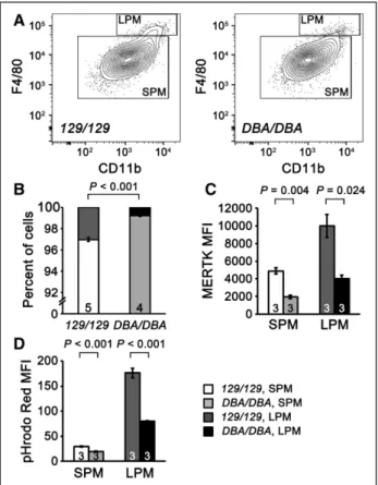
thioglycollate, which induce infiltration of SPMs into the peri-toneal cavity from myeloid progenitor cells in the bone mar-row. We found that 3 days after stimulation by thioglycollate, the percentage of LPM was significantly smaller in Aath4aDBA/ DBA than in control mice (0.8% versus 3.0% in Aath4aDBA/ DBA and control mice, respectively; P<0.001; Figure 5A and
5B). Both SPMs and LPMs express MERTK, although in Aath4aDBA/DBA macrophages, MERTK was markedly reduced in
both SPMs and LPMs (Figure 5C). Consistent with a previous report, phagocytosis was higher in LPMs than in SPMs,17 but
importantly, both LPMs and SPMs showed similarly reduced phagocytosis in Aath4aDBA/DBA macrophages (Figure 5D).
MERTK Amino Acid Differences Do Not Affect Efferocytosis
There are 9 amino acid substitutions between 129S6 and DBA/2J MERTK, 3 of which (W25G, T80E, and S479R) are predicted to potentially alter protein function (Figure 6A).4
The S479R substitution located adjacent to the ADAM17 cleavage site does not seem to have a significant effect on the shedding of MERTK because the proportion of sMER:MER was not altered between control and Aath4aDBA/DBA
macro-phages (Figure 3B). Likewise, when plasmids carrying Mertk cDNA of 129S6 or DBA/2J were transfected into HEK293T (human embryonic kidney 293T) cells, MERTK protein expression was similar, and comparable amounts of sMER were detected in the respective culture medium (Figure 6B). These results suggest that W25G, which is located close to the cleavage site of the secretion signal peptide, does not affect the protein maturation process.
We next examined the ability of the HEK293T cells expressing MERTK to phagocytose apoptotic Jurkat T cells. Overexpression of MERTK increased phagocytosis of apop-totic Jurkat T cells compared with the empty vector-trans-fected control cells, but no significant difference was observed between the 129S6 and the DBA/2J (empty vectors: 0.8±0.5%, 129S6-MERTK: 28.4±1.5%, and DBA/2J-MERTK: 29.5±1.5%; Figure 6C). Addition of protein S, which bridges apoptotic cells and MERTK, stimulated phagocytosis in all of them, but a genetic effect was not observed (empty vectors: 4.1±1.6%, 129S6-MERTK: 45.3±1.5%, and DBA/2J-MERTK: 48.7±2.7%; Figure 6C). Thus, the T80E substitution in the first immunoglobulin-like domain, which contains a ligand binding site, does not affect the binding to bridging protein S. These results indicate that the DBA/2J-specific amino acid substitu-tions in MERTK do not affect phagocytotic function.
Differences in Mertk Expression Levels Modulate Efferocytosis
Previous works have shown that mice lacking Mertk and Apoe (Mertk−/−Apoe−/−) have increased accumulation of
apop-totic cells and expanded necrotic cores in their plaques when compared with Mertk+/+Apoe−/− mice,12 and that peritoneal
macrophages from Mertk-deficient mice (Mertk−/−)
phagocy-tose apoptotic cells at a much reduced level (<20% compared with Mertk+/+ macrophages).6 This raises the question whether
differences in the level of expression of MERTK affect effe-rocytosis in a graded manner. To test this, we examined phago-cytosis of apoptotic Jurkat T cells by peritoneal macrophages isolated from wild-type (Mertk+/+) and Mertk heterozygous Figure 3. Expression of MERTK is decreased in Aath4aDBA/DBA. A, Mertk mRNA expression in thioglycollate-elicited peritoneal macrophages
from control Aath4a129/129 (129/129) and Aath4aDBA/DBA (DBA/DBA) mice measured by quantitative RT-PCR (n=4). B, MERTK protein
expres-sion in cultured peritoneal macrophages and soluble form of MERTK (sMER) in their medium detected by western blotting (left). Signal intensity ratios of sMER in the conditioned medium and cell-associated MERTK (right). C, Cell-surface expression of MERTK in perito-neal macrophages analyzed by flow cytometry. An overlay of representative histograms showing MERTK-positive cells is displayed (left). Median fluorescence intensity (MFI) of MERTK in CD11b-positive macrophages (right). D,sMER in the plasma detected by Western blot-ting. Equal amounts of plasma (2.5 μL per lane) were loaded in each lane. Data are shown as the mean±SE. Numbers in the bar diagrams indicate the number of animals analyzed. N.S. indicates not significant; and RT-PCR, reverse transcription-polymerase chain reaction.
(Mertk+/−) mice. We found that Mertk heterozygous
macro-phages, which are supposed to express 50% of MERTK, show
≈70% phagocytosis compared with wild-type macrophages (P=0.002; Figure 6D), indicating that the amount of MERTK modulates efficiency of phagocytosis.
In agreement, when the amount of Mertk-plasmid trans-fected to HEK293 cells was gradually decreased, cell sur-face 129-MERTK and DBA-MERTK and phagocytosis were equally reduced (Figure 6E). We conclude that the amount of MERTK determines the efficiency of phagocytosis in a dose-dependent, not an all-or-none manner, and that decreased Mertk expression in Aath4aDBA/DBA macrophages is the cause
of the reduced phagocytosis.
Transcription Activity of the Proximal Promoter Region of Mertk
There are a large number of nucleotide polymorphisms throughout the Mertk gene, which could affect steady state levels of mRNA by affecting its stability. Accordingly, to clar-ify the cause of reduced Mertk mRNA in Aath4aDBA/DBA
mac-rophages, we asked whether the degradation of Mertk mRNA is enhanced in Aath4aDBA/DBA macrophages. We found that the
estimated half-life of Mertk mRNA was 5.6 hours in the control Aath4a129/129 macrophages and 5.7 hours in the Aath4aDBA/DBA
macrophages, suggesting that the lower amount of Mertk mRNA in Aath4aDBA/DBA macrophages is likely caused
by reduced Mertk transcription, rather than by faster Figure 4. Efferocytosis is reduced in Aath4aDBA/DBA macrophages.
A, Phagocytosis of apoptotic Jurkat T cells by peritoneal mac-rophages. Apoptotic Jurkat T cells labeled with pHrodo Red were added to macrophages isolated from control Aath4a129/129
(129/129) and Aath4aDBA/DBA (DBA/DBA) mice and incubated for 1
h at 37°C. Apoptosis of Jurkat T cells was confirmed as shown in Figure IIA in the online-only Data Supplement. B, Representative flow cytometry panels showing phagocytosis of apoptotic Jur-kat T cells by peritoneal macrophages from control Aath4a129/129
(129/129) and Aath4aDBA/DBA (DBA/DBA) mice. Detailed gating
strategy is shown in Figure IIB in the online-only Data Supple-ment. Apoptotic Jurkat T cells and macrophages were labeled with pHrodo Red and anti–CD11b-FITC, respectively. Gates indicate pHrodo Red–positive macrophages phagocytosing apoptotic cells. C, An overlay of representative histograms show-ing pHrodo Red–positive phagocytosshow-ing cells in the CD11b-positive macrophages (left). Phagocytosis assessed by the median fluorescent intensity (MFI) of pHrodo Red (right, n=4). Data are shown as the mean±SE. FITC indicates fluorescein isothiocyanate.
Figure 5. Small peritoneal macrophages (SPMs) and large perito-neal macrophages (LPMs) in Aath4aDBA/DBA mice. A,
Representa-tive flow cytometry panels showing SPM and LPM populations in control Aath4a129/129 (left) and Aath4aDBA/DBA (right) mice. SPMs are
defined as a F4/80lowCD11blow and LPMs as a F4/80highCD11bhigh
population. B, Percentage of SPMs and LPMs in control
Aath4a129/129 (129/129) and Aath4aDBA/DBA (DBA/DBA)
macro-phages. Data are representative of 5 independent experiments each using 4 to 5 mice of each genotype. C, Cell-surface expres-sion of MERTK on SPMs and LPMs measured by flow cytometry (n=3). D, Phagocytosis in SPMs and LPMs assessed by MFI of pHrodo Red (n=3). Data are shown as the mean±SE.
degradation of Mertk mRNA (Figure VII in the online-only Data Supplement).
We next searched for DBA/2J-specific differences in the promoter region of the Mertk gene that could affect the efficiency of transcription. Within the 1.1 kb immediately upstream of the Mertk transcription start site, there are 10 SNPs and 1 deletion that are unique to DBA compared with B6 and 129 sequences (Figure VIIIA in the online-only Data Supplement). The Mertk gene has a CpG island promoter and does not contain a typical TATA box or CAAT box.
To test whether the sequence differences in the proxi-mal promoter region of Mertk affect the transcription level, we compared promoter activity of 129S6 and DBA/2J by a luciferase reporter assay. Mertk promoters of both 129S6 and DBA/2J enhanced luciferase expression in HEK293T cells ≈5× compared with empty vector, but the transcription
activities of the Mertk promoter from 129S6 and DBA/2J were not significantly different (Figure VIIIB in the online-only Data Supplement). Furthermore, addition of factors that are thought to regulate Mertk expression18 including
granu-locyte-macrophage colony-stimulating factor, macrophage colony-stimulating factor, and interleukin-4 did not signifi-cantly change the promoter activities (Figure VIIIC in the online-only Data Supplement). Our experiments suggest that the genomic variants affecting the expression of Mertk must reside outside of this proximal 1.1 kb promoter region.
Indeed, when we searched for eQTLs associated with the expression levels of Mertk from Hybrid Mouse Diversity Panel by UCLA (https://systems.genetics.ucla.edu/data/hmdp),19
more than 15 SNPs within the Aath4a interval were signifi-cantly associated with Mertk expression levels (Table I in the online-only Data Supplement). The SNPs that are shared by Figure 6. Phagocytosis of apoptotic cells is not affected by amino acid substitutions in DBA MERTK, but is modulated by the amount of MERTK. A, Schematic structure of mouse MERTK indicating the 9 amino acids, which differ between 129S6 and DBA/2J proteins. Sub-stitutions are shown as 129S6-amino acid position-DBA/2J. B, Expression of MERTK in HEK293T (human embryonic kidney 293T) cells and sMER in the conditioned medium detected by Western blotting. C, Phagocytosis of apoptotic Jurkat T cells by Mertk-transfected HEK293T cells with or without 25 nmol/L of protein S (n=3). D, Phagocytosis of apoptotic Jurkat T cells by peritoneal macrophages iso-lated from Mertk+/+ and Mertk+/− mice (n=4). Data are shown as the mean±SE. E, Correlation between the MERTK levels and phagocytosis
in HEK293T cells transfected with varying amounts of Mertk cDNA. 129 indicates 129S6-Mertk transfected cells; DBA, DBA/2J-Mertk transfected cells; EV, empty vector control; FN-III, fibronectin type-III domains; Ig-like, immunoglobulin-like domains; N.S., not significant; TK, tyrosine kinase domain; TM, transmembrane domain; and UT, untransfected cells.
129 and B6 but unique in DBA, and located between 63 kb and 2.6 Mb upstream of the Mertk gene, seem to regulate its expression in the liver and adipose tissue, whereas SNPs located between 2.3 and 2.5 Mb downstream were broadly associated with the expression levels in macrophages (Table I in the online-only Data Supplement). Two SNPs at −325 and −202 kb upstream also showed strong association with the Mertk expression in macrophages, but 129 and DBA share the same variants at these positions. Together, it is likely that distant genetic variants affect the expression of Mertk.
Discussion
Each QTL identified in the crosses of inbred mice generally spans a large genomic distance, sometimes almost an entire chromosome. In complex phenotypes such as atherosclerosis, where a large number of genes are involved, transferring a tar-get region onto an inbred background and creating congenic line is a powerful step toward identifying causative genes. Here we have analyzed the effect of the atherosclerosis QTL Aath4 by establishing a congenic line (Aath4aDBA/DBA), where the 5′
region of DBA Aath4 was backcrossed onto a 129S6-Apoe−/−
background. As expected, the resulting Aath4aDBA/DBA males had
significantly larger plaques, and macrophages isolated from these mice exhibited reduced efferocytosis as a consequence of allele-specific decrease in MERTK expression. Together, our results provide strong evidence that the increased susceptibil-ity to atherosclerosis determined by the DBA allele of Aath4 is, at least in part, due to decreased MERTK expression.
MERTK is known to play a significant role in efferocytosis and the resolution of inflammation during atherosclerosis.12,13 In
this report, we have demonstrated that 9 nonsynonymous SNPs in Mertk, which are uniquely different in DBA/2J compared with C57BL/6J and 129S6, are not responsible for the reduced effero-cytosis observed in peritoneal macrophages from Aath4aDBA/DBA.
Notably, our experiments demonstrated that the level of MERTK expression controls phagocytosis in Mertk+/− and Aath4aDBA/ DBA macrophages in a dose-dependent, not an all-or-none
man-ner. This is consistent with many published reports that disease-related SNPs identified by GWAS (genome-wide association study) are usually located in introns or intergene regions rather than in coding sequences. Unlike diseases in which mutations in a single gene cause a drastic phenotype, the pathogenesis of athero-sclerosis is more complex and involves numerous factors. SNPs or other alterations in regulatory regions typically lead to small changes in gene expression, which cumulatively influence suscep-tibility to disease. Our experiments show that Mertk expression is likely reduced at the transcription level because the stability of Mertk mRNA was unchanged in the Aath4aDBA/DBA macrophages.
Furthermore, our reporter assay tests show that the genetic differ-ences that cause different Mertk expression in DBA/2J and 129S6 are likely to be outside the proximal 1.1 kb promoter region, and search of eQTL database suggests that the expression of Mertk is affected by distant genetic variants. In humans, an SNP rs869016 in intron 1 of MERTK is associated with decreased risk of carotid atherosclerosis, although it is unknown whether the SNP modu-lates Mertk mRNA expression.20 Further investigation of the
caus-ative variants is clearly required.
Efferocytosis, the phagocytosis of apoptotic cells by macrophages, is critical in preventing progression of
athero-sclerosis.21 Consistently, it has been shown that Mertk−/−Apoe−/−
mice fed a Western-type diet have increased numbers of apop-totic cells and expanded necrosis in advanced lesions.12 In
early plaques, removal of apoptotic cells is normally efficient; although, we have observed signs of basophilic (dystrophic) calcium deposits even in the early raised plaques of Aath4aDBA/ DBA mice. Our attempt to detect apoptotic cells in vivo in plaques
by TUNEL assay was not productive to make a comparison because TUNEL-positive nuclei were few per section in both Aath4a129/129 and Aath4aDBA/DBA. This is because mice were fed
with normal chow to eliminate additional effects caused by high-fat diet, and most of the lesions we observed were at their early stages. However, the calcium deposits are likely the rem-nants of dead/dying cells via apoptotic and necrotic processes, giving strong evidence that the normal process of removing dead cells is restricted in the Aath4aDBA/DBA mice. Delayed
apoptotic cell clearance is expected to cause acceleration of plaque development and necrotic core formation. The cal-cium deposits in the early plaques, however, seem to be short-lived because larger calcified areas in advanced Aath4aDBA/DBA
plaques are not different from the control plaques and are seen mostly deeper in the intima near the internal elastic lamina. Although the thickness of fibrous caps was not significantly different, the Aath4aDBA/DBA plaques contained less interstitial
matrix proteins than similar-sized plaques in the control mice. In advanced plaques, efferocytosis by macrophages becomes less effective than in early plaques, but an alteration in the balance between synthesis and degradation of matrix protein would be expected to contribute to vulnerable plaque mor-phology. Although we have not encountered any premature deaths associated with the Aath4aDBA/DBA mice, a more detailed
examination of later stage plaques would be worthwhile. Despite the well-known heterogeneity of mouse perito-neal macrophages,15,16 we found that MERTK is expressed in
both LPMs and SPMs, and that it is similarly reduced in both populations of Aath4aDBA/DBA macrophages. We also observed
that the LPMs, which have higher phagocytic activity than SPMs,17 were consistently fewer in Aath4aDBA/DBA than in
con-trols. Fewer LPMs and more SPMs could be another factor contributing to the reduced phagocytosis in Aath4aDBA/DBA
peritoneal macrophages. SPMs are induced from circulat-ing monocytes by inflammatory stimuli, whereas LPMs are thought to be tissue-resident macrophages, maintained locally by differentiation from precursor cells. Macrophages in atherosclerotic plaques are similarly heterogeneous,22,23
and the role of circulating monocyte-derived macrophages in the development of plaques has been well-documented. Evidence is also beginning to accumulate that tissue-resident macrophages originating from the adventitial precursor cells contribute to atherogenesis. Factors that control the relative balance of these populations are not known, but a shift in the population of the Aath4aDBA/DBA macrophages in their plaques
is conceivable. Whether MERTK expression directly affects this shift or not requires further study.
Although we identified Aath4 as a QTL specific to the arch lesion, the Aath4aDBA/DBA mice have significantly larger plaques
in both the aortic arch and the root. This is in agreement with our prediction that the reduced efferocytosis of Aath4aDBA/DBA
macrophages should equally affect both the aortic arch and the
root. Our previous studies of F2 population between 129S6- Apoe−/− and C57BL/6-Apoe−/− indicated that 129S6 genome
carries the sequence that affects the aortic arch geometry, which are also associated with increased aortic arch atherosclerosis.2
Therefore, the variant in Mertk expression is a risk factor that is additional to the 129S6 sequences that determine the aor-tic arch development because the Aath4aDBA/DBA strain is based
upon the129S6 genome. Moreover, the contribution of Mertk expression to the 50% increase in root lesion is, although sig-nificant, relatively small, considering that the parental DBA/2J show 10× larger plaques in the root than 129S6 mice.
The genomic region of DBA/2J carried in the Aath4aDBA/DBA
mice is large, and the effect of DNA variants in other loca-tions in this region must be considered. For example, Siglec1 at 131 Mb, which encodes sialoadhesin (CD169), is involved in the retention of hematopoietic stem cells in the bone mar-row niche.24 It is also expressed in a subset of
tissue-resi-dent macrophages, preventing excessive inflammation upon injury.25 Similarly, CD169 may also play a role in
phagocy-tosis.26,27 Altered expression of Bcl2l11 at 128 Mb, encoding
for a proapoptotic protein Bim, could affect early cell death in the Aath4aDBA/DBA plaques. In addition, as the backcrossed
region in the Aath4aDBA/DBA line is the proximal half of Aath4,
the distal half may include genes that are proatherogenic only in the arch, or are athero-suppressive specifically in the root. One of the candidates in distal Aath4 is Cd93 at 148 Mb, a transmembrane glycoprotein which is expressed in endothe-lial and myeloid cells. CD93-deficient mice show a defect in the clearance of apoptotic cells in vivo,28 and an SNP in Cd93
gene is associated with an increased risk of coronary heart disease.29 Analysis of a congenic line carrying the distal half
of Aath4 of DBA/2J is currently under way.
We also note that the plaque size difference was observed only in Aath4aDBA/DBA males, whereas the male/female
dif-ferences in plaque size were not seen in the parental strains: DBA/2J-Apoe−/− mice show larger root lesions compared
with 129S6-Apoe−/− in both males and females3; arch lesion
size was comparable between the 2 strains in both males and females.4 The QTLs on Chr 2 were detected in both males
and females in the F2 population from DBA/2J-Apoe−/− and
129S6-Apoe−/− mice.3,4 Because no QTL was detected on Chr
2 for plasma lipids in the intercross between the 2 strains, the plasma lipid differences between males and females are not likely the strong determinant of the plaque size differences.3
The major determinants for the plaque sizes in females must, therefore, lie outside of the Aath4a interval. The sex dimor-phism in the Aath4aDBA/DBA mice requires further investigation.
In summary, our experiments have shown that the DBA allele of the 5′ region of Aath4 causes inefficient efferocy-tosis via lower expression of MERTK, and that difference contributes to the enhanced plaque development observed in Aath4aDBA/DBA mice. Because the effects of reduced MERTK
and efferocytosis are seen in the both aortic arch and root areas, further studies will be required to fully understand the factors that cause location specificity in atherosclerosis.
Acknowledgments
We thank Dr Glenn Matsushima and Akhil Patel for Mertk+/- mice; Dr
Hyung-Suk Kim for quantitative RT-PCR; Longquan Xu, Svetlana
Zhilicheva, Sylvia Hiller, and Jennifer Wilder for technical assis-tance; and the UNC CGIBD Histology Core, Microscopy Services Laboratory, and Flow Cytometry Core Facilities for technical support. We also thank Drs Marlon Lawrence, Glenn Matsushima, Jonathon Homeister, and Oliver Smithies for critical reading of the article; and Dr Robert Reddick at the University of Texas Health Science Center at San Antonio for discussions on evaluating atherosclerotic plaques.
Sources of Funding
This research was supported by a National Institutes of Health grant HL042630. The UNC Flow Cytometry Core Facility is supported, in part, by P30 CA016086 Cancer Center Core Support Grant to the UNC Lineberger Comprehensive Cancer Center.
Disclosures
None.
References
1. Maeda N, Johnson L, Kim S, Hagaman J, Friedman M, Reddick R. Anatomical differences and atherosclerosis in apolipoprotein E-deficient mice with 129/SvEv and C57BL/6 genetic backgrounds. Atherosclerosis. 2007;195:75–82. doi: 10.1016/j.atherosclerosis.2006.12.006.
2. Tomita H, Zhilicheva S, Kim S, Maeda N. Aortic arch curvature and ath-erosclerosis have overlapping quantitative trait loci in a cross between 129S6/SvEvTac and C57BL/6J apolipoprotein E-null mice. Circ Res. 2010;106:1052–1060. doi: 10.1161/CIRCRESAHA.109.207175. 3. Kayashima Y, Tomita H, Zhilicheva S, Kim S, Kim HS, Bennett BJ, Maeda
N. Quantitative trait loci affecting atherosclerosis at the aortic root iden-tified in an intercross between DBA2J and 129S6 apolipoprotein E-null mice. PLoS One. 2014;9:e88274. doi: 10.1371/journal.pone.0088274. 4. Kayashima Y, Makhanova NA, Matsuki K, Tomita H, Bennett BJ, Maeda
N. Identification of aortic arch-specific quantitative trait loci for athero-sclerosis by an intercross of DBA/2J and 129S6 apolipoprotein E-deficient mice. PLoS One. 2015;10:e0117478. doi: 10.1371/journal.pone.0117478. 5. Graham DK, Dawson TL, Mullaney DL, Snodgrass HR, Earp HS. Cloning
and mRNA expression analysis of a novel human protooncogene, c-mer.
Cell Growth Differ. 1994;5:647–657.
6. Scott RS, McMahon EJ, Pop SM, Reap EA, Caricchio R, Cohen PL, Earp HS, Matsushima GK. Phagocytosis and clearance of apoptotic cells is mediated by MER. Nature. 2001;411:207–211. doi: 10.1038/35075603. 7. Seitz HM, Camenisch TD, Lemke G, Earp HS, Matsushima GK.
Macrophages and dendritic cells use different Axl/Mertk/Tyro3 receptors in clearance of apoptotic cells. J Immunol. 2007;178:5635–5642. 8. Paul SR, Merberg D, Finnerty H, Morris GE, Morris JC, Jones SS, Kriz R,
Turner KJ, Wood CR. Molecular cloning of the cDNA encoding a recep-tor tyrosine kinase-related molecule with a catalytic region homologous to c-met. Int J Cell Cloning. 1992;10:309–314. doi: 10.1002/stem.5530100509. 9. Graham DK, Bowman GW, Dawson TL, Stanford WL, Earp HS,
Snodgrass HR. Cloning and developmental expression analysis of the murine c-mer tyrosine kinase. Oncogene. 1995;10:2349–2359.
10. Uehara H, Shacter E. Auto-oxidation and oligomerization of protein S on the apoptotic cell surface is required for Mer tyrosine kinase-mediated phagocytosis of apoptotic cells. J Immunol. 2008;180:2522–2530. 11. Duncan JL, LaVail MM, Yasumura D, Matthes MT, Yang H, Trautmann N,
Chappelow AV, Feng W, Earp HS, Matsushima GK, Vollrath D. An RCS-like retinal dystrophy phenotype in mer knockout mice. Invest Ophthalmol
Vis Sci. 2003;44:826–838.
12. Thorp E, Cui D, Schrijvers DM, Kuriakose G, Tabas I. Mertk receptor mutation reduces efferocytosis efficiency and promotes apoptotic cell accumulation and plaque necrosis in atherosclerotic lesions of apoe-/- mice. Arterioscler Thromb
Vasc Biol. 2008;28:1421–1428. doi: 10.1161/ATVBAHA.108.167197. 13. Ait-Oufella H, Pouresmail V, Simon T, Blanc-Brude O, Kinugawa K,
Merval R, Offenstadt G, Lesèche G, Cohen PL, Tedgui A, Mallat Z. Defective mer receptor tyrosine kinase signaling in bone marrow cells promotes apoptotic cell accumulation and accelerates atherosclero-sis. Arterioscler Thromb Vasc Biol. 2008;28:1429–1431. doi: 10.1161/ ATVBAHA.108.169078.
14. Thorp E, Vaisar T, Subramanian M, Mautner L, Blobel C, Tabas I. Shedding of the Mer tyrosine kinase receptor is mediated by ADAM17 protein through a pathway involving reactive oxygen species, protein kinase Cδ, and p38 mitogen-activated protein kinase (MAPK). J Biol
Chem. 2011;286:33335–33344. doi: 10.1074/jbc.M111.263020.
15. Ghosn EE, Cassado AA, Govoni GR, Fukuhara T, Yang Y, Monack DM, Bortoluci KR, Almeida SR, Herzenberg LA, Herzenberg LA. Two physi-cally, functionally, and developmentally distinct peritoneal macrophage subsets. Proc Natl Acad Sci U S A. 2010;107:2568–2573. doi: 10.1073/ pnas.0915000107.
16. Cassado Ados A, D’Império Lima MR, Bortoluci KR. Revisiting mouse peritoneal macrophages: heterogeneity, development, and function. Front
Immunol. 2015;6:225. doi: 10.3389/fimmu.2015.00225.
17. Cain DW, O’Koren EG, Kan MJ, Womble M, Sempowski GD, Hopper K, Gunn MD, Kelsoe G. Identification of a tissue-specific, C/EBPβ -dependent pathway of differentiation for murine peritoneal macrophages.
J Immunol. 2013;191:4665–4675. doi: 10.4049/jimmunol.1300581. 18. Zizzo G, Hilliard BA, Monestier M, Cohen PL. Efficient clearance of
early apoptotic cells by human macrophages requires M2c polarization and MerTK induction. J Immunol. 2012;189:3508–3520. doi: 10.4049/ jimmunol.1200662.
19. Bennett BJ, Farber CR, Orozco L, et al. A high-resolution association mapping panel for the dissection of complex traits in mice. Genome Res. 2010;20:281–290. doi: 10.1101/gr.099234.109.
20. Hurtado B, Abasolo N, Muñoz X, García N, Benavente Y, Rubio F, García de Frutos P, Krupinski J, Sala N. Association study between polymor-phims in GAS6-TAM genes and carotid atherosclerosis. Thromb Haemost. 2010;104:592–598. doi: 10.1160/TH09-11-0787.
21. Tabas I. Consequences and therapeutic implications of macrophage apoptosis in atherosclerosis: the importance of lesion stage and phago-cytic efficiency. Arterioscler Thromb Vasc Biol. 2005;25:2255–2264. doi: 10.1161/01.ATV.0000184783.04864.9f.
22. Moore KJ, Sheedy FJ, Fisher EA. Macrophages in atherosclerosis: a dynamic balance. Nat Rev Immunol. 2013;13:709–721. doi: 10.1038/ nri3520.
23. Peled M, Fisher EA. Dynamic aspects of macrophage polarization during atherosclerosis progression and regression. Front Immunol. 2014;5:579. doi: 10.3389/fimmu.2014.00579.
24. Chow A, Lucas D, Hidalgo A, Méndez-Ferrer S, Hashimoto D, Scheiermann C, Battista M, Leboeuf M, Prophete C, van Rooijen N, Tanaka M, Merad M, Frenette PS. Bone marrow CD169+ macrophages promote the retention of hematopoietic stem and progenitor cells in the mesenchymal stem cell niche. J Exp Med. 2011;208:261–271. doi: 10.1084/jem.20101688. 25. O’Neill AS, van den Berg TK, Mullen GE. Sialoadhesin - a
macrophage-restricted marker of immunoregulation and inflammation. Immunology. 2013;138:198–207. doi: 10.1111/imm.12042.
26. Guo M, Härtlova A, Dill BD, Prescott AR, Gierliński M, Trost M. High-resolution quantitative proteome analysis reveals substantial differences between phagosomes of RAW 264.7 and bone marrow derived macro-phages. Proteomics. 2015;15:3169–3174. doi: 10.1002/pmic.201400431. 27. Xiong YS, Yu J, Li C, Zhu L, Wu LJ, Zhong RQ. The role of Siglec-1
and SR-BI interaction in the phagocytosis of oxidized low density lipo-protein by macrophages. PLoS One. 2013;8:e58831. doi: 10.1371/journal. pone.0058831.
28. Norsworthy PJ, Fossati-Jimack L, Cortes-Hernandez J, Taylor PR, Bygrave AE, Thompson RD, Nourshargh S, Walport MJ, Botto M. Murine CD93 (C1qRp) contributes to the removal of apoptotic cells in vivo but is not required for C1q-mediated enhancement of phagocytosis. J Immunol. 2004;172:3406–3414.
29. van der Net JB, Oosterveer DM, Versmissen J, Defesche JC, Yazdanpanah M, Aouizerat BE, Steyerberg EW, Malloy MJ, Pullinger CR, Kastelein JJ, Kane JP, Sijbrands EJ. Replication study of 10 genetic polymorphisms associated with coronary heart disease in a specific high-risk population with familial hypercholesterolemia. Eur Heart J. 2008;29:2195–2201. doi: 10.1093/eurheartj/ehn303.
Highlights
• Aath4aDBA/DBA, a congenic line of an atherosclerosis QTL Aath4 on distal Chr 2, was generated by transferring DBA/2J alleles of Aath4 to the129S6-Apoe−/− strain.
• Aath4aDBA/DBA males develop larger plaques and peritoneal macrophages isolated from Aath4aDBA/DBA showed reduced phagocytosis of apoptotic cells.
• Lower Mertk transcription, rather than DBA/2J-specific amino acid substitutions, causes restricted efferocytosis in Aath4aDBA/DBA.