ISSN(Online): 2320-9801
ISSN (Print): 2320-9798
I
nternational
J
ournal of
I
nnovative
R
esearch in
C
omputer
and
C
ommunication
E
ngineering
(An ISO 3297: 2007 Certified Organization)
Vol. 3, Issue 7, July 2015
Medical Image Edge Detection Based on Soft
Computing Approach
J.Mehena1, M. C. Adhikary2
Research Scholar, Fakir Mohan University, Balasore, Odisha, India1 Professor, Fakir Mohan University, Balasore, Odisha, India2
ABSTRACT: Edge detection is an important pre-processing step in image analysis and computer vision. Medical image edge detection is an important work for object recognition of the human organs. Edge detection of medical images is one of the most important applications for image segmentation. It refers to the process of identifying and locating sharp discontinuities in medical images. In this paper, a soft computing approach based on fuzzy logic is introduced to detect the edges for noisy images. Here, an image is considered as a fuzzy set and pixels are taken as elements of fuzzy set. The performance of the proposed edge detector is evaluated on different medical images and compared with popular edge detection algorithm. From the experimental results it is clear that the proposed approach has better performance than those of other competing edge detection algorithm for noisy medical images.
KEYWORDS: Medical image,edge detection, image segmentation, soft computing, fuzzy logic. I. INTRODUCTION
Image segmentation is one of the most difficult tasks in image analysis and computer vision [1]. Edge detection is a discontinuity based approach used for image segmentation. Medical image edge detection is an important work for object recognition of the human organs, and it is an essential pre-processing step in medical image segmentation. The work of the edge detection decides the result of the final processed image. Classical methods of edge detection involve convolving the image with an operator (a 2-D filter), which is constructed to be sensitive to large gradients in the image while returning values of zero in uniform regions [2]. There are an extremely large number of edge detection operators available, each designed to be sensitive to certain types of edges. Variables involved in the selection of an edge detection operator include edge orientation, noise environment and edge structure. There are two main types of edge detector: one is the first derivative- based edge detection operator to detect image edges by computing the image gradient values, such as Roberts, Sobel and Prewitt operator; the other one is the second derivative –based edge detection operator, by seeking in the second derivative zero-crossing to edge detection such as LOG and Canny operator [3].
Conventionally, edge is detected according to the first order derivative like Sobel, Prewitt and Laplacian of Gaussian operator, but in theory they belong to the high pass filtering, which are not fit for noise medical image edge detection because noise and edge belong to the scope of high frequency. In real world applications, medical images contain object boundaries and object shadows and noise. Therefore, they may be difficult to distinguish the exact edge from noisy medical images. These detectors are simple to implement but they are usually inaccurate and highly sensitive to noise. The second order derivative operator has fixed detection characteristics in all directions but they are very sensitive to noise too. The Gaussian edge detectorsreduce the undesirable negative effects of noise by smoothing the image before performing edge detection. Hence, they exhibit much superior performance over other operators especially in noisy conditions. The Canny detector [4], which is a Gaussian edge detector, is one of the most popular edge detectors and it has been widely used in many applications. Although the Gaussian detectors exhibit relatively better performance, they are computationally much more complex than classical derivative based edge detectors. Furthermore, their performances quickly decrease as the density of the corrupting noise increases.
ISSN(Online): 2320-9801
ISSN (Print): 2320-9798
I
nternational
J
ournal of
I
nnovative
R
esearch in
C
omputer
and
C
ommunication
E
ngineering
(An ISO 3297: 2007 Certified Organization)
Vol. 3, Issue 7, July 2015
and morphological transformations which derived from addition and subtraction are defined to extract features in images. As the performance of classic edge detectors degrades with noise, morphological edge detector has been studied. It is a better method for edge information detecting and noise filtering than differential operation, which is sensitive to noise. This method is a better compromise between noise smoothing and edge orientation [7], but the computation is more complex than general edge detection algorithms. Therefore, a novel edge detector that is capable of extracting edges from medical images corrupted by noise is highly desirable.
Soft computing can deal with the ambiguity and uncertainty in image processing in better way as compared to the traditional approaches. When soft computing is used for edge detection gives better result compare to the classical approach [8]. The classical techniques like Sobel, Prewitt, Roberts, Canny edge detector have limitations of using fixed value of parameters or threshold. While the nature of edges is not constant due to which few edges left by being detected. Fuzzy logic a branch of soft computing provides us flexibility by allowing the values without any such restrictions. Fuzzy logic has IF-THEN rules and has simple structure to implement [9-12]. Simply by changing or adding few more fuzzy rules result can be changed and some techniques are knowledge based in which training is required. Fuzzy logic is a logical system which is an extension of multi valued logic. It is a propositional calculus in which there are more than two truth values. By using fuzzy techniques edges having different thickness can also be detected. Fuzzy logic is conceptually easy to understand and is flexible and is tolerant of imprecise data. Fuzzy logic is to map an input space to an output space and for doing this a list of if then statements called rules are evaluated in parallel. These Rules are useful because they use variables and adjectives that describe those variables. In this paper, a soft computing approach based on fuzzy logic is introduced to detect the edges for noisy medical images. Results show that the proposed algorithm works well in noise condition and detect more edges as compared to the traditional approaches.
The rest of this research paper is organized as follows. In Section II, morphological approach is described in details. Section III describes the proposed approach for medical image edge detection. Experimental results and comparison with existing edge detection methods are presented in Section IV. Finally, conclusions followed by references come in Section V.
II. MORPHOLOGICAL APPROACH
Mathematical morphology [5] commonly refers to a broad set of image processing operations that process images based on shapes. Morphological operations selects appropriate structuring element of the processed image and makes use of the basic theory of morphology including erosion, dilation, opening and closing operation and the synthesization operations of them to get clear medical image edge. In the process, the synthesized modes of the operations and the feature of structuring element decide the result of the processed image. Detailed saying, the synthesized mode of the operation reflects the relation between the processed image and origin image, and the selection of structuring element decides the effect and precision and the result. Therefore, the keys of morphological operations can be generalized for the design of morphological filter structure and the selection of structuring element [6]. We must select appropriate structuring element by texture features of the image and the size, shape and direction of structuring element must been considered roundly. Usually, except for special demand, we select structuring element by 3 × 3 square. By the operation features of morphology, erosion and dilation operations satisfy:
𝐹 ⊝ 𝐵 ⊆ 𝐹 ⊆ 𝐹⨁𝐵 1
Where, 𝐹 denote a gray scale medical image,𝐵 denote structuring element. Opening and closing operations satisfy:
𝐹 ∘ 𝐵 ⊆ 𝐹 ⊆ 𝐹 ∙ 𝐵 (2) The edge of medical image 𝐹, which is denoted by 𝐸𝑑(𝐹),is defined as the difference set of the dilation domain of 𝐹
and the domain of 𝐹. This is also known as dilation residue edge detector:
ISSN(Online): 2320-9801
ISSN (Print): 2320-9798
I
nternational
J
ournal of
I
nnovative
R
esearch in
C
omputer
and
C
ommunication
E
ngineering
(An ISO 3297: 2007 Certified Organization)
Vol. 3, Issue 7, July 2015
Accordingly, the edge of medical image 𝐹, which is denoted by 𝐸𝑒(𝐹),can also be defined as the difference set of the
domain of 𝐹 and the erosion domain of 𝐹.This is also known as erosion residue edge detector:
𝐸𝑒 𝐹 = 𝐹 − 𝐹 ⊝ 𝐵 (4)
The dilation and erosion often are used to compute the morphological gradient of the medical image F, denoted by
𝐺(𝐹):
𝐺 𝐹 = 𝐹⨁𝐵 − 𝐹 ⊝ 𝐵 (5)
The morphological gradient highlights sharp gray-level transition in the medical image. The opening top-hat transformation of medical image 𝐹, which is denoted by 𝑇𝐻𝑜(𝐹), is defined as the difference set of the domain of F
and the opening domain of 𝐹.It is defined as
𝑇𝐻𝑜 𝐹 = 𝐹 − 𝐹 ∘ 𝐵 (6)
Similarly, the closing top-hat transformation of medical image 𝐹, which is denoted by 𝑇𝐻𝑐(𝐹),can also be defined as
the difference set of the closing domain of 𝐹and the domain of 𝐹, it is defined as
𝑇𝐻𝑐 𝐹 = 𝐹 ⋅ 𝐵 − 𝐹 (7)
The top-hat transformation, which owes its original name to the use of a cylindrical structuring element function with a flat top, is useful for enhancing detail in the presence of shading. The effect of erosion and dilation operations is better for medical image edge by performing the difference between processed image and original image, but they are worse for noise filtering. As opposed to erosion and dilation, opening and closing operations are better for filtering. But because they utilize the complimentarily of erosion and dilation, the result of processed image is only correlative with the convexity and concavity of the image edge. Accordingly what we get is only the convex and concave features of the image by performing the difference between processed image and original image, but not all the features of image edge.
Mathematical morphological approach is proposed here for finding the edges of the medical image. Opening-closing operation is firstly used as preprocessing to filter noise. Then smooth the image by first closing and then dilation. The perfect medical image edge will be got by performing the difference between the processed image by above process and the image before dilation, the following is the morphological approach [7]:
𝑀 ⋅ 𝐵 ⨁ 𝐵 − 𝑀 ∙ 𝐵 (8) Where, 𝑀 = 𝐹 ∙ 𝐵 ∘ 𝐵 (9)
III. PROPOSED APPROACH
The proposed approach uses fuzzy IF- THEN rules for the detection of edges in medical MR images. As we know, edges in images are constituted significant gray level changing. In the other words, the edges are high frequency components of an image. That is to say a high-pass filter can detect edges in an image. But working on frequency domain can load the task with additional complexity. So commonly edge detection techniques based on spatial domain working. On the spatial domain, an edge can be classically detected via first or second order derivative. Because of second order derivative has high sensitivity towards noises, first order derivative has been used for edge detection. Commonly and especially gradient based approach which is based on first order derivative is popular [8]. Before examining the first order derivative, we keep in mind our fuzzy system designed based on the relationship between each pixel and its eight closest neighbor pixels.
Images are two-dimensional. Hence, the gradient vector of 𝑓 𝑥, 𝑦 is also two dimensional. The gradient of an image
ISSN(Online): 2320-9801
ISSN (Print): 2320-9798
I
nternational
J
ournal of
I
nnovative
R
esearch in
C
omputer
and
C
ommunication
E
ngineering
(An ISO 3297: 2007 Certified Organization)
Vol. 3, Issue 7, July 2015
∇𝑓 𝑥, 𝑦 = 𝑔𝑔𝑥 𝑦 =
𝜕𝑓(𝑥, 𝑦) 𝜕𝑥 𝜕𝑓(𝑥, 𝑦)
𝜕𝑦
(10)
Where, 𝑔𝑥 and 𝑔𝑦 are the gradient along x-axis and y-axis respectively. The magnitude of this vector, generally
referred to as the gradient ∇𝑓 is
∇𝑓 𝑥, 𝑦 = 𝑚𝑎𝑔 ∇𝑓 𝑥, 𝑦 = 𝑔𝑥2+ 𝑔𝑦2 (11)
Edge strength is indicated by the edge magnitude. The common practice is to approximate the gradient with absolute values that are simpler to implement as
∇𝑓 𝑥, 𝑦 = 𝑔𝑥 + 𝑔𝑦 (12)
∇𝑓 𝑥, 𝑦 can be an edge, a line or a point at x and/or y direction. The first order numerical derivative calculated as
𝑔𝑥= 𝑓 𝑥 − 𝑓 𝑥 − 1 = 𝑓 𝑥 + 1 − 𝑓 𝑥 , 𝑔𝑦= 𝑓 𝑦 − 𝑓 𝑦 − 1 = 𝑓 𝑦 + 1 − 𝑓 𝑦 (13)
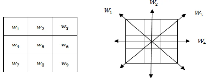
If we assign centre and its neighbor pixels in a sample 3 × 3 matrix to 𝑤 coefficients with 𝑤5 is at origin, the first
derivative values for all directions shown in Fig.1.
Fig. 1. Coefficients and possible edge directions for 3x3 matrix
The first order derivative values of Fig.1 for all directions and the possible edge value are calculated as
∇𝑊1= 𝑤5− 𝑤1 + 𝑤9− 𝑤5 , ∇𝑊2= 𝑤5− 𝑤2 + 𝑤8− 𝑤5 , ∇𝑊3= 𝑤5− 𝑤3 + 𝑤7− 𝑤5 ,
∇𝑊4= 𝑤5− 𝑤4 + 𝑤6− 𝑤5 (14)
∇W = ∇𝑊1+ ∇𝑊2+ ∇𝑊3+ ∇𝑊4 (15)
In this paper, we have calculated ∇𝑊1, ∇𝑊2, ∇𝑊3 and ∇𝑊4 and put into the fuzzy system that uses the triangular
membership function and uses singleton fuzzifier and Sugeno Inference System. The input membership functions are generalized triangular type
𝜇𝑡𝑟𝑖 𝑥 = 𝑥 − 𝑎
𝑏 − 𝑎 , 𝑎 ≤ 𝑥 < 𝑏 𝑐 − 𝑥
𝑐 − 𝑏 , 𝑏 ≤ 𝑥 < 𝑐
ISSN(Online): 2320-9801
ISSN (Print): 2320-9798
I
nternational
J
ournal of
I
nnovative
R
esearch in
C
omputer
and
C
ommunication
E
ngineering
(An ISO 3297: 2007 Certified Organization)
Vol. 3, Issue 7, July 2015
Here the parameters 𝑎, 𝑏 and 𝑐 are constants that characterize the shape of the membership functions. The five inputs are applied to the fuzzy system those are very low (VL) ,low(LO) ,medium (MD) ,high(HI) and very high(VH).In this work the Sugeno constant values are 𝐾 = [0,16,32,64,128]. The fuzzy rules are applied to the inputs and then the output can be found by calculating the weighted average of the individual rule output to obtain all possible direction components as edges.
𝑌 = 𝜇𝑉𝐿 ∇𝑊𝑘 𝑅𝑘+ 𝜇𝐿𝑂 ∇𝑊𝑘 𝑅𝑘+ 𝜇𝑀𝐷 ∇𝑊𝑘 𝑅𝑘+ 𝜇𝐻𝐼 ∇𝑊𝑘 𝑅𝑘+ 𝜇𝑉𝐻 ∇𝑊𝑘 𝑅𝑘 𝟒
𝒌=𝟏
𝜇𝑉𝐿 ∇𝑊𝑘 + 𝜇𝐿𝑂 ∇𝑊𝑘 + 𝜇𝑀𝐷 ∇𝑊𝑘 + 𝜇𝐻𝐼 ∇𝑊𝑘 + 𝜇𝑉𝐻 ∇𝑊𝑘 4
𝒌=𝟏
(17)
Where, 𝑅𝑘 denotes the output of the 𝑘𝑡 rule.
IV. EXPERIMENTAL RESULTS
In this section experiments are made to verify the performance of soft computing approach based on fuzzy logic on different test medical images. The images that are corrupted by any noise are considered here. All test images are 8-bit grey level images. The noisy images used in the edge detection experiments are obtained by corrupting the original test images by impulse noise with 5% noisy density. The noisy test images are shown in Fig.2.Proposed approach; the experimental results are shown in Fig.3 and compared to various edge detection methods such as Sobel, Canny and Morphological approaches.
It is observed from the figure that the performance of the Sobel operator is very poor. For all test medical images, its output images are severely degraded by noise, most noise pulses are incorrectly detected as edges. The canny edge detector demonstrates a considerably better performance than the Sobel detector. It correctly detects most of the noise pulses. However, the effect of noise is still clearly visible as real edges are significantly distorted by the noise. The morphological edge detector removes most of the noise, but the edges are not clearly identified. On the other hand, the proposed approach exhibit very good detection performance and successful detect most of the edges in all test medical images.
ISSN(Online): 2320-9801
ISSN (Print): 2320-9798
I
nternational
J
ournal of
I
nnovative
R
esearch in
C
omputer
and
C
ommunication
E
ngineering
(An ISO 3297: 2007 Certified Organization)
Vol. 3, Issue 7, July 2015
noisy image used in the edge detection experiments.
Sobel Canny Morphological Proposed
Fig. 3. Comparison of the proposed approach with Sobel, Canny and Morphological approaches V. CONCLUSIONS
This paper presented a soft computing approach based on fuzzy logic to detect the edges of noisy medical images. In this approach, heuristic rules applied the system and results were observed for different medical images. The experimental results show that the proposed approach is more efficient for medical image denoising and edge detection than the usually edge detection methods such as Sobel, Canny and Morphological approaches. From the experimental results it is clear that the proposed approach can be used for efficient extraction of edges in medical images corrupted by impulse noise.
REFERENCES
1. M.I. Rajab, M.S. Woolfson, and S.P. Morgan, “Application of region-based segmentation and neural network edge detection to skin lesions”, Computerized Medical Imaging and Graphics, Vol. 28, pp.61-68, 2004.
2. Raman Maini and Himanshu Aggarwal, “Study and Comparison of Various Image Edge Detection Techniques”, International Journal of Image Processing (IJIP), Vol. 3, 20, pp.1-12, 2010.
3. R. C. Gonzalez, R.E. Woods and S.L.Eddins, “Digital Image Processing Using MATLAB”, 2nd Edn., Mc Graw Hill, New Delhi, 2010. 4. J. Canny, “A Computational Approach to Edge Detection”, IEEE Transactions on Pattern Analysis and Machine Intelligence,Vol. 8, pp.
679-687, 1986.
5. Jing Xiao-jun, Yu Nong, and Shang Yong, “Image Filtering Based on Mathematical Morphology and Visual Perception Principle”, Chinese Journal of Electronics, Vol. 13, pp. 612-616, 2004.
6. Richard A P, “A New Algorithm for Image Noise Reduction Using Mathematical morphology”, IEEE Transaction on Image Processing, Vol. 4, pp. 554-568, 1995.
7. J. Mehena, “Medical Image edge detection based on mathematical morphology”, International Journal of Computer and communication technology, Vol. 2, pp.45-48, 2011.
ISSN(Online): 2320-9801
ISSN (Print): 2320-9798
I
nternational
J
ournal of
I
nnovative
R
esearch in
C
omputer
and
C
ommunication
E
ngineering
(An ISO 3297: 2007 Certified Organization)
Vol. 3, Issue 7, July 2015
9. Liang, L.R., Looney, C.G., “Competitive fuzzy edge detection”, Applied Soft Computing, Vol. 3, pp. 132-137, 2003.
10. Choi YS, Krishnapuram R., “A robust approach to image enhancement based on fuzzy logic”, IEEE Trans. Image Processing, Vol.6, pp.808-825, 1997.
11. Abdallah A. Alshennawy, and Ayman A. Aly., “Edge Detection in Digital Images Using Fuzzy Logic Technique”,World Academy of Science, Engineering and Technology, pp-178-186,2009.
12. Pushpajit A. Khaire and Nileshsingh V. Thakur, “A Fuzzy Set Approach for Edge Detection”, International Journal of Image Processing, Vol.6, pp.403-412, 2012.
13. Russo,F., “Edge Detection in Noisy Images Using Fuzzy Reasoning”, IEEE Trans. On Inst. and Meas., Vol.47, pp.802-808, 1998.
BIOGRAPHY
Mr. J.Mehena, Research scholar in I & CT, Fakir Mohan University, Balasore, Odisha. He has 15 years of teaching and research experience. He received his M. Tech in Electronics Engg. From Visvesvaraya National Institute of Technology (VNIT), Nagpur and continuing his Ph.D in the area of medical Image segmentation. He has published many research papers in national and international journals. His areas of interest include Medical Image Processing, VLSI Design and Signal Processing.
.