The dramatic physiological responses of seals to forced submersion are well documented (Scholander, 1940; Elsner, 1965; Zapol et al., 1979; Blix et al., 1983). Often referred to as the dive response, this combination of severe bradycardia (often below 10 beats min−1), vasoconstriction and circulatory
isolation of peripheral organs preserves the large blood oxygen store of the seal for use primarily by the obligate aerobic organs of the animal, the heart and brain. However, studies of unrestrained, trained and free-diving seals have shown that the degree of bradycardia is variable and often not as severe as during forced submersions (Elsner, 1965; Kooyman and Campbell, 1973; Hill et al., 1987; Fedak et al., 1988; Thompson and Fedak, 1993; Andrews et al., 1997; Ponganis et al., 1997a,b; Hindell and Lea, 1998). In those dives with higher heart rates, the degrees of peripheral vasoconstriction and end organ ischemia have not been directly measured. In harbor seals (Phoca vitulina), aortic blood flow is markedly reduced despite higher heart rates during trained dives (Elsner et al., 1966). However, constant inulin and Indocyanine Green
clearance rates in foraging Weddell seals (Leptonychotes
weddellii) are consistent with the maintenance of renal and
hepatic blood flow during such dives (Davis et al., 1983). Decreased clearances of radiolabeled metabolites during isolated dives by Weddell seals, however, suggest reduced organ perfusion during diving (Guppy et al., 1986). Other studies have collected indirect evidence suggesting some degree of muscle blood flow during diving (Guppy et al., 1986; Ponganis et al., 1993; Guyton et al., 1995). It is essential to know the magnitude of organ and muscle perfusion during diving as these may be critical to the depletion rate of the blood O2 store and the duration of aerobic metabolism (Davis and
Kanatous, 1999).
To examine physiological responses during submersions with heart rates greater than those observed during typical forced submersion studies, we attempted to habituate or train yearling harbor seals to short forced submersions, as has been done with ducks (Gabrielsen, 1985; Gabbott and Jones, 1987). If a less intense bradycardia could be elicited during trained JEB3572
In several pinniped species, the heart rates observed during unrestrained dives are frequently higher than the severe bradycardias recorded during forced submersions. To examine other physiological components of the classic ‘dive response’ during such moderate bradycardias, a training protocol was developed to habituate harbor seals (Phoca vitulina) to short forced submersions. Significant changes were observed between physiological measurements made during naive and trained submersions (3–3.5 min). Differences were found in measurements of heart rate during submersion (naive 18±4.3 beats min−1 versus trained 35±3.4 beats min−1), muscle blood flow measured using laser-Doppler flowmetry (naive 1.8±0.8 ml min−1100 g−1 versus trained 5.8±3.9 ml min−1100 g−1), change in venous P
O∑ (naive
−0.44±1.25 kPa versus trained −1.48±0.76 kPa) and muscle deoxygenation rate (naive −0.67±0.27 mvd s−1 versus trained −0.51±0.18 mvd s−1, a relative measure of muscle oxygenation provided by the Vander Niroscope, where mvd are milli-vander units). In contrast to the naive
situation, the post-submersion increase in plasma lactate levels was only rarely significant in trained seals. Resting eupneic (while breathing) heart rate and total oxygen consumption rates (measured in two seals) were not significantly different between the naive and trained states. This training protocol revealed that the higher heart rate and greater muscle blood flow in the trained seals were associated with a lower muscle deoxygenation rate, presumably secondary to greater extraction of blood O2 during trained submersions. Supplementation of muscle oxygenation by blood O2 delivery during diving would increase the rate of blood O2 depletion but could prolong the duration of aerobic muscle metabolism during diving. This alteration of the dive response may increase the metabolic efficiency of diving.
Key words: dive response, heart rate, laser-Doppler, muscle blood flow, myoglobin, near-infrared spectroscopy, oxygen store, harbour seal, Phoca vitulina.
Summary
Introduction
Effects of training on forced submersion responses in harbor seals
P. D. Jobsis*, P. J. Ponganis and G. L. Kooyman
Center for Marine Biotechnology and Biomedicine, Scripps Institution of Oceanography, University of California, San Diego, La Jolla, CA 92093-0204, USA
*Present address: Loras College, 1450 Alta Vista, Dubuque, IA 52004-0178, USA (e-mail: [email protected])
forced submersions, we wished to determine the effects of this on muscle blood flow, muscle oxygen depletion rates and post-submersion blood lactate levels. This is the first simultaneous measurement of these three variables in breath-holding animals.
Materials and methods
Four harbor seals Phoca vitulina L. (two male, two female) were brought from the Rehabilitation Program of Sea World Califonia to the holding tanks of the Center for Marine Biotechnology and Biomedicine at Scripps Institution of Oceanography under the conditions and guidelines of US National Marine Fisheries Service marine mammal permit no. 779. The seals were 6–10 months old and ranged in mass from 25 to 33 kg. They were maintained on a diet of herring (10 % of body mass per day) and vitamin supplements, and were released at sea following completion of this study.
Submersion training protocol
Once the seals had acclimated to the holding facilities, a submersion training protocol was begun. A submersion duration of 3 min was selected for seals lighter than 30 kg and 3.5 min for those above 30 kg. Both values are lower than the calculated aerobic dive limits for harbor seals and are within the common dive duration of free-diving harbor seals (Fedak et al., 1988; Kooyman, 1989; Jobsis, 1998). The training protocol consisted of conducting five forced submersions per day, each with a 20 min resting period, for approximately 10 (9–11) days over a 14-day period. The first session was always fully monitored (all recorded variables are described below), and all data collected during this first session were designated as naive. The last session was also fully monitored, and if the submersion heart rate (fH) response changed significantly, the data collected were designated as trained. In all intervening training sessions, only the electrocardiogram (ECG) and fH
were recorded. Data obtained during naive and trained sessions were compared using a paired t-test (significance, P<0.05) and expressed as mean ± standard deviation (S.D.) unless noted otherwise. Following the fifth submersion of the trained session, one seal was subjected to a final 5 min submersion to detect changes in the submersion response when taken beyond the trained submersion duration.
At the start of each training session, a seal was weighed and restrained on a ‘U’-shaped restraining board. A clear Lucite ‘diving’ helmet was placed over the head of the animal and loosely secured to the restraining board with elastic cord. The helmet used a two-layer neoprene gasket around the neck of the seal to prevent leaks. Room air was pumped through the helmet at 60 l min−1. To avoid overheating, the seals were kept
wet and cooled by air from an electric fan. To facilitate training, the durations of submersions and resting periods remained constant throughout all training sessions; visual and auditory cues were given to signal the beginning and end of each submersion, and all efforts were made to reduce or eliminate any discomfort experienced by the seals. To initiate
a submersion, the draining valve of the helmet was closed (visual cue), the air pump was turned off (auditory cue) and fresh water, at ambient temperature, was poured into the helmet. When ending the submersion, the drain valve was opened and the air pump was turned on. The period from the initial visual cue to the start of submersion was approximately 15 s and that from opening the valve to the first breath at the end of submersion was approximately 10 s.
Oxygen consumption
In two seals, the rate of oxygen consumption was continuously monitored during naive and trained sessions by sampling the exhausted air from the helmet. The open-flow respirometry system was similar to the system described by Davis et al. (1985).
Heart rate
Three surface ECG electrodes were attached to each seal as described previously (Ponganis et al., 1997b). Recordings of ECG and fHwere made during each submersion for all training sessions with a UFI (Morro Bay, California, USA) ECG/fH
monitor and an Astro-Med eight-channel recorder. Heart rate was calculated as the total number of ‘qrs’ waveforms in a given period divided by the duration of that period. Resting or pre-submersion heart rate was measured for a 1 min period 3 min before submersion. Heart rate during submersion is the mean rate during the entire submersion. The post-submersion period consisted of a 1 min period immediately following the end of the submersion period. After completion of the training protocol, the ECG electrodes were removed.
Anesthesia and blood sampling
All probe, catheter and electrode placements were conducted under local anesthesia (2 % Xylocaine and 0.25 % Bupivacaine) using aseptic techniques. All insertions were percutaneous, with a standard catheter-over-needle technique. For intramuscular probe insertions, the same technique was used with a peel-away catheter; this allowed removal of the catheter after insertion of the probe through it. At the time of catheterization, 1 g of cefazolin was given intravenously as an antibiotic prophylaxis. In addition, oral antibiotics, 1 g of cefalexin per day, were continued for 1 week following a monitored session. The insertion sites were monitored daily, and no seal showed any symptoms of infection following the procedure.
The extradural vein was catheterized at the start of naive and trained sessions using a 12.7 cm 14 gauge catheter. A heparinized saline (2 u.i. ml−1in 0.9 % NaCl) flush was used to
this time may give a more accurate measure of pre-submersion venous oxygenation than sample A, which does not reveal the effects of pre-submersion hyperventilation by the seals. All blood samples were analyzed for PO∑with a Radiometer ABL2
blood gas analyzer. Plasma lactate and glucose concentrations were determined with a YSI lactate/glucose analyzer (YSI model 2300, Yellowsprings, Ohio, USA).
Muscle blood flow
Muscle blood flow was measured using two laser-Doppler blood flow (LDBF) meters. The first was a Transonics (Ithaca, New York, USA) ALF-21 LDBF monitor used for all four naive sessions and two trained sessions. Since this monitor was unavailable for two trained sessions, an Oxford Optronix (Oxford, UK) MPM 35 LDBF monitor was used. The catheter-like fiber-optic probe of the LDBF monitor was percutaneously placed into the ilioicostalis lumborum/longissimus dorsi (ILLD) muscle group 5 cm to the left of the spinal column, 2.5 cm above the crest of the ilium. Since two dissimilar LDBF meters were used and because the LDBF meters could not be calibrated in situ, muscle blood flow is given both as the absolute value provided by the factory calibrations and as a percentage of mean resting muscle blood flow (%RMBF), measured over 1 min, 3 min prior to each submersion. Muscle blood flow during submergence was measured by averaging over the entire submersion period. The post-submergence muscle blood flow was measured over the first minute following the submersion period.
Muscle oxygenation
Muscle oxygenation was monitored during naive and trained sessions by near-infrared spectroscopy (NIRS) (Jöbsis-VanderVliet et al., 1987). The Niroscope (Vander Corp., Durham, North Carolina, USA) uses NIRS to provide trend monitoring of combined oxyhemoglobin (HbO) and oxymyoglobin (MbO), combined deoxyhemoglobin (Hb) and deoxymyoglobin (Mb) and total hemoglobin (tHb). The data provided by the Niroscope show the change in the amount of HbO(+MbO), Hb(+Mb) and tHb in milli-vander units (mvd). The milli-vander unit is a measure of the relative change in the amount of HbO, Hb and tHb within the volume of tissue measured by the optical probes of the Niroscope. The standard surface probes of the instrument were replaced with a 17 gauge catheter-like fiber-optic probe with four emitter fibers at the tip and eight detector fibers recessed 0.5 cm from the tip (Jobsis, 1998). The probe was percutaneously placed 5 cm to the right side of the spinal column and 2.5 cm above the crest of the ilium into the ILLD muscle group. The blunt tip of the probe was used to puncture the fascia of the muscle group and was inserted approximately 1 cm into the muscle. To reduce the possibility of interference between the Niroscope and LDBF light-emitting probes, the probes were placed on opposite sides of the spinal column.
Spontaneous apneas
During resting periods, the seals voluntarily performed
spontaneous apneas, designated in this study as a voluntary apnea greater than 30 s. Heart rate, muscle blood flow and muscle oxygenation were recorded during spontaneous apneas and compared with those of naive and trained submersions as well as resting conditions. Blood sampling occasionally corresponded with these spontaneous apneas, but the sampling protocol was not altered. If a scheduled submersion occurred during a spontaneous apnea, the seal was ‘awakened’ and allowed to resume a normal respiratory pattern before the scheduled submersion. Results are presented as means ± S.D. unless stated otherwise.
Results
Submersion training protocol
Three of the four seals modified their response to submersion with training. The three seals that did modify their responses appeared to do so gradually over the training period. Seal L did not habituate to the submersion protocol. Seal L is included in results that are not influenced by its lack of habituation to the training procedure; where it is included, it is specifically cited (e.g. seals L, A, S and W; or N=4).
O2consumption
The mean oxygen consumption rate of two seals during a naive submersion session was 8.0±1.7 ml O2min−1kg−1. After
training, the oxygen consumption rate of these two seals was 7.8±0.7 ml O2min−1kg−1 (N=2), which was not statistically
significantly different.
Heart rate response
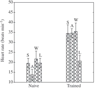
Heart rate during submersion showed a marked increase with training in three of the four seals (Fig. 1). In these three habituated seals, the mean fH during naive submersions, 18± 4.3 beats min−1, was significantly different from the mean fH
during trained submersions, 35±3.4 beats min−1(Table 1). The
onset as well as the overall fHof the submersion bradycardia showed differences between naive and trained submersions. During naive submersions, the drop in fH was nearly instantaneous, often with the lowest fHearly in the submersion. During trained submersions, the fHdecreased more gradually, and the lowest fH values were usually recorded at the end of the submersion. This is demonstrated by comparing the mean
fHduring the first 30 s of submersion. In naive submersions, the fH for this period was 18±4.8 beats min−1, and in trained
submersions the rate was 47±12.3 beats min−1. The mean
submersion fH increased steadily over the training period (Fig. 2), with the first submersion of each training session often having the lowest fH. The fHwhile resting prior to submersion and the tachycardia observed after submersion showed no statistical difference between naive and trained sessions, and fH
was 111±22 beats min−1while resting and 154±6.1 beats min−1
during the post-submersion tachycardia. Seal L, which did not significantly change its fH with training, had a submerged fH of 20±2.4 beats min−1 during naive sessions
consistently showed the ‘naive’ response in other variables as well.
Muscle blood flow
There was a significant difference between naive and trained muscle blood flow for the three habituated seals. The mean muscle blood flow during naive submersions was 1.8±0.8 ml min−1100 g−1 (12.3±5.8 %RMBF), and during
trained submersions it was 5.8±3.9 ml min−1100 g−1
(23.3±15.7 %RMBF) (Table 1). Eupneic muscle blood flow levels as measured by the LDBF meter differed between naive conditions when flow was 14.9±4.9 ml min−1100 g−1
(100 % naive RMBF) and trained conditions when it was 24.7±9.0 ml min−1100 g−1 (100 % of trained RMBF). After
each submersion, a hyperemic period was observed averaging 25.7±4.2 ml min−1100 g−1 (175.4±58.8 %RMBF) for naive
animals and 41.5±13.2 ml min−1100 g−1(163.6±36.7 %RMBF)
for trained animals.
In seal L, no significant differences were found between naive and trained submerged muscle blood flow; muscle blood flow was 1.2±0.5 ml min−1100 g−1 (4.8±2.0 %RMBF)
during naive submergence and 1.1±0.4 ml min−1100 g−1
(6.5±2.4 %RMBF) during trained submergence. The post-submersion muscle blood flow values for this animal were unique in that they were lower than the resting level; 8.8±4.2 ml min−1100 g−1 (35.5±16.9 %RMBF) during the
Naive Trained
10 15 20 25 30 35 40 45 50
S
A W
L
S W
L A
Heart rate (beats min
[image:4.612.76.268.75.250.2]-1)
[image:4.612.309.561.96.404.2]Fig. 1. Mean heart rate (mean ±S.D.) for each of the four seals (S, A, W and L) during five submersions during naive and trained sessions. Seals S, A and W are considered to have adapted to the training protocol and were grouped together for later comparison and analysis. Seal L did not increase its heart rate and was excluded from the habituated group.
Table 1. Physiological variables during submersion of three naive and trained harbor seals
Naive Trained
Heart rate (beats min−1)
Resting 111±22 110±13
Submerged 18±4.3 35±3.4*
First 30 s submerged 18±4.8 47±12.3*
Post-submersion 156±5.8 152±5.8
Spontaneous apnea 60±7.5 56±4.2*
Muscle blood flow Resting
%RMBF‡ 100 100
ml min−1100 g−1 14.9±4.9 24.7±9.0*
Submerged
%RMBF 12.3±5.8 23.3±15.7*
ml min−1100 g−1 1.8±0.8 5.8±3.9*
Post-submersion
%RMBF 175.4±59 163.6±36.7
ml min−1100 g−1 25.7±4.2 41.5±13.2*
Spontaneous apnea
%RMBF 30.9±14.1 46.6±26.7
ml min−1100 g−1 5.4±2.1 12.3±6.6*
Other variables
Muscle deoxygenation rate −0.67±0.27 −0.51±0.18* (mvd s−1)‡‡
Submerged ∆PvO2(kPa)‡‡‡ −0.44±1.25 −1.48±0.76*
∆[Lactate] (mmol l−1) 0.51±0.57 0.31±0.36
Data were collected from five naive and five trained submersions for each seal (N=15). Values for spontaneous apneas are the mean of 72 occurring during the naive sessions and 173 during the trained session.
Asterisks indicate values obtained during naive and trained submersions are significantly different, P<0.05, using a paired t-test.
‡Muscle blood flow is given as a percentage of resting muscle
blood flow (%RMBF) and in the factory-calibrated units of ml min−1100 g−1.
‡‡The muscle deoxygenation rate is in milli-vanders s−1 (see text for clarification).
‡‡‡Submerged change in PvO
2from 1 min before the submersion
(sample A) to 30 s before the end of submersion (sample B) (see text for further clarification).
1 3 5 7 9
5 10 15 20 25 30 35 40 45
*
Heart rate (beats min
-1)
Training session number
2 4 6 8
[image:4.612.329.550.587.738.2]first submersion session and 16.2±4.4 ml min−1100 g−1
(95.3±25.9 %RMBF) during the last.
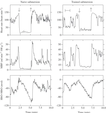
During the reduced muscle blood flow of naive submersions, small spikes in the muscle blood flow profile corresponded to individual heart beats. The muscle blood flow was low, continuous and variable during trained submersions and did not appear to be intermittently pulsatile (Fig. 3). In the rest period between submersions, muscle blood flow showed marked oscillations that corresponded to the breathing pattern of the animal. Perfusion increased during inspiration and decreased during expiration. The absence of this pattern was useful in identifying periods of spontaneous apnea.
Muscle oxygenation
Muscle oxygenation declined steadily during submersion and increased promptly after the end of the submersion (Figs 3, 4). A majority of the submersions were preceded by a small increase in muscle oxygenation (HbO+MbO). This increase occurred during pre-submersion hyperventilation (as the helmet was filled with water) and was quickly reversed at the onset of submersion. This increase in oxygenation was countered by a decrease in the amount of Hb+Mb observed. The total amount of Mb and Hb (tHb)
measured by the Niroscope showed a slight increase during both naive and trained submersions.
The three habituated seals showed a significant decrease in the rate of muscle deoxygenation during trained submersions compared with naive submersions. The rate of muscle deoxygenation during naive submersion measured by the Niroscope was −0.67±0.27 mvd s−1;
this was significantly different from the value of −0.51±0.18 mvd s−1found
during trained submersions. Seal L showed no significant difference in the
rate of change in muscle oxygenation during naive and trained submersions.
Extradural vein PO∑
Considerable variation was found when analyzing extradural vein samples taken during the naive and trained sessions for venous PO∑(PvO∑). This variation appeared to be caused by the
respiratory state of the animal since some samples were taken during spontaneous apneas while others were taken during the normal eupneic cycle. Indeed, the lowest PvO∑value (4.12 kPa)
obtained occurred during a resting period while the seal was undergoing a spontaneous apnea and not during a submersion. As a result of this variation, the PvO∑ values during naive
and trained sessions at rest or at the end of submersion did not differ significantly. However, the difference in PvO∑between
pre-submersion (sample A) and the sample taken during submersion (sample B) was significant. These decreases during submersion averaged −0.44±1.25 kPa for naive sessions and −1.48±0.76 kPa for trained sessions. This decrease in PvO∑may
[image:5.612.211.568.357.739.2]not represent the full drop in venous oxygen since sample A was taken before the pre-submersion hyperventilation and tachycardia. In one seal (seal S), a small sample was taken at
Fig. 3. Comparison of two submersions, the fourth of the naive session and the third of the trained session, for seal S. Arrows indicate the start and end of submersions. A spontaneous apnea is shown before submersion during the trained session and is indicated by the solid line. Note that heart rate and muscle blood flow (MBF) are higher and the change in muscle oxygenation (oxygenated hemoglobin and myoglobin, HbO+MbO) is lower during trained submersions. Units for Niroscope data are milli-vanders (mvd), which represent the change in concentration within the observed volume of tissue (see Materials and methods).
0 10 20 30 40 50 0 50 100 150
Time (min)
0 2.5 5.0 7.5 10.0
-120 -80 -40 0 0 10 20 30 40 50 0 50 100 150
Naive submersion Trained submersion
H
ear
t
ra
te (
b
ea
ts
min
-1)
MBF
(m
l min
-1
1
00
g
-1)
H
b
O+
M
b
O
(m
v
d
)
Time (min)
0 2.5 5.0 7.5 10.0
the start of submersion during the trained session, and the PvO∑of these five samples was consistently higher
than that of the ‘A’ samples.’
Plasma lactate levels
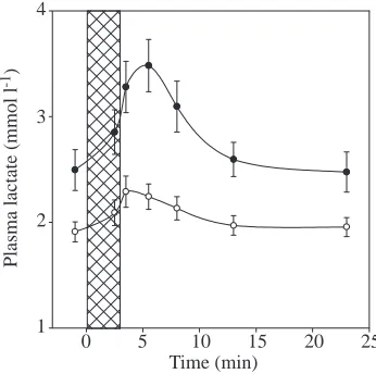
Plasma lactate concentrations increased and peaked early in the post-submersion period (Fig. 5). A significant increase in plasma lactate from the resting level to the submersion peak (2.5 min post-submersion) occurred during naive submersions for each seal except seal S. During trained submersion sessions, only seal W showed a significant increase in plasma lactate levels between resting levels and the post-submersion peak (30 s post-submersion). Although the concentrations were lower than those found in forced submersion studies with longer submersion durations (Scholander, 1940), the overall appearance of the lactate washout is similar.
Spontaneous apneas
During the 20 min post-submersion resting period, the seals performed intermittent spontaneous apneas, possibly including occasional episodes of sleep apnea as described by Castellini et al. (1994) or quiescent apneas as described by Ridgway et al. (1975). During the majority of these events, the eyes were closed and the seal was completely motionless. In total, 245 spontaneous apneas were recorded during the resting periods. For three of the seals (W, S, L), there was no difference in the duration of these events (50±20 s) between naive and trained sessions. In seal A, which had the longest recorded spontaneous apnea (243 s), the mean length of a spontaneous apnea during the naive session, 102±64 s, was more than twice that found during the trained session, 42±13 s. In all seals, more spontaneous apneas occurred during trained sessions, with a mean of 18 per naive session and 43 per trained session.
The most consistent indicator of spontaneous apnea was a decrease in fH from the eupneic level of 110 beats min−1 to
the apneic level of less than 80 beats min−1(Fig. 3). For the
three trained seals (W, A and S), the mean fH during spontaneous apnea was 60±7.5 beats min−1 during naive
sessions, which was significantly different from the value of 56±4.2 beats min−1occurring during trained sessions. In seal
L, fH during spontaneous apneas of the naive session was 78±4 beats min−1 and 66±6 beats min−1 during spontaneous
apneas of the trained session.
Muscle blood flow showed marked oscillations during the 20 min resting periods that reflected the pattern seen in the heart rate trace (Fig. 3) and corresponded to the breathing pattern. Spontaneous apneic muscle blood flow during naive and trained sessions of the three trained seals differed: during the naive sessions, it was 5.4±2.1 ml min−1100 g−1
(30.9±14.1 %RMBF) and during the trained sessions it was 12.3±6.6 ml min−1100 g−1 (46.6±26.7 %RMBF). For seal L,
the spontaneous apneic muscle blood flows during naive and
trained sessions were not significantly different. During both monitored sessions for all four seals, the spontaneous apneic muscle blood flow was always greater than muscle blood flow during submersion. Muscle deoxygenation rates during spontaneous apneas were lower in the trained sessions than in the naive sessions for the three trained seals. There was,
-50 0 50 100
-150 -100 -50 0
Time (min)
0 6 10
0 50 100 150
Hb+Mb
Oxyg
en
at
ed
Hb
O
+Mb
O
T
o
ta
l
Hb
2 4 8
0 2 4 6 8 10
Time (min)
0 5 10 15 20 25
1 2 3 4
Plasma lactate (mmol l
[image:6.612.281.561.70.315.2]-1)
[image:6.612.351.524.414.586.2]Fig. 5. The mean plasma lactate concentration in blood samples from seals during and after naive (filled circles) and trained (open circles) submersions. The cross-hatched area represents a 3.0 min submersion period. Values are means ±S.E.M. (N=15). For simplicity, 3.0 and 3.5 min submersion values are plotted together following the 3.0 min time line.
however, considerable variation, and muscle oxygenation increased slightly during some spontaneous apneic events.
Discussion
Heart rate
The heart rates during submersion of the trained seals were approximately 100 % greater than the corresponding value during naive sessions and approached levels measured during spontaneous apneic events in this study as well as the fHlevels recorded during trained and unrestrained diving and reported by several authors (Elsner, 1965; Harrison and Ridgway, 1975; Ridgway et al., 1975; Williams et al., 1991; Fedak et al., 1988; Thompson and Fedak, 1993). The fH response of naive and trained seals not only differed in overall mean fH, but also in the fH profile during the submersion, where the naive seals showed a rapid reduction in fHand the trained seals showed a slower drop in fH. The fH of the trained seals increased progressively throughout the training sessions, showing a gradual decrease in the degree of bradycardia rather than a reflexive switch from one level to another. It is also notable that, in both naive and trained sessions, the seals often showed an increase in heart rate immediately prior to the first breath ending the submersion (as the water level in the helmet was lowered). This anticipatory tachycardia has been observed in previous studies of unrestrained seals (Fedak, 1986; Fedak et al., 1988; Hill et al., 1987; Williams et al., 1991). The similarities in the fHresponse of the trained seals in this study and unrestrained seals suggest that this protocol may serve as a useful model for cardiovascular responses that occur during short-duration dives of seals at sea.
Muscle blood flow
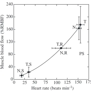
Muscle blood flow showed an obvious correlation with fH
over the whole range of mean fHand muscle blood flow in this study (Fig. 6). Both linear and curvilinear regression analyses show a highly significant relationship (P<0.05). A second-order regression fitted the data best. This may be due to a difference between apneic and eupneic stroke volume (Ponganis et al., 1990) and to peripheral vascular regulation. However, the high degree of correlation may be misleading, for if one tests the relationship between fHand muscle blood flow taken during just one condition, such as post-submersion, there is a low degree of correlation and a significantly different regression line. This suggests that other factors, such as the intensity of local arterial constriction, may modify muscle blood flow during periods of similar heart rate.
The presence of substantial muscle blood flow during trained submersions differs from the observations made during typical forced submersion studies (Grinnell et al., 1942; Murdaugh et al., 1966; Zapol et al., 1979; Blix et al., 1983). The maintenance of muscle blood flow during apnea found in the present study is consistent with past studies of free-diving Weddell seals, which implied some degree of muscle blood flow during aerobic diving. Guppy et al. (1986) recorded an increase in lactate levels prior to the end of dives, suggesting
that muscle blood flow may persist and allow lactate washout during the dive. Ponganis et al. (1993) found that muscle temperature during dives was constant or decreased, which suggests that the working muscles are not isolated from cooler blood in the circulation. Finally, Guyton et al. (1995) measured lower than expected muscle deoxygenation rates, which suggested some level of perfusion.
The presence of a dense sympathetic innervation of proximal arterioles in pinnipeds (White et al., 1973) and the persistent muscle ischemia found in the presence of O2 depletion and
lactate accumulation during forced submersions (Scholander, 1940) suggest that regulation of muscle blood flow in seals is largely independent of local tissue metabolite control at the capillary level. Gooden and Elsner (1985) have reviewed this concept of an extramuscular throttle for muscle blood flow. These authors also proposed that arteriolar smooth muscle perfusion and smooth muscle metabolite accumulation might be more critical regulators of arteriolar tone during intense sympathetic vasoconstriction. They hypothesized that local ischemia and metabolite accumulation in the more proximal vascular smooth muscle wall could result in an intermittent, pulsatile blood flow to the tissues even in the presence of continuous, intense sympathetic nerve activity. Such intermittent pulsations were not observed in this study, but have been reported in seal myocardial flow during forced submersions (Kjekshus et al., 1982; Elsner et al., 1985). Gooden and Elsner (1985) also emphasized that a ‘trickle’ of blood flow to the periphery could maintain perfusion of the arteriolar wall during severe (but incomplete) ischemia induced by sympathetic vasoconstriction. The muscle blood
0 40 80 120 160 200 240
T N
PS N,R T,R
T,S N,S
Muscle
blood
flo
w
(%
R
MBF)
Heart rate (beats min-1)
[image:7.612.351.533.71.253.2]0 25 50 75 100 125 150 175
flow pattern, especially the persistent low degree of muscle blood flow found in naive seals with this protocol, is consistent with this latter mechanism. The even greater degree of muscle blood flow observed during trained submersions should not only be adequate for vascular smooth muscle perfusion, but should also be compatible with some degree of oxygen transport to skeletal muscle. Although the increased muscle blood flow during trained submersions appears to enhance the rate of blood oxygen depletion, this may actually maximize the duration of whole-body aerobic metabolism during diving, as suggested by Jobsis (1998) and proposed in a theoretical model of O2store utilization by Davis and Kanatous (1999).
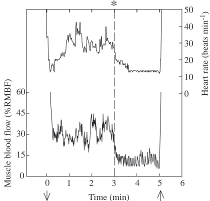
Remarkable regulation of muscle blood flow is depicted during the final 5 min submersion (Fig. 7) of seal S. This seal had been trained for 3 min submersions, but on the final submersion, the duration was extended to 5 min. During the first 3 min, fH and muscle blood flow were typical of the trained response. No cues were provided to the seal. However, at exactly 3 min of submersion, fH and muscle blood flow were lowered to levels found in the naive state and then maintained at that level until the end of the submersion. Such changes in peripheral flow probably also occur when fHchanges abruptly during free dives (Andrews et al., 1997; Thompson and Fedak, 1993). In this situation, blood O2is maximally conserved for aerobic metabolism by
the heart and central nervous system.
Muscle oxygenation
If muscle metabolic rate remained constant during the training protocol (as suggested by the lack of change in whole-body rate of O2 consumption), then increased muscle blood
flow should be reflected by a decrease in the rate of muscle desaturation. The data from naive and trained submersions show a 24 % reduction in the rate of muscle desaturation during trained submersions. The reduction in the rate of muscle deoxygenation coincided with an increase in muscle blood flow and an increase in mixed venous deoxygenation measured from the extradural vein. This suggests greater blood O2extraction
and supplementation of the muscle O2 store during the
habituated state, which could delay the onset of anaerobic metabolism, thereby increasing metabolic efficiency while diving (Jobsis, 1998). These findings are consistent with the model proposed by Davis and Kanatous (1999), in which optimal utilization of blood and muscle O2stores (maximum
aerobic dive time) requires supplementation of muscle metabolism with blood O2.
Spontaneous apnea
Spontaneous apnea provides an interesting comparison with the naive and trained submersions and provides a monitoring situation in which the apnea duration is under the control of the seal. Heart rate during spontaneous apneas was consistently higher than that found during trained submersions. The measured variables show a pattern that is consistent with an even greater peripheral perfusion during spontaneous apneas than during trained submersions. These
measured variables include higher muscle blood flow and lower muscle oxygen depletion rates, suggesting a greater reliance on blood oxygen during spontaneous apneas than during trained submersions.
In conclusion, the training method used in this study was effective in habituating three out of four harbor seals to short forced submersions in which the heart rate response is more like that of free-diving harbor seals than that of typical forced submersion protocols. Although application of this technique is valuable because of the ability to make other physiological measurements, it must be remembered that it still differs from free diving by not mimicking the energy demands associated with swimming and foraging and by not duplicating the ability of the seal to determine submersion duration.
The seals that modified their responses showed significantly greater fHand muscle blood flow during trained versus naive submersions, but both variables were still reduced compared with the spontaneous apneic and eupneic levels. The increased muscle blood flow during submersion found in habituated seals was associated with a reduced rate of muscle deoxygenation and an increased rate of venous deoxygenation. In free-diving seals, such supplementation of the muscle oxygen store would delay the onset of anaerobic metabolism in the muscle and, thereby, decrease the metabolic cost of dives within the aerobic dive limit of the animal. These adaptations would potentially benefit the seals by increasing both the metabolic and foraging efficiencies during diving, as previously suggested by a mathematical model (Davis and Kanatous, 1999).
0 10 20 30 40 50
Time (min)
0 4 5 6
0 15 30 45 60
Muscl
e
blood
flo
w
(
%RMBF
)
H
eart
rat
e (
b
eat
s min
-1)
1 2 3
[image:8.612.330.540.74.275.2]*
We would like to thank M. Horning, L. Starke and L. Winter for their assistance with this project and anonymous reviewers for their constructive comments. This research was supported by US Health Service Grant NHLBF 5-P01-HL-17731.
References
Andrews, R. D., Jones D. R., Williams, J. D., Thorson, P. H., Oliver G. W., Costa, D. P. and Le Boeuf, B. J. (1997). Heart rates of northern
elephant seals diving at sea and resting on the beach. J. Exp. Biol. 200, 2083–2095.
Blix, A. S., Elsner, R. W. and Kjekshus, J. K. (1983). Cardiac output and
its distribution through capillaries and A-V shunts in diving seals. Acta
Physiol. Scand. 118, 109–116.
Castellini, M. A., Milsom, W. K., Berger, R. J., Costa, D. P., Jones, D. R., Castellini, J. M., Rea, L. D., Bharma, S. and Harris, M. (1994). Patterns
of respiration and heart rate during wakefulness and sleep in elephant seal pups. Am. J. Physiol. 266, R863–R869.
Davis, R. W., Castellini, M. A., Kooyman, G. L. and Maue, R. (1983). GFR
and hepatic blood flow during voluntary diving in Weddell seals. Am. J.
Physiol. 245, R743–R748.
Davis, R. W. and Kanatous, S. B. (1999). Convective oxygen transport and
tissue oxygen consumption in Weddell seals during aerobic dives. J. Exp.
Biol. 202, 1091–1113.
Davis, R. W., Williams, T. M. and Kooyman, G. L. (1985). Swimming
metabolism of yearling and adult Harbor seals (Phoca vitulina). Physiol.
Zool. 58, 590–596.
Elsner, R. W. (1965). Heart rate response in forced versus trained
experimental dives in pinnipeds. Hvalråd. Skr. 48, 24–29.
Elsner, R. W., Franklin, D., Van Citters, R. L. and Kenney, D. W. (1966).
Cardiovascular defense against asphyxia. Science 153, 941–949.
Elsner, R. W., Millard, R. W., Kjekshus, J. K. and White, F. (1985).
Coronary blood flow and myocardial segment dimensions during simulated dives in seals. Am. J. Physiol. 249, H1119–H1126.
Fedak, M. A. (1986). Diving and exercise in seals: interactions of behavior
and physiology. In Behavioral Ecology of Underwater Organisms. Report
of the 19th Symposium of the Underwater Association, March 22–23,
London. Prog. Underwater Sci. 11, 155–169.
Fedak, M. A., Pullen, M. R. and Kanwisher, J. (1988). Circulatory
responses of seals to periodic breathing: heart rate and breathing during exercise and diving in the laboratory and open sea. Can. J. Zool. 66, 53–60.
Gabbott, G. R. J. and Jones, D. R. (1987). Habituation of the cardiac
response to involuntary diving in diving and dabbling ducks. J. Exp. Biol.
131, 403–415.
Gabrielsen, G. W. (1985). Free and forced diving in ducks: habituation of the
initial dive response. Acta Physiol. Scand. 123, 67–72.
Gooden, B. A. and Elsner, R. W. (1985). What diving animals might tell us
about blood flow regulation. Perspect. Biol. Med. 28, 465–474.
Grinnell, S. W., Scholander, P. F. and Irving, L. (1942). Experiments on
the reaction between blood flow and heart rate in the diving seal. J. Cell.
Comp. Physiol. 19, 341–350.
Guppy, M., Hill, R. D., Schneider, R. C., Qvist, J., Liggins, G. C., Zapol, W. M. and Hochachka, P. W. (1986). Microcomputer-assisted metabolic
studies of voluntary diving of Weddell seals. Am. J. Physiol. 250, R175–R187.
Guyton, G. P., Stanek, K. S., Schneider, R. C., Hochachka, P. W., Hurford, W. E., Zapol, D. G., Liggins, G. C. and Zapol, W. M. (1995).
Myoglobin saturation in free-diving Weddell seals. J. Appl. Physiol. 70, 1148–1155.
Harrison, R. J. and Ridgway, S. H. (1975). Restrained and unrestrained
diving in seals. Rapp. Pev. Réun. Cons. Int. Expl. Mer. 169, 76–80.
Hill, R. D., Schneider, R. C., Liggins, G. C., Schuette, A. H., Elliott, R. L., Guppy, M., Hochachka, P. W., Qvist, J., Falke, K. J. and Zapol, W. M.
(1987). Heart rate and body temperature during free diving of Weddell seals.
Am. J. Physiol. 253, R344–R351.
Hindell, M. A. and Lea, M. A. (1998). Heart rate, swimming speed and
estimated oxygen consumption of free-ranging southern elephant seal.
Physiol. Zool. 71, 74–84.
Jobsis, P. D. (1998). Muscle oxygenation and blood flow during submersion
in ducks (Anas platyrhynchos) and seals (Phoca vitulina). PhD dissertation, University of California at San Diego. 142pp.
Jöbsis-VanderVliet, F. F., Fox, E. and Sugioka, K. (1987). Monitoring of
cerebral oxygenation and cytochrome aa3redox state. Int. Anesthesiol. Clin. 25, 209–230.
Kjekshus, J. K., Blix, A. S., Elsner, R., Hol, R. and Amundsen, E. (1982).
Myocardial blood flow and metabolism in the diving seal. Am. J. Physiol.
242, R97–R104.
Kooyman, G. L. (1989). Diverse Divers. Berlin, New York, London:
Springer-Verlag. 201pp.
Kooyman, G. L. and Campbell, W. B. (1973). Heart rate in freely diving
Weddell seals (Leptonychotes weddellii). Comp. Biochem. Physiol. 43, 31–36.
Murdaugh, H. V., Robin, E. D., Miller, J. E., Drewry, W. F. and Weiss, E. (1966). Adaptations to diving in the harbor seal: cardiac output during
diving. Am. J. Physiol. 210, 176–180.
Ponganis, P. J., Kooyman, G. L., Baranov, E. A., Thorson, P. H. and Stewart, B. S. (1997a). The aerobic submersion limit of Baikal seals. Can. J. Zool. 75, 1323–1327.
Ponganis, P. J., Kooyman, G. L., Castellini, M. A., Ponganis, E. P. and Ponganis, K. V. (1993). Muscle temperature and swim velocity profiles
during diving in a Weddell seal (Leptonychotes weddellii). J. Exp. Biol. 183, 341–346.
Ponganis, P. J., Kooyman, G. L., Winter, L. M. and Starke, L. N. (1997b).
Heart rate and plasma lactate responses during submerged swimming and trained diving in California sea lions (Zalophus californianus). J. Comp.
Physiol. B 167, 9–16.
Ponganis, P. J., Kooyman, G. L., Zornow, M. H., Castellini, M. A. and Croll, D. A. (1990). Cardiac output and stroke volume in swimming harbor
seals. J. Comp. Physiol. B 160, 473–482.
Ridgway, S. H., Harrison, R. J. and Joyce, P. L. (1975). Sleep and cardiac
rhythm in gray seals. Science 187, 553–555.
Scholander, P. F. (1940). Experimental investigations on the respiratory
function in diving mammals and birds. Hvalråd. Skr. 22, 1–131.
Thompson, D. and Fedak, M. A. (1993). Cardiac responses of grey seals
during diving at sea. J. Exp. Biol. 174, 139–154.
White, F. N., Ikeda, M. and Elsner, R. W. (1973). Adrenergic innervation
of large arteries in the seal. Comp. Gen. Pharmac. 4, 271–276.
Williams, T. M., Kooyman, G. L. and Croll, D. A. (1991). The effect of
submergence on heart rate and oxygen consumption of swimming seals and sea lions. J. Comp. Physiol. 160, 637–644.
Zapol, W. M., Liggins, G. C., Schneider, R. C., Qvist, J., Snider, M. T., Creasy, R. K. and Hochachka, P. W. (1979). Regional blood flow during