Received 1 December 2016 Accepted 28 February 2017
Edited by A. J. Lough, University of Toronto, Canada
Keywords:powder diffraction; Pigment Red 254; diketopyrrolo-pyrrole (DPP) pigments; simulated annealing; Rietveld refinement.
CCDC reference:1517793
Supporting information:this article has supporting information at journals.iucr.org/e
Crystal structure of Pigment Red 254 from X-ray
powder diffraction data
Svetlana N. Ivashevskaya*
Institute of Geology, Karelian Research Centre, Russian Academy of Sciences, Pushkinskaya 11, 185910 Petrozavodsk, Russian Federation. *Correspondence e-mail: [email protected]
The crystal structure of Pigment Red 254 [P.R. 254, C18H10Cl2N2O2; systematic
name: 3,6-bis(4-chlorophenyl)-2,5-dihydropyrrolo[3,4-c]pyrrole-1,4-dione] was solved from laboratory X-ray powder diffraction data using the simulated annealing method followed by Rietveld refinement because the very low solubility of the pigment in all solvents impedes the growth of single crystals suitable for X-ray analysis. The molecule lies across an inversion center. The dihedral angle between the benzene ring and the pyrrole ring in the unique part of the molecule is 11.1 (2). In the crystal, molecules are linkedviaN—H O hydrogen bonds, forming chains along [110] incorporatingR22(8) rings.
1. Chemical context
Within the range of diketopyrrolo-pyrrole (DPP) pigments presently offered to the market, P.R. 254 plays the most important role (Herbst & Hunger, 2004), this commercially available type the pigment being widely used in industrial paints, for example for automotive finishes, and plastics which are processed at high temperature. P.R. 254 affords medium shades of red in full shades, while reductions made with a white paint are somewhat bluish red. The pigment demon-strates excellent fastness to organic solvents and weather-fastness, as well as good coloristic and fastness properties. It also shows good hiding power and high tinctorial strength.
The pigment exhibits very low solubility in all solvents, impeding the growth of single crystals suitable for X-ray analyses. Pigments are not dissolved in their application media, but finely dispersed. Consequently the final product properties depend on the crystal structure of the pigments. The crystal structure was successfully solved from laboratory X-ray powder diffraction data using the simulated annealing method followed by Rietveld refinement.
2. Structural commentary
The molecule of the title compound (Fig. 1) lies across an inversion center. The dihedral angle between the benzene (C1–C6) and pyrrole (N1/C7–C9/C8 rings is 11.1 (2). In the
crystal, molecules are linkedviaN—H O hydrogen bonds, forming one-dimensional chains along [110] incorporating R22(8) rings.que part of the molecule (C1/C2/C3/C4/C5/C6) and
[image:2.610.47.294.72.181.2]the pyrrole ring [C7/C9/N1/C8/C8(x+ 1,y+ 1,z+ 1)] is 11.1 (2). An intramolecular C—H O hydrogen bond occurs
(Table 1).
3. Supramolecular features
In the crystal, molecules are linked viaN—H O hydrogen bonds, forming chains along [110] incorporating R2
2(8) rings
(Fig. 2). In addition, – stacking interactions between symmetry-related benzene rings with a centroid–centroid distance of 3.871 (2)connect these chains along [100] (Fig. 3).
4. Synthesis and crystallization
The technical product P.R. 254 (TR008.052.11-F) supplied by Clariant Produkte (Deutschland) GmbH was taken as is.
5. Refinement
Crystal data, data collection and structure refinement details are summarized in Table 2. Rietveld refinement was carried out withTOPAS(Coelho, 2007) using all diffraction data. The TOPAS input file (including all crystallographic constraints and chemical restraints) was generated automatically by the DASH-to-TOPASlink.
Simulated annealing method (SA) was used to solve the crystal structure from the powder pattern in direct space. The starting molecular geometry was built from known crystal structure of similar compound from the Cambridge Structural Database (CSD; Groomet al., 2016). The half of the molecule has three flexible torsion angles, which combined with three
508
Ivashevskaya and Ivashevskaja C [image:2.610.311.566.82.175.2] [image:2.610.44.297.451.569.2]18H10Cl2N2O2 Acta Cryst.(2017). E73, 507–510
research communications
Table 1
Hydrogen-bond geometry (A˚ ,).
D—H A D—H H A D A D—H A
N1—H7 O1i 0.985 (17) 1.904 (18) 2.884 (5) 173 (2)
C2—H2 O1ii 1.04 (2) 2.542 (18) 3.489 (6) 151.2 (16) C2—H2 N1iii 1.04 (2) 2.55 (2) 3.255 (6) 124.8 (11)
C6—H6 O1 1.062 (14) 2.28 (2) 2.959 (6) 119.9 (14)
Symmetry codes: (i) xþ2;yþ2;zþ1; (ii) x1;y1;z; (iii)
xþ1;yþ1;zþ1.
Figure 2
Part of the crystal structure of the title compound (viewed along thea
[image:2.610.287.560.483.712.2]axis). Hydrogen bonds are shown as dashed lines. Figure 1
The molecular structure of the title compound. Unlabelled atoms are related by the symmetry code (x+ 1,y+ 1,z+ 1). The atoms are represented by spheres of arbitrary size.
Figure 3
Layered arrangement in the crystal structure of the title compound. Numerical values refer to distances in A˚ .
Figure 4
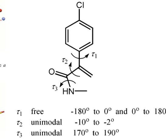
[image:2.610.46.298.617.701.2]translational and three orientational degrees of freedom corresponds to a total of nine degrees of freedom. The program DASH(David et al., 2006) was used for structure solution.DASHallows the torsion angles to be restricted to intervals that significantly reduce the search space. These three flexible torsion angles and their allowed ranges are shown in Fig. 4. The powder pattern was truncated to a real space resolution of 2.6 A˚ , which for Cu K1 radiation
corresponds to 34.6 in 2. The background was subtracted
with a Bayesian high-pass filter (David & Sivia, 2001). The number of SA runs was increased to 50 to get better statistics regarding reproducibility. The background subtraction, peak fitting, Pawley refinement and SA algorithms were used as implemented in the programDASH.
Accurate peak positions for indexing were obtained by fitting 20 manually selected peaks with an asymmetry-corrected Voigt function. Indexing was done with the program DICVOL91(Boultif & Loue¨r, 1991). A triclinic unit cell was determined withM(20)= 40.7 (de Wolffet al., 1968),F(20)= 82.8 (Smith & Snyder, 1979). From volume considerations, the unit cell can contain one molecule of P.R. 254 (Z = 1). The molecule has an inversion centre, which means the asymmetric unit must consist of a one half of the molecule.
Pawley refinement (Pawley, 1981) was carried out for refining the background, unit-cell parameters, zero-point error, peak-width and peak-asymmetry parameters. It allows extracting integrated intensities and their correlations. All intensities were refined without reference to a structural model and the result is the best fit that is theoretically possible: Rwp= 13.57,Rexp = 11.20,
2
= 1.467.
Suitable chemical restraints were applied for all bond lengths, valence angles and the planarity of the aromatic ring systems (including the five-membered condensed system). Anisotropic peak broadening was included to allow the peak profiles to be described accurately. The discrepancies between the observed and the calculated profile appeared to system-atically depend on thehklindices of the reflections, indicating preferred orientation in the [001] direction. The March– Dollase formula (Dollase, 1986) was used. The diffraction profiles and the differences between the measured and calculated profiles are shown in Fig. 5.
Acknowledgements
Professor Dr Martin Schmidt (Frankfurt University) is grate-fully acknowledged for the technical product P.R. 254 (TR008.052.11-F). Dr Lothar Fink and Edith Alig (Frankfurt University) are gratefully acknowledged for the collection of the powder diffraction patterns.
Funding information
Funding for this research was provided by: Deutscher Akademischer Austauschdienst, Forschungsaufenthalte fu¨r Hochschullehrer und Wissenschaftler.
References
Boultif, A. & Loue¨r, D. (1991).J. Appl. Cryst.24, 987–993.
Coelho, A. A. (2007).TOPAS Academic 4.1. http://members.optus-net.com.au/alancoelho
David, W. I. F., Shankland, K., van de Streek, J., Pidcock, E., Motherwell, W. D. S. & Cole, J. C. (2006).J. Appl. Cryst.39, 910– 915.
David, W. I. F. & Sivia, D. S. (2001).J. Appl. Cryst.34, 318–324. Dollase, W. A. (1986).J. Appl. Cryst.19, 267–272.
Groom, C. R., Bruno, I. J., Lightfoot, M. P. & Ward, S. C. (2016).Acta Cryst.B72, 171–179.
[image:3.610.315.565.69.231.2]Herbst, W. & Hunger, K. (2004). In Industrial Organic Pigments: Production, Properties, Applications, 3rd ed. Weinheim: Wiley-VCH.
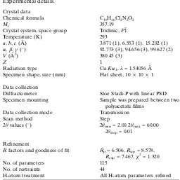
Table 2
Experimental details.
Crystal data
Chemical formula C18H10Cl2N2O2
Mr 357.19
Crystal system, space group Triclinic,P1
Temperature (K) 293
a,b,c(A˚ ) 3.871 (1), 6.553 (1), 15.292 (1)
,,() 92.773 (3), 94.656 (3), 99.627 (2)
V(A˚3) 380.45 (3)
Z 1
Radiation type CuK1,= 1.54056 A˚
Specimen shape, size (mm) Flat sheet, 10101
Data collection
Diffractometer Stoe Stadi-P with linear PSD
Specimen mounting Sample was prepared between two polyacetate films
Data collection mode Transmission
Scan method Step
2values (
) 2min= 2.00 2max= 60.00
2step= 0.01
Refinement
Rfactors and goodness of fit Rp= 6.506,Rwp= 8.578,
Rexp= 7.467,2= 1.320
No. of parameters 115
No. of restraints 44
H-atom treatment All H-atom parameters refined
Computer programs:WINXPOW(Stoe & Cie, 2004),TOPAS-Academic(Coelho, 2007),
[image:3.610.41.297.83.340.2]DASH3.1(Davidet al., 2006) andMercury(Macraeet al., 2008).
Figure 5
Macrae, C. F., Bruno, I. J., Chisholm, J. A., Edgington, P. R., McCabe, P., Pidcock, E., Rodriguez-Monge, L., Taylor, R., van de Streek, J. & Wood, P. A. (2008).J. Appl. Cryst.41, 466–470.
Pawley, G. S. (1981).J. Appl. Cryst.14, 357–361.
Smith, G. S. & Snyder, R. L. (1979).J. Appl. Cryst.12, 60–65. Stoe & Cie (2004).WINXPOW. Stoe & Cie, Darmstadt, Germany. Wolff, P. M. de (1968).J. Appl. Cryst.1, 108–113.
510
Ivashevskaya and Ivashevskaja Csup-1 Acta Cryst. (2017). E73, 507-510
supporting information
Acta Cryst. (2017). E73, 507-510 [https://doi.org/10.1107/S2056989017003309]
Crystal structure of Pigment Red 254 from X-ray powder diffraction data
Svetlana N. Ivashevskaya
Computing details
Data collection: WINXPOW (Stoe & Cie, 2004); cell refinement: TOPAS-Academic (Coelho, 2007); data reduction:
DASH3.1 (David et al., 2006); program(s) used to solve structure: DASH3.1 (David et al., 2006); program(s) used to
refine structure: TOPAS-Academic (Coelho, 2007); molecular graphics: Mercury (Macrae et al., 2008).
3,6-Bis(4-chlorophenyl)-2,5-dihydropyrrolo[3,4-c]pyrrole-1,4-dione
Crystal data
C18H10Cl2N2O2 Mr = 357.19 Triclinic, P1
a = 3.871 (1) Å
b = 6.553 (1) Å
c = 15.292 (1) Å
α = 92.773 (3)°
β = 94.656 (3)°
γ = 99.627 (2)°
V = 380.45 (3) Å3
Z = 1
Dx = 1.57 Mg m−3
Cu Kα1 radiation, λ = 1.54056 Å T = 293 K
Particle morphology: powder red
flat_sheet, 10 × 10 mm
Data collection
Stoe Stadi-P with linear PSD diffractometer
Radiation source: sealed X-ray tube Primary focussing, Ge 111 monochromator
Specimen mounting: sample was prepared between two polyacetate films
Data collection mode: transmission Scan method: step
2θmin = 2.00°, 2θmax = 60.00°, 2θstep = 0.01°
Refinement
Refinement on Inet
Least-squares matrix: full with fixed elements per cycle
Rp = 6.506 Rwp = 8.578 Rexp = 7.467
5800 data points
Excluded region(s): none
Profile function: fundamental parameters 115 parameters
44 restraints 0 constraints
All H-atom parameters refined
Weighting scheme based on measured s.u.'s w = 1/σ[Yobs)2
(Δ/σ)max = 0.001
Background function: Chebyshev function with 20 terms
Preferred orientation correction: March–Dollase formula (Dollase, 1986), (001) direction
Fractional atomic coordinates and isotropic or equivalent isotropic displacement parameters (Å2)
x y z Uiso*/Ueq
C1 0.8418 (7) 0.4738 (6) 0.6911 (3) 0.05373
supporting information
sup-2 Acta Cryst. (2017). E73, 507-510
C3 0.8543 (9) 0.1967 (6) 0.7905 (3) 0.05373
C4 1.0452 (8) 0.3351 (7) 0.8528 (3) 0.05373
C5 1.1403 (8) 0.5402 (7) 0.8377 (3) 0.05373
C6 1.0380 (9) 0.6065 (7) 0.7573 (3) 0.05373
C7 0.7380 (7) 0.5404 (6) 0.6038 (3) 0.05373
C8 0.4818 (10) 0.5527 (8) 0.4587 (4) 0.05373
C9 0.8702 (6) 0.7478 (4) 0.5715 (2) 0.05373
H2 0.606 (5) 0.157 (2) 0.6614 (15) 0.06447
H3 0.796 (4) 0.039 (2) 0.8095 (13) 0.06447
H5 1.286 (4) 0.644 (2) 0.8892 (16) 0.06447
H6 1.113 (4) 0.767 (2) 0.7484 (14) 0.06447
H7 0.774 (5) 0.879 (2) 0.4529 (17) 0.06447
O1 1.0679 (7) 0.8859 (6) 0.6108 (3) 0.05373
N1 0.7025 (7) 0.7478 (5) 0.4806 (3) 0.05373
Cl1 1.1601 (7) 0.2385 (4) 0.9528 (3) 0.05373
Geometric parameters (Å, º)
C1—C2 1.411 (6) C3—H3 1.08 (2)
C1—C6 1.388 (5) C5—H5 1.07 (2)
C1—C7 1.470 (6) C6—H6 1.06 (1)
C2—C3 1.392 (6) C9—O1 1.186 (4)
C3—C4 1.362 (6) C9—N1 1.485 (5)
C4—C5 1.369 (6) N1—C8 1.422 (5)
C5—C6 1.374 (6) N1—H7 0.98 (2)
C7—C9 1.492 (5) C4—Cl1 1.736 (6)
C2—H2 1.04 (2)
C2—C1—C6 117.3 (4) C2—C3—H3 125 (1)
C2—C1—C7 119.3 (3) C4—C3—H3 115 (1)
C6—C1—C7 123.4 (3) C6—C5—H5 122 (1)
C1—C2—C3 120.0 (3) C4—C5—H5 119 (1)
C1—C6—C5 122.5 (4) C7—C9—O1 126.8 (4)
C1—C7—C9 124.4 (3) C7—C9—N1 106.4 (2)
C2—C3—C4 119.8 (4) C9—N1—C8 108.8 (4)
C6—C5—C4 118.5 (4) O1—C9—N1 126.8 (3)
C3—C4—C5 121.8 (4) C3—C4—Cl1 116.6 (4)
C1—C2—H2 120 (1) C5—C4—Cl1 121.6 (3)
C3—C2—H2 119.7 (9) C9—N1—H7 113 (1)
C1—C6—H6 121 (1) C8—N1—H7 138 (1)
C5—C6—H6 116 (1)
Hydrogen-bond geometry (Å, º)
D—H···A D—H H···A D···A D—H···A
N1—H7···O1i 0.985 (17) 1.904 (18) 2.884 (5) 173 (2)
sup-3 Acta Cryst. (2017). E73, 507-510
C2—H2···N1iii 1.04 (2) 2.55 (2) 3.255 (6) 124.8 (11)
C6—H6···O1 1.062 (14) 2.28 (2) 2.959 (6) 119.9 (14)