ORIGINAL RESEARCH
FUNCTIONAL
Preoperative Mapping of the Supplementary Motor Area in
Patients with Brain Tumor Using Resting-State fMRI with
Seed-Based Analysis
X J. Wongsripuemtet,X A.E. Tyan,XA. Carass,XS. Agarwal,XS.K. Gujar,XJ.J. Pillai, andXH.I. Sair
ABSTRACT
BACKGROUND AND PURPOSE: The supplementary motor area can be a critical region in the preoperative planning of patients under-going brain tumor resection because it plays a role in both language and motor function. While primary motor regions have been successfully identified using resting-state fMRI, there is variability in the literature regarding the identification of the supplementary motor area for preoperative planning. The purpose of our study was to compare resting-state fMRI to task-based fMRI for localization of the supplementary motor area in a large cohort of patients with brain tumors presenting for preoperative brain mapping.
MATERIALS AND METHODS: Sixty-six patients with brain tumors were evaluated with resting-state fMRI using seed-based analysis of hand and orofacial motor regions. Rates of supplementary motor area localization were compared with those in healthy controls and with localization results by task-based fMRI.
RESULTS:Localization of the supplementary motor area using hand motor seed regions was more effective than seeding using orofacial motor regions for both patients with brain tumor (95.5% versus 34.8%,P⬍.001) and controls (95.2% versus 45.2%,P⬍.001). Bilateral hand motor seeding was superior to unilateral hand motor seeding in patients with brain tumor for either side (95.5% versus 75.8%/75.8% for right/left,P⬍.001). No difference was found in the ability to identify the supplementary motor area between patients with brain tumors and controls.
CONCLUSIONS: In addition to task-based fMRI, seed-based analysis of resting-state fMRI represents an equally effective method for supple-mentary motor area localization in patients with brain tumors, with the best results obtained with bilateral hand motor region seeding.
ABBREVIATIONS:DCS⫽direct cortical stimulation; rs-fMRI⫽resting-state fMRI; SMA⫽supplementary motor area; tb-fMRI⫽task-based fMRI
P
recise brain function localization is critical for neurosurgical procedures in which lesions are near the eloquent cortex. While direct cortical stimulation (DCS) is the criterion standard for assessing brain function, preoperative noninvasive functional brain imaging can aid in operative planning and decrease opera-tive time.1-4Task-based fMRI (tb-fMRI), a noninvasive technique for examining brain function, demonstrates good correlation with DCS for identification of eloquent brain regions, particu-larly in areas of sensorimotor and language representation.5,6Thus, fMRI is a valuable clinical tool for preoperative brain function localization.
There are, however, limitations to tb-fMRI, including dedi-cated hardware and software, trained personnel to administer tasks, designing a suitable task to interrogate a specific brain func-tion, and, most important, patient cooperafunc-tion, which may be difficult for subsets of patients with cognitive impairment or vanced neurologic disease. Resting-state fMRI (rs-fMRI) can ad-dress some of these limitations.7Instead of limiting interrogation to a specific brain function interrogated by a particular task, mul-tiple brain networks can be assessed with a single rs-fMRI acqui-sition. Because no specific task needs to be performed, rs-fMRI can be more readily applied in patients with neurologic deficits or cognitive disturbance and in young children. Finally, intrinsic brain networks have been shown to be present even during sleep or anesthesia, opening the possibility of using this technique in patients who otherwise may not be able to tolerate MR imaging.8,9 The sensorimotor network has been well-studied using rs-fMRI in the preoperative setting,10 with good agreement of
Received January 26, 2018; accepted after revision May 8.
From the Russell H. Morgan Department of Radiology and Radiological Sciences (J.W., A.E.T., S.A., S.K.G., J.J.P., H.I.S.) and Department of Neurosurgery (J.J.P.), Johns Hopkins University School of Medicine, Baltimore, Maryland; Department of Radi-ology (J.W.), Siriraj Hospital, Mahidol University, Bangkok, Thailand; and Depart-ment of Computer Science and DepartDepart-ment of Electrical and Computer Engineer-ing (A.C.), Johns Hopkins University, Baltimore, Maryland.
Please address correspondence to Haris Sair, MD, Russell H. Morgan Department of Radiology and Radiological Science, 600 N Wolfe St, Phipps B-100, Baltimore, MD 21287; [email protected]; @hsairmd
rs-fMRI with both tb-fMRI11and DCS.12While the sensorimotor network proper is readily characterized in these studies, there is, however, variability in depiction of the supplementary motor area (SMA) in these studies; in the study of Kokkonen et al,11the SMA was not reliably demonstrated in all subjects. In clinical tb-fMRI, often there are specific requests to localize the SMA due to adja-cent lesions.
Many postoperative brain tumor studies have shown that the degree of neurologic deficit after tumor resection correlates with the extent of resection of SMA regions.9,13Findings of transient contralateral weakness and transient speech deficits, including mutism (in the case of SMA resection in the dominant hemi-sphere) after resection or manipulation are well-established in the neurosurgical literature.14
To date, there are limited studies specifically evaluating the feasibility of rs-fMRI for SMA localization in patients with brain tumors. Vassal et al15recently published a study characterizing the pre- and postoperative SMA in 6 patients with brain tumors. With small numbers of subjects however, the effects of intersub-ject variability are limited; indeed, while early reports of rs-fMRI for preoperative language localization showed promise in a small number of subjects,162 subsequent independent analyses with larger sample sizes demonstrated a wide range of accuracy across subjects.3,17We therefore aimed to evaluate the utility of rs-fMRI using seed-based analysis in localizing the SMA in a larger cohort of patients with brain tumors. We hypothesized that the SMA can be readily identified using rs-fMRI in patients with brain tumors and is comparable with tb-fMRI.
MATERIALS AND METHODS
SubjectsIn this institutional review board–approved retrospective study, 113 patients with brain lesions who underwent preoperative func-tional MR imaging between May 2012 and June 2016 were iden-tified using the Johns Hopkins Hospital radiology information system. Forty-one patients were excluded due to lack of hand motor tb-fMRI and rs-fMRI performed during the same scanning session. Two patients were excluded due to artifacts. Four patients were excluded due to diagnoses other than brain tumors. After exclusion, data from 66 patients were used for analysis.
Additionally, 21 healthy subjects from the Kirby 21 Multi-Modal Reproducibility Study dataset (F.M. Kirby Center, Ken-nedy Krieger Institute, Baltimore, Maryland) were included in the study for comparison.18
Handedness
A laterality index for patients was calculated using the Edinburgh Handedness Inventory questionnaire.19
Lesion Characterization
Tumor location for each patient was determined from structural MR imaging. The World Health Organization histologic grade was recorded. Lesion volume (in cubic millimeters) was calcu-lated by manual ROI drawing on T2 fluid-attenuated inversion recovery images using the Medical Image Processing, Analysis and Visualization application.20
Image Acquisition
A 3T Trio Tim system (Siemens, Erlanger, Germany) was used with a 12-channel head coil. Structural images included a 3D T1 sequence (TR⫽2300 ms, TI⫽900 ms, TE⫽3.5 ms, flip angle⫽ 9°, FOV⫽24 cm, acquisition matrix⫽256⫻256⫻176, slice thickness⫽1 mm) and a 2D T2 FLAIR sequence (TR⫽9310 ms, TI⫽2500 ms, TE⫽116 ms, flip angle⫽141°, FOV⫽24 cm, acquisition matrix⫽320⫻240⫻50, slice thickness⫽3 mm).
2D gradient-echo-planar imaging was used for blood oxygen level– dependent functional imaging (TR⫽2000 ms, TE⫽30 ms, flip angle⫽90°, FOV⫽24 cm, acquisition matrix⫽64⫻64⫻ 33, slice thickness ⫽ 4 mm, slice gap ⫽ 1 mm, interleaved acquisition).
Given brain shifts due to mass lesions, tb-fMRI was performed in the brain tumor patient group to improve localization of the SMA. All patients underwent bilateral sequential finger-tapping tasks, and 50/66 patients additionally underwent a tongue motor task involving repetitive vertical tongue movements because their lesions were more in proximity to the ventral motor region. A block design of a 30-second alternation of task and rest was used for both tasks with 90 volumes acquired.
One hundred eighty volumes were acquired for rs-fMRI (⬃6 minutes). All patients were instructed to not move, keep their eyes closed, and not to think of anything during the acquisition.
Imaging details for the Kennedy Krieger Institute dataset are previously described.18
Image Analysis and Processing
FMRI was analyzed using Statistical Parametric Mapping (SPM; (http://www.fil.ion.ucl.ac.uk/spm/software/spm12) as well as custom Matlab (MathWorks, Natick, Massachusetts) scripts.
Both tb-fMRI and rs-fMRI underwent slice-timing correction, motion-correction, coregistration to the T1-weighted images, normalization to the Montreal Neurological Institute-152 tem-plate, and spatial smoothing using a 6-mm full width at half max-imum Gaussian kernel. For rs-fMRI, additional processing in-cluded tagging of outlier volumes based on large shifts in global average signal using the ArtRepair toolbox (http://cibsr.stanford. edu/tools/human-brain-project/artrepair-software.html) in SPM21 after motion correction, linear detrending, physiologic nuisance regression using CompCor,22bandpass filtering (0.01– 0.1 Hz) after normalization, and finally removal of outlier volumes tagged by ArtRepair (“scrubbing”).
Standard general linear model analysis was used for tb-fMRI with the canonical hemodynamic response function convolved with a boxcar function. No derivatives, global intensity normal-ization, or confound matrix was used. A 128-second high-pass filter was used. An autoregressive model was used to account for temporal autocorrelations. A design matrix was constructed to detect activation during the active task state compared with rest. SPM T-contrast maps were generated without clustering or mul-tiple comparison correction as is customary for single-subject clinical data at our institution.
mean time-varying signal across time within the ROI, this time course was used to generate Pearson correlation coefficients with time courses of every other voxel, generating a 3D correlation coefficient map across the entire brain:
1) Hand motor functional areas in bilateral brain hemispheres of each subject were determined by identifying the middle of the hand knob (⍀-shaped) of the primary motor cortex within the precentral gyri.
2) Bilateral orofacial motor functional areas were identified in the inferolateral part of the precentral gyrus, superior to the Syl-vian fissures, and below the imaginary line from the intersection of the inferior frontal sulcus and precentral sulcus on the sagittal view.
The anatomic coordinates were placed by a neuroradiologist with 3 years of experience following subspecialty training and supervised by a neuroradiologist with 6 years of experience, in-cluding in functional brain imaging.
SMA Positive-Connectivity Evaluation
For each patient, 6 whole-brain correlation maps were generated with ROIs in the following areas: 1) the left hand motor region, 2) the right hand motor region, 3) the left orofacial region, 4) the right orofacial region, 5) bilateral hand motor regions, and 6) bilat-eral orofacial regions. Final correlation maps were registered to the anatomic image to identify areas of functional connectivity with the SMA region. The SMA was defined as the area in the medial portion of the superior frontal gyrus in front of the primary motor cortex and superior to the cingulate sulcus. Its anterior boundary was defined by a line passing perpendicu-larly through the rostrum of the corpus callosum.23 Identifica-tion of the SMA was evaluated by 2 neuroradiologists with specific expertise in fMRI with consensus. Tb-fMRI was used to localize the SMA for confirmation in the patient group.
Statistical Analysis
The number of cases (as well as the percentage of total cases) in which the SMA was identified from rs-fMRI was recorded and reported for each of the 6 ROI schemes. Considering the de-pendent design, we used the McNemar2test to assess differ-ences in positive SMA identification when the ROI went from single to bilateral regions. All analysis was performed in R statistical and computing software (Version 3.2.4; http://www. r-project.org).
RESULTS
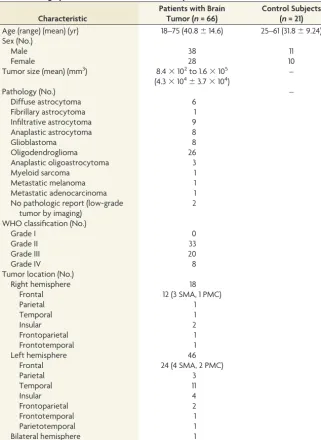
SubjectsOf the 66 patients, 38 were men and 28 were women, ranging from 18 to 75 years of age (mean, 40.8⫾14.6 years). For the 21 healthy control subjects, there were 11 men and 10 women, from 25 to 61 years of age (mean, 31.8⫾9.2 years).
Handedness
By means of the laterality index cutoff points of⫾0.2 (less than
⫺0.2⫽left-handedness,⬎0.2⫽right-handedness, and⫺0.2– 0.2⫽ambidexterity), in the patient group, the laterality index ranged from⫺0.72 to 1 (mean⫽0.696). Sixty-three patients
demonstrated right-handedness, and 3 patients demonstrated left-handedness.
For control patients, on the basis of the Kirby 21 Multi-Modal Reproducibility Study data base, 19 demonstrated right-handed-ness and 2 subjects demonstrated left-handedright-handed-ness. No laterality index information was available from this dataset.
Lesions
Of 66 patients, 46 had tumors in the left hemisphere (24 involving the left frontal lobe); 18, in the right hemisphere (12 involving the right frontal lobe); 1, in both frontal lobes; and 1 had multiple lesions throughout both hemispheres. In 7 patients, there was direct tumor involvement of the expected region of the SMA (4 on the left and 3 on the right side); in 3 patients, there was involve-ment of the primary motor cortex.
Fifty-three percent (35/66) of tumors were low-grade gliomas (6 diffuse astrocytomas, 1 fibrillary astrocytoma, 26 oligodendro-gliomas, and 2 that had not been histologically confirmed but appeared to be low-grade gliomas on imaging). Forty-two percent (28/66) of the lesions were high-grade gliomas (9 infiltrative as-trocytomas, 8 anaplastic asas-trocytomas, 3 anaplastic oligoastrocy-tomas, and 8 glioblastomas). The remaining patients were diag-nosed with myeloid sarcoma, metastatic melanoma, or metastatic adenocarcinoma. The tumor sizes ranged from 8.4⫻102to 1.6⫻ 105mm3(mean⫽4.3⫻104⫾3.7⫻104mm3).
The demographics of subjects, location, histopathology, and World Health Organization grading of the lesions are listed in
Table 1.
SMA Identification
In both the control and patient groups, the SMA was identified more frequently using seeding location from hand motor areas than from orofacial areas, with use of bilateral ROIs superior to the unilateral right or left motor ROI (FigureandTable 2).
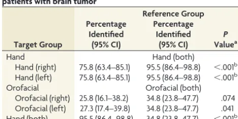
In the patients with brain tumors, the SMA was identified in 50/66 (75.8%) using the left hand motor ROI, 50/66 (75.8%) using the right hand motor ROI, 63/66 (95.5%) using the bilateral hand motor ROIs, 18/66 (27.3%) using the left orofacial ROI, 17/66 (25.8%) using the right orofacial ROI, and 23/66 (34.8%) using the bilateral orofacial ROIs (Table 3). A significant difference was seen between using bilateral hand motor ROIs versus either unilateral hand motor ROI (P⬍.001). In addition, a significant difference was seen between using bilateral hand motor ROIs versus bilateral orofa-cial ROIs (P⬍.001) (Table 3).
In the control group, the SMA was identified in 34/42 (81.0%) scans using the left hand motor ROI, 38/42 (90.5%) scans using the right hand motor ROI, 40/42 (95.2%) scans using the bilateral hand motor ROIs, 9/42 (21.4%) scans using the left orofacial ROI, 14/42 (33%) scans using the right orofacial ROI, and 19/42 (45.2%) scans using the bilateral orofacial ROIs. While no signif-icant difference was found between unilateral and bilateral ROIs for either hand or orofacial motor ROIs after multiple-compari-son correction, there was a significant difference between bilateral hand motor ROIs versus bilateral orofacial ROIs (Table 4).
DISCUSSION
There are operative challenges for brain tumors near the SMA, a functional brain region that is not uniformly anatomically bound according to well-established landmarks in contrast to the pri-mary motor cortex. Resection of the SMA, an area primarily in-volved in voluntary motor movement,24can result in motor and language deficits.25The degree of neurologic deficit corresponds to the extent of resection: Aphasia without motor impairment was seen with anterior SMA resection, likely extending into language-related regions termed “pre-SMA”; and manipulation extending to the posterior SMA was correlated with contralateral paresis, with a small percentage of cases having permanent deficits.13Because of these potential deficits, tb-fMRI has been used to help localize the SMA for operative planning, supplemented with DCS if necessary, though the SMA is often difficult to localize even with DCS.
A recent study using seed-based rs-fMRI was able to demon-strate the SMA in patients with gliomas using seeds placed in the
hand motor region.26We extended the investigation in a larger cohort of sub-jects by comparing the accuracy of SMA detection in patients with brain lesions with controls and used different sites of ROI placement, because the hand motor region may not always be feasible for ROI placement depending on the vol-ume and distribution of brain lesions. We demonstrate that the SMA can be identified in both patients with brain tu-mor and healthy controls using rs-fMRI with seed-based analysis. In a recent study comparing tb-fMRI primary hand motor activation with rs-fMRI using the task-based activation as the seed region for seed-based analysis, the authors re-ported that the SMA could be identified from the rs-fMRI but not from the tb-fMRI.27In our study, the seed location was blinded to tb-fMRI, thus represent-ing a possible scenario in which rs-fMRI may be used in isolation without tb-fMRI activation maps.
[image:4.594.56.378.55.496.2]Between the patients with brain tu-mor and healthy controls, there was no statistical difference in the rates of SMA detection with the use of hand motor seeding for rs-fMRI. Within the larger cohort of patients with brain tumor, the detection rate of SMA activation was sig-nificantly higher when selecting the hand motor region as the seeding area instead of the orofacial motor region. This result indicates stronger correla-tion between hand motor areas and the SMA compared with the orofacial mo-tor areas. This difference may be due to the variable connectivity of the dorsal-versus-ventral motor systems; indeed, when independent component analysis is performed, these 2 systems routinely are separated into differ-ent compondiffer-ents with currdiffer-ent acquisition and analysis methods. Underlying differences in dynamic connectivity may drive this phenomenon, with the ventral motor system demonstrating higher homotopic connectivity compared with the dorsal motor system.28In addition, our study demonstrated higher rates of SMA detection when using seeds from the bilateral hand motor areas compared with the unilateral hand motor areas, conceivably also due to potential differences in dynamic connectivity between the homotopic regions, which, when averaged, result in improved detection of the SMA. While the SMA is known to lateralize with lateralizing tasks, signal from the unilateral ROIs may be less robust to demonstrate the SMA with the standard methods used in our study. Further studies may investigate whether incorporating dynamic connectivity can improve SMA detec-tion, as well as determine whether the SMA may be lateralized Table 1: Demographic and clinical characteristics of patients and controls
Characteristic
Patients with Brain Tumor (n= 66)
Control Subjects (n= 21) Age (range) (mean) (yr) 18–75 (40.8⫾14.6) 25–61 (31.8⫾9.24) Sex (No.)
Male 38 11
Female 28 10
Tumor size (mean) (mm3) 8.4⫻102to 1.6⫻105 (4.3⫻104⫾3.7⫻104)
–
Pathology (No.) –
Diffuse astrocytoma 6
Fibrillary astrocytoma 1
Infiltrative astrocytoma 9
Anaplastic astrocytoma 8
Glioblastoma 8
Oligodendroglioma 26
Anaplastic oligoastrocytoma 3
Myeloid sarcoma 1
Metastatic melanoma 1
Metastatic adenocarcinoma 1
No pathologic report (low-grade tumor by imaging)
2
WHO classification (No.)
Grade I 0
Grade II 33
Grade III 20
Grade IV 8
Tumor location (No.)
Right hemisphere 18
Frontal 12 (3 SMA, 1 PMC)
Parietal 1
Temporal 1
Insular 2
Frontoparietal 1
Frontotemporal 1
Left hemisphere 46
Frontal 24 (4 SMA, 2 PMC)
Parietal 3
Temporal 11
Insular 4
Frontoparietal 2
Frontotemporal 1
Parietotemporal 1
Bilateral hemisphere 1
Multiple lesions 1
using unilateral ROIs with improved scanning, processing, and analysis methods.
Regarding various rs-fMRI techniques, prior studies have demonstrated high correspondence in overall connectivity results between seed-based analysis and independent component analy-sis methods.29Seed-based analysis requires a priori selection of ROIs, which can be somewhat challenging and may slightly differ
between study protocols. In particular, brain shifts or gyral expan-sion in patients with large brain tumors may cause a large amount of distortion that precludes accurate placement of seed ROIs. In-dependent component analysis overcomes this specific problem; however, identification of specific network subcomponents may be limited due to the number of targeted independent component analysis components. This may be the limiting factor in another study in which the SMA was inconsistently found using indepen-dent component analysis, which was thought to be due to network fragmentation related to independent component analysis or-der.11Our approach demonstrates that despite possible mass ef-fect, the SMA can be readily identified in nearly all patients using well-known anatomic landmarks that are not in the immediate vicinity of the area of interest. The question of SMA localization for operative planning is relevant when tumors are within or ad-jacent to the expected location of the SMA. Therefore, in most cases, the primary motor cortex may still be effectively used for ROI placement as shown in this study.
[image:5.594.54.285.47.228.2]There are several limitations to our study, one being that find-ings were not compared directly with the criterion standard of direct cortical stimulation. However, SMA evaluation using DCS is more challenging compared with evaluation of the primary mo-tor regions; indeed, at our institution, neurosurgical requests for primary motor function localization using fMRI are far less com-mon compared with requests for SMA localization, due to the difficulty of obtaining accurate results for the latter using DCS. For identification of the SMA, no independent assessment was performed between the 2 neuroradiologists, and consensus eval-uation was used. In addition, no quantitative analysis was per-formed; while there have been attempts to standardize threshold-ing of fMRI (eg, usthreshold-ing a percentage of the maximum local FIGURE. Mean group correlation maps across different seed
loca-tions for patients with brain tumor (bilateral hand motor seed in red, left hand motor in blue, right hand motor in green, left tongue motor in yellow, and right tongue motor in pink; please note that left and right denote the cerebral hemisphere of the seeds and thus represent contralateral functional regions). The greatest correlation in the supplementary motor area (arrowon the axial image) is seen with bilateral hand motor seeds. To a much lesser extent in this group visualization, left hand and right hand motor seeds demonstrate correlation in the SMA; however, neither tongue motor seed does so. R indicates right; L, left.
Table 2: Number of cases with identifiable SMA by seeding areas
ROI
Patient Control
+SMA (No.) (%)
−SMA (No.) (%)
+SMA (No.) (%)
−SMA (No.) (%) Hand (right) 50 (75.8) 16 (24.2) 38 (90.5) 4 (9.5) Hand (left) 50 (75.8) 16 (24.2) 34 (81.0) 8 (19.0) Hand (both) 63 (95.5) 3 (4.5) 40 (95.2) 2 (4.8) Orofacial (right) 17 (25.8) 49 (74.2) 14 (33.3) 28 (66.7) Orofacial (left) 18 (27.3) 48 (72.7) 9 (21.4) 33 (78.6) Orofacial (both) 23 (34.8) 43 (65.2) 19 (45.2) 23 (54.8)
[image:5.594.300.533.66.185.2]Note:—⫹SMA indicates that the SMA was identified;⫺SMA, the SMA was not identified.
Table 3: Identifiable SMA percentages compared across ROIs in patients with brain tumor
Target Group
Percentage Identified
(95% CI)
Reference Group Percentage
Identified (95% CI)
P
Valuea
Hand Hand (both)
Hand (right) 75.8 (63.4–85.1) 95.5 (86.4–98.8) ⬍.001b Hand (left) 75.8 (63.4–85.1) 95.5 (86.4–98.8) ⬍.001b
Orofacial Orofacial (both)
Orofacial (right) 25.8 (16.1–38.2) 34.8 (23.8–47.7) .074 Orofacial (left) 27.3 (17.4–39.8) 34.8 (23.8–47.7) .041 Hand (both) 95.5 (86.4–98.8) 34.8 (23.8–47.7) ⬍.001b aFrom the McNemar2test.
b
[image:5.594.302.533.226.335.2]Significant.
Table 4: Identifiable SMA percentages compared across ROIs in the control group
Target Group
Percentage Identified
(95% CI)
Reference Group Percentage
Identified (95% CI)
P
Valuea
Hand Hand (both)
Hand (right) 90.5 (76.4–96.9) 95.2 (82.6–99.2) .480 Hand (left) 81.0 (65.4–90.8) 95.2 (82.6–99.2) .041
Orofacial Orofacial (both)
Orofacial (right) 33.3 (20.0–49.6) 45.2 (30.2–61.2) .074 Orofacial (left) 21.4 (10.8–37.2) 45.2 (30.2–61.2) .004b Hand (both) 95.2 (82.6–99.2) 45.2 (30.2–61.2) ⬍.001b a
From the McNemar2
test.
b
[image:5.594.53.285.359.456.2]Significant.
Table 5: Number of cases with identifiable SMA across ROIs, control vs brain tumor group
SMA Identified
(No.) (%) P
Valuea Control Brain Tumor
Total 42 (100.0) 66 (100.0)
Hand (right) 38 (90.5) 50 (75.8) .055
Hand (left) 34 (81.0) 50 (75.8) .527
Hand (both) 40 (95.2) 63 (95.5) 1.000
Orofacial (right) 14 (33.3) 17 (25.8) .513 Orofacial (left) 9 (21.4) 18 (27.3) .649 Orofacial (both) 19 (45.2) 23 (34.8) .315 a
[image:5.594.52.286.504.621.2]signal),30there are no current established standards. Our qualita-tive method, however, actually represents a more real-world sce-nario, in which localization and thresholding of the SMA in tb-fMRI are performed using clinical judgment. Finally, the images of the patients with brain tumor were acquired on different scan-ners than for the healthy controls, potentially introducing scanner variability as a confounder in our results.
CONCLUSIONS
We demonstrate that rs-fMRI can be reliably used to identify the SMA in most patients with brain tumors, to the same degree as subjects with no brain lesions. We also demonstrate that the use of bilateral hand motor regions for seed placement is superior in identifying the SMA compared with unilateral hand motor or orofacial motor region seeding.
Disclosures: Jay J. Pillai—UNRELATED:Royalties: Springer Science & Business Media, Elsevier,Comments: royalty for books.
REFERENCES
1. Ojemann G, Ojemann J, Lettich E, et al.Cortical language localiza-tion in left, dominant hemisphere: an electrical stimulalocaliza-tion map-ping investigation in 117 patients. 1989. J Neurosurg2008;108: 411–21CrossRef Medline
2. Matthews PM, Honey GD, Bullmore ET.Applications of fMRI in translational medicine and clinical practice.Nat Rev Neurosci2006; 7:732– 44CrossRef Medline
3. Sair HI, Yahyavi-Firouz-Abadi N, Calhoun VD, et al.Presurgical brain mapping of the language network in patients with brain tu-mors using resting-state fMRI: Comparison with task fMRI.Hum Brain Mapp2016;37:913–23CrossRef Medline
4. Schneider FC, Pailler M, Faillenot I, et al.Presurgical assessment of the sensorimotor cortex using resting-state fMRI.AJNR Am J Neu-roradiol2016;37:101– 07CrossRef Medline
5. Branco P, Seixas D, Deprez S, et al.Resting-state functional mag-netic resonance imaging for language preoperative planning.Front Hum Neurosci2016;10:11CrossRef Medline
6. Bizzi A, Blasi V, Falini A, et al.Presurgical functional MR imaging of language and motor functions: validation with intraoperative elec-trocortical mapping.Radiology2008;248:579 – 89CrossRef Medline
7. Biswal B, Yetkin FZ, Haughton VM, et al.Functional connectivity in the motor cortex of resting human brain using echo-planar MRI. Magn Reson Med1995;34:537– 41CrossRef Medline
8. Palanca BJ, Mitra A, Larson-Prior L, et al.Resting-state functional magnetic resonance imaging correlates of sevoflurane-induced un-consciousness.Anesthesiology2015;123:346 –56CrossRef Medline
9. Sa¨mann PG, Wehrle R, Hoehn D, et al.Development of the brain’s default mode network from wakefulness to slow wave sleep.Cereb Cortex2011;21:2082–93CrossRef Medline
10. Dierker D, Roland JL, Kamran M, et al.Resting-state functional magnetic resonance imaging in presurgical functional mapping: sensorimotor localization.Neuroimaging Clin N Am2017;27:621–33
CrossRef Medline
11. Kokkonen SM, Nikkinen J, Remes J, et al.Preoperative localization of the sensorimotor area using independent component analysis of resting-state fMRI.Magn Reson Imaging2009;27:733– 40CrossRef Medline
12. Liu H, Buckner RL, Talukdar T, et al.Task-free presurgical mapping using functional magnetic resonance imaging intrinsic activity. J Neurosurg2009;111:746 –54CrossRef Medline
13. Fontaine D, Capelle L, Duffau H.Somatotopy of the supplementary motor area: evidence from correlation of the extent of surgical re-section with the clinical patterns of deficit.Neurosurgery2002;50: 297–303, discussion 303– 05Medline
14. Russell SM, Kelly PJ.Incidence and clinical evolution of postopera-tive deficits after volumetric stereotactic resection of glial neo-plasms involving the supplementary motor area. Neurosurgery
2003;52:506 –16; discussion 515–16CrossRef Medline
15. Vassal M, Charroud C, Deverdun J, et al.Recovery of functional connectivity of the sensorimotor network after surgery for diffuse low-grade gliomas involving the supplementary motor area.J Neu-rosurg2017;126:1181–90CrossRef Medline
16. Mitchell TJ, Hacker CD, Breshears JD, et al.A novel data-driven approach to preoperative mapping of functional cortex using rest-ing-state functional magnetic resonance imaging. Neurosurgery
2013;73:969 – 82, discussion 982– 83CrossRef Medline
17. Cochereau J, Deverdun J, Herbet G, et al.Comparison between rest-ing state fMRI networks and responsive cortical stimulations in gli-oma patients.Hum Brain Mapp2016;37:3721–32CrossRef Medline
18. Landman BA, Huang AJ, Gifford A, et al.Multi-parametric neuro-imaging reproducibility: a 3-T resource study.Neuroimage2011;54: 2854 – 66CrossRef Medline
19. Oldfield RC.The assessment and analysis of handedness: the Edin-burgh inventory.Neuropsychologia1971;9:97–113CrossRef Medline
20. McAuliffe MJ, Lalonde FM, McGarry D, et al.Medical image pro-cessing, analysis and visualization in clinical research.In: Proceed-ings of the IEEE Symposium on Computer-Based Medical Systems, Bethesda, Maryland. July 26 –27, 2001:381– 86
21. Mazaika PK, Fumiko H, Glover GH, et al.Methods and software for fMRI analysis for clinical subjects. In:Proceedings of the Annual Meeting of the Organization for Human Brain Mapping, San Francisco, California; June 15, 2009
22. Behzadi Y, Restom K, Liau J, et al.A component based noise correc-tion method (CompCor) for BOLD and perfusion based fMRI. Neu-roimage2007;37:90 –101CrossRef Medline
23. Chauvel PY, Rey M, Buser P, et al.What stimulation of the supple-mentary motor area in humans tells about its functional organiza-tion.Adv Neurol1996;70:199 –209Medline
24. Fried I.Electrical stimulation of the supplementary sensorimotor area.Adv Neurol1996;70:177– 85Medline
25. Ibe Y, Tosaka M, Horiguchi K, et al.Resection extent of the supple-mentary motor area and post-operative neurological deficits in gli-oma surgery.Br J Neurosurg2016;30:323–29CrossRef Medline
26. Qiu TM, Yan CG, Tang WJ, et al.Localizing hand motor area using resting-state fMRI: validated with direct cortical stimulation.Acta Neurochir (Wien)2014;156:2295–302CrossRef Medline
27. Hou BL, Bhatia S, Carpenter JS.Quantitative comparisons on hand motor functional areas determined by resting state and task BOLD fMRI and anatomical MRI for pre-surgical planning of patients with brain tumors.Neuroimage Clin2016;11:378 – 87CrossRef Medline
28. Syed MF, Lindquist M, Pillai JJ, et al.Dynamic functional connectiv-ity states between the dorsal and ventral sensorimotor networks revealed by dynamic conditional correlation analysis of resting state functional magnetic resonance imaging.Brain Connect2017;7: 635– 42CrossRef Medline
29. Rosazza C, Minati L, Ghielmetti F, et al.Functional connectivity during resting-state functional MR imaging: study of the corre-spondence between independent component analysis and region-of-interest-based methods.AJNR Am J Neuroradiol2012;33:180 – 87
CrossRef Medline