ABSTRACT
RHONDA M. TERRELL. The Potential Applications of Laser Raman Spectroscopy in the Field of Industrial Hygiene. (Under the Direction of DR. DAVID A. ERASER)
The feasability of using a portable laser Raman spectrometer in the field of industrial hygiene as a tool for the evaluation of environmental contaminants in an occupational setting was
completed. It was determined that three primary criteria must be met prior to development of a portable laser Raman system. These
conditions consist of technical criteria, need criteria and
resource criteria. The technical criteria was demonstrated, and
TABLE OF CONTENTS
Chapter Page
INTRODUCTION ii I. THEORY OF LASER RAMAN SPECTROSCOPY 1
Electromagnetic Radiation The Raman Effect
Type of Information Gained
Instrumentation
Laser Theory
Laser Applications in Raman Systems Comparison to Infrared Spectroscopy
II. APPLICATIONS OF LASER RAMAN SPECTROSCOPY 21
General Applications
Industrial Hygiene Related Applications Analysis of Specific Contaminants
III. FUTURE APPLICATIONS OF LASER RAMAN SPECTROSCOPY 33 IV. SUMMARY 36
11
INTRODUCTION
Raman spectroscopy has been a useful tool for chemists, physicists, physicians and scientists in many other disciplines since it was developed in the first half of the twentieth century. With the advent of laser technology in the last thirty
years, Raman spectroscopy has evolved into an extremely practical technique for investigation into the fundamental
structure and characteristics of molecules. As laser technology has become more efficient, inexpensive and reliable, laser Raman spectroscopy has developed into a potential tool for concentration determination and identification of various atmospheric
contaminants. The possibility of utilizing a laser as the single frequency light source for the stimulation of Raman emissions from
airborne gases, vapors and particulates, and the potential of such a system for the detection and analysis of contaminants warrant investigation into the feasability of using laser Raman
spectroscopy in the field of industrial hygiene and other related disciplines.
Raman spectroscopy is based on a light scattering process that was first predicted by A. Smekal, and later demonstrated by C.V. Raman in 1928(7). In general, the Raman effect is the
inelastic scattering of single frequency light by molecules and is indicative of the vibrational and rotational levels of the
molecules(12). A very weak phenomenon, Raman spectroscopy is often considered less significant than many of the other spectral
techniques, such as infrared spectroscopy. Its diverse
Ill
is a valuable technique.
In recent years, laser Raman spectroscopy has been used in
various ways. It has been used extensively in a variety of
scientific fields including chemistry, physics, biology, and
medicine. It has also been utilized in combustion diagnostics of
various engine exhaust emissions, vapor concentrationmeasurements, particulate and vapor identification, and pollution
monitoring. The purpose of this paper is to describe the theory
necessary for understanding Raman spectroscopy, state of the art
instrumentation, the past and current applications of this
technique and future potential applications of laser Raman
systems. Special emphasis is placed on the feasability of
developing a portable laser Raman spectrophotometer for use as an
analytical tool for the evaluation of environmental contaminants
THE POTENTIAL APPLICATIONS OF LASER RAMAN SPECTROSCOPY IN THE FIELD OF INDUSTRIAL HYGIENE
Rhonda M, Terrell
I. THEORY OF LASER RAMAN SPECTROSCOPY
Spectroscopy is an analytical technique based on the
exposure of a sample to various types of electromagnetic radiation
and interpretation of the radiation absorbed and emitted by the
substance. The absorption and emission of radiation is
indicative of the energy levels of the molecules, specifically,
the vibrational, rotational and electronic energy levels of the
molecules composing the substance(7). There are many different
kinds of spectroscopic techniques that utilize various ranges of
electromagnetic radiation to obtain specific spectra for a sample
under examination. Raman spectroscopy is one of many spectral
disciplines and detailed spectra have been compiled for aid in
i identification of any molecules considered. Figure 1 illustrates
typical Raman spectra for several common molecules.
Electromagnetic Radiation
Electromagnetic radiation consists of a spectrum of
frequencies which includes, but is not limited to, infrared,
visible and ultraviolet light. Light is considered to be both
wavelike and particulate at the same time(ll).
As a wave, electromagnetic radiation can be described as "the
process whereby energy is transferred from one oscillating dipole
to another at a distance"(12). The transmission of light through
A space occurs as fluctuating electric and magnetic fields oscillate
VEHTAHt
3^»o zrroo
SHIFT IN CM"'
lioo
IBiX>
ETHriEHt
—»---f-i—t---Pi^oFYLFNE"
'^^l
L ^^
i4
r- 1
\
^—-iSoa ifk>c
Sf4/F7 IN CM''
(>£p >JJ
BE"N7tN^
..U™..™
t—+—-TOLtt^MF
I §{iQ I ZOO '^OiJ 6ot>
SHtP'T 1{J cm''
&?«
i SC'O
SHIFT (Ki c-M'*
fe-C„; 3oo Butane
-,...,;...:-—-^ ͣ •ͣͣ...:'-'...-"'-ͣ
•...-:.-,---_„„ͣ.:,„-«->- ^ͣ».= .^
-1 -1 , '. : ,
1I.
J „ --J„.-^.
(S'Po iz^o "3oo too .ibo
SH'Tf ;Kj J'\'
FIGURE 1(33)
The Raman spectra of several hydrocarbons. The spectral_].ines
represent shifted frequency in the scattered light in cm or
The transmission of light through space is described as electric and magnetic fields oscillating at right angles to each other.
OM£ CSCLE
a ^ycies -per secor-K:i, Chcv-tz)
ye\ocii'\j of
i/ghf (^3 K 10'- do./.^^)
The wave theory equation for electromagnetic radiation;
V A = c
by the equation :
V A = c
where v equals the frequency of oscillation in hertz, A equals the
wavelength in centimeters and c equals the velocity of light. In
the infrared part of the spectrum, the frequency in hertz is a
large number and the wavenumber v, or reciprocal wavelength which
equals -r in units cm , is used to characterize a light wave.
In Raman spectroscopy the wavenumber is always used instead of the
frequency(7).
In particulate theory, light is described as packets of
energy called photons. This can be related by Planck's constant
as in the following equation:
E = h V
where E equals the energy of the photons, h equals Planck's
constant and v equals the frequency of oscillation. Molecules
absorb or emit this photon energy, thereby changing the energy of
the molecule. This change in energy is denoted by AE and is
commonly used in the form:
V = A E he
This equation can be expressed as "the wavenumber of the absorbed
or emitted photon is equal to the change in the molecular energy
term expressed in cm "(7).
The Raman Effect
scattering, absorption, or transmission. Several kinds of
scattering effects have been observed including Tyndall, Rayleigh
and Raman. In the Tyndall effect, electromagnetic radiation is
scattered by particulate matter suspended in air. Rayleigh
scattering occurs when molecules scatter light incident upon
it. In each of these effects, the scattering is considered to be
elastic; the wavelength of individual photons remains unchanged.
Raman scattering differs from Tyndall and Rayleigh scattering
because it is the inelastic scattering of electromagnetic
radiation. This means that light incident upon a molecule changes
the vibrational or rotational energy of the molecule. Becauseenergy is always conserved, the energy of the scattered photon
differs from the energy of the incident photon. The scattered
light varies in energy and frequency from its original state,
and it is this variance that is measured in Raman spectroscopy(7).
Consider a stream of photons colliding with a molecule. The
molecule temporarily absorbs these energy packets and is raised to
a higher unstable energy state. As the molecule returns to a more
stable state, it again interacts with a photon and scatters light.
If the molecule returns to its original energy level, the energy
of light scattered is equal to the light that excited the molecule
and Rayleigh scattering occurs. If however, the molecule returns
to an energy state different from its original level, the
scattered light has a different energy level and frequency, and
Raman scattering occurs(11). (See Figure 3.)
V:^ I
v=o>
R4YLt")6N
SCATTERING
STofctS
Line
FIGURE 3(11)
Molecules exist at an initial energy level. Upon exposure to
single frequency light, absorption followed by scattering occurs,
momentarily changing the existing energy level of the molecule.
In Rayleigh scattering, the emitted photons are equal to the
incident photons, the light that is scattered is the same
frequency as the incident light, and the molecules are returned to
their initial energy state without a net energy gam or loss. In
Raman scattering, the energy of the photons emitted is unequal to
the energy of the incident photons, the scattered light is at a
different frequency than the incident light, and the molecules are
returned to an energy level that is higher (Stokes) or lower
frequency shift depending on whether the molecule has a net energy
loss or gain following Raman emissions. If the molecule
experiences a net energy gain, it returns to a higher energy level
than its initial state. This is called Stokes scattering, and the
wavelength of the emitted light is longer than the wavelength of
the incident light. In contrast, anti-Stokes scattering occurs
when the molecule is returned to an energy level lower than its
initial state, and the light emitted has a shorter wavelength
than the incident light. Stokes scattering results in molecular
transitions from ground to excited states and are more frequent
and intense than anti-Stokes scattering. Raman scattering is an
inherently weak effect with low signal intensity, and for this
reason, the more intense Stokes scattering, representing an energy
gain to the molecule, is more commonly studied(23). Figure 3 also
illustrates Stokes versus anti-Stokes scattering.
Type of Information Gained
Raman spectroscopy provides both quantitative and qualitative
information about materials studied. The information gained is
generally at the molecular level and is characteristic of certain
atomic groups and chemical bonds. This information is indicated
by specific frequencies in the Raman spectrum. Functional groups
in a compound can be identified by group frequencies in the
spectrum to aid in chemical identification. Additionally, the
intensity of Raman spectrum bands are indicative of the
concentration of a compound of interest. This is even detectable
a substance can also be gained using micro-Raman spectroscopy,
which incorporates a microscope objective into the
spectrophotometer system. Micro-Raman spectroscopy can
distinguish between different geometric forms of a compound,
determine the degree of crystallinity in solids and define the
symmetry of polymers(23).
Instrumentation
A Raman spectrophotometer basically consists of a source of
single frequency illumination and a device for recording the
spectrum emitted(23). Raman's original experiments utilized
filtered sunlight as the illumination source, the scattered beam
was then filtered, and observed using the naked eye. Further
developments led to the use of the mercury lamp as the exciting
radiation and the scattered light was filtered and detected with a
simple spectrograph. With the development of the laser in the
early sixties, the Raman technique experienced a renaissance as a
good single frequency light source became commercially available.
"Modern instrumentation typically includes a laser as the source
of illumination and a double spectrometer, positioned at 90* to
the excitation beam, for analysis of the scattered light
spectrum"(23). Figure 4 shows a schematic drawing of a
typical Raman system.
Lasers provide intense, monochromatic light sources for
Raman spectroscopy. Coupled with this light source must be an
l^^/irr ^g' LA^Ee]—
ͣ
JAJ.V. SUPfVi
PHoTCt'-l
CoatJTi M 6
SY-ST£/v\
SV,5T£'M
M- Mirror
XKIT£(2FEJ?.ENt£
-4—--'
ͣ
.'M,DEWlc^
I / f
I ,/ , L£H5 i /O
^1^
I
lEHS
I
1 1/ M
/
1/ '
1/
l/
fe ll % J
If ' ' Mc
Do(^ble S-raima Specrirome^r-„,...j
FIGURE 4(21)
10
capabilities to obtain Raman spectra in the presence of the more
intense Rayleigh scatter. (Remember that Rayleigh scatter is
elastic, i.e. the scattering is identical to the frequency of the
exciting beam, and must be filtered out for Raman emissions to be
detected.) Generally, a double spectrometer with two gratings and
two sets of entrance and exit slits is used to minimize the
unwanted radiation from Rayleigh scattering, and maximize the
Raman signal. Single spectrometers are feasable when the sample
under consideration has intense Raman emissions, and the frequency
of the Raman scatter is significantly different from the frequency
of the incident radiation. Additionally, improved spectrometer
gratings provide greater stray light rejection and performance in
the frequency region close to the exciting line(13). For samples
that have weak Raman emissions and a minimal frequency shift,
however, the converse is true and triple spectrometers are
required for detection(20).
The resulting filtered radiation is usually detected by a
red sensitive photomultiplier tube (PMT). Multialkali or gallium
arsenide PMT's with high quantum efficiency and thermoelectrically
cooled housing to reduce background noise are often used.
Microcomputers are usually used for storage and analysis of the
detector signal. Many commercially available systems combine
spectrometer control with data analysis in the microcomputer
system(23).
A recent innovation in Raman spectroscopy is the modification
of the Raman instrument to incorporate a microscope as the sample
11
investigation of microscopic samples non-destructively, and has
been used in various applications including geochemistry, thesemiconductor industry and corrosion analysis(23). Figure 5
illustrates the schematic drawing of a typical Raman microprobe
system. Other innovations include the development of a new
optical filter element called the crystalline colloidal Bragg
diffraction device. This new filter accurately rejects the
excitation wavelength, but passes the Raman scattered light. It
has been noted of these filters that "The experimental
sensitivity, the signal-to-noise ratios, and the detection limits
of these new instruments closely approach the theoretical limit.
The resulting simplification of the instrumentation, the decreased
cost, and the increased sensitivity should play a pivotal role in
making Raman spectroscopy one of the routine optical spectroscopic
techniques"(2). Other future innovations in Raman spectroscopy
could include the use of fiber optics to provide Raman stimulation
at remote areas and return Raman scattered light to thecorresponding spectrophotometer(20).
There are currently several manufacturers of Raman system.s in
the United States. Three such companies include Spex Industries,
Edison, New Jersey, EG&G Princeton Applied Research, Princeton,
New Jersey, and SA Instruments, Metuchen, New Jersey. These firms
supply Raman systems primarily to laboratories and industry. A
complete Raman system is estimated to cost in the $100,000
12
TV
i-OPTICS Q oiii;cr/v£s
^
^
-
€
)
miHk LBN^
^
BMN
iK'aDt.MT
sT/te^" o
0 0—
T£AWSMjtT&D
iLUJMfMATloN
' MlA?.(<^o^^.
FIGURE 5(24)
13
Laser Theory
The laser, which is an acronym for "light amplification by
stimulated emission of radiation", was first developed in the
early sixties. Basically filled with a medium such as a crystal,
gas or semiconductor, a laser consists of a chamber of specific
length bounded on both ends with parallel mirrors. These mirrors
reflect light waves back and forth and excite the atoms of the
medium. The laser chamber is designed to be a specific length
dependent on the active medium used. The atoms of the medium give
off light as light hits them thus amplifying the light source. By
making one mirror partially transparent, the amplified lightemerges as a high power single frequency laser beam(5). Figure 6
provides a schematic drawing of a typical laser source.At the atomic level, stimulated emission "is initiated by a
photon whose energy equals the exact difference in energy between
two energy states or levels of an electron. Such a photon can
cause an electron in the upper excited level to fall to the lower
level and radiate its energy as a new photon so there are now two
photons instead of one"(7). If enough electrons are brought to
this excited level, and new photons are continually created, the
intensity of this energy increases at a given wavelength of light
and a single frequency laser beam emerges(7).
Lasers are typically classified by two properties, source
type and wavelength at which maximum power is provided. Lasers
are either continuous wave or pulsed source and maximum power
wavelength is often called the principal line. Laser selection is
14
PRISM
ruBE
[RESONATOR
REFLECTOR APERTURE
OUTPUT COUPLER
spLirreR
POWER SUPPLY PHOTO
HARD-SEAL
CRYSTALLINE QUARTZ
BREWSTER WINDOW
DOUBLE COOLED MAGNET
GAS
RESERVOIR
F-ORMED COPPER :up ION SHIELD
TUNGSTEN DISK
GAS RETURN HOLES
FIGURE 6(6)
15
lasers include argon ion, krypton ion, and helium-neon. Pulsed lasers can be include dye lasers which offer tunable excitation frequencies. Dye lasers utilize a set of interchangeable dyes to provide continuous selection of laser frequencies from the
ultraviolet through the infrared. Figure 7 provides a table of lasers commonly used in Raman systems and their principal
lines(23)•
Laser Applications in Raman Systems
The advent of laser technology provided an excellent source of radiation for spectroscopic studies. Lasers are "extremely
intense, sharply focusable and virtually single frequency" and
offer an excellent means for exploring molecular structure(5). The Raman effect provides for the effects of the rotation and vibration of molecules to be transferred from the infrared to the visible range of the spectrum where less complex sources
facilitate examination and analysis(5).
Utilization of lasers as the single frequency light source in Raman spectroscopy provides for several advantages over
conventional light sources. These advantages include:
1) observation of the Raman scattered light which would be far too weak to be seen using incoherent sources,
2) better resolution of Raman lines (lasers provide extremely narrow linewidth as compared to large linewidth from
conventional sources),
3) increase in the Raman scattering (which increases as light
16
LASER EMISSION WAVELENGTHS COMMONLY USED TO EXCITE RAMAN SPECTRA
Lasing Medium Wavelength (s), nm
He-Ne 632.8 Ar"*" 4 88.0
514.5 Kr"*" 5 30.9
647.1
FIGURE 7(13)
17
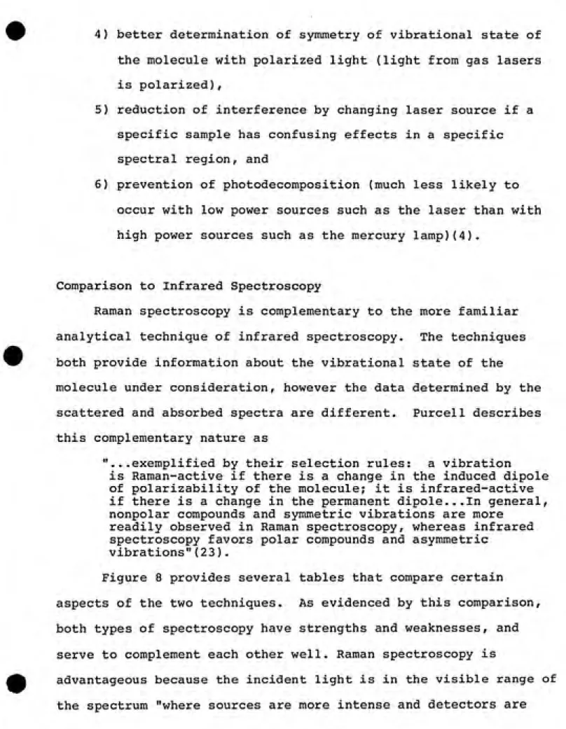
4) better determination of symmetry of vibrational state of the molecule with polarized light (light from gas lasers
is polarized),
5) reduction of interference by changing laser source if a specific sample has confusing effects in a specific spectral region, and
6) prevention of photodecomposition (much less likely to occur with low power sources such as the laser than with
high power sources such as the mercury lamp)(4).
Comparison to Infrared Spectroscopy
Raman spectroscopy is complementary to the more familiar
analytical technique of infrared spectroscopy. The techniques both provide information about the vibrational state of the
molecule under consideration, however the data determined by the scattered and absorbed spectra are different. Purcell describes
this complementary nature as
"...exemplified by their selection rules: a vibration
is Raman-active if there is a change in the induced dipole of polarizability of the molecule; it is infrared-active if there is a change in the permanent dipole...In general, nonpolar compounds and symmetric vibrations are more
readily observed in Raman spectroscopy, whereas infrared spectroscopy favors polar compounds and asymmetric
vibrations"(23).
Figure 8 provides several tables that compare certain
aspects of the two techniques. As evidenced by this comparison,
both types of spectroscopy have strengths and weaknesses, and
serve to complement each other well. Raman spectroscopy is
advantageous because the incident light is in the visible range of
18
COMPARISON OF INSTRUMENTATION FOR RAMAN AND INFRARED SPECTROSCOPY
Raman Infrared
Complexity Source Detector
Resolution (cm )
Principal limitation Wave number range
Purge Requirement Photometry moderate laser photomultiplier tube 0.15 energy 10-4000 no scattering single slightly greater blackbody or diode
laser thermal, pyroelectric bolometers 0.05 energy 180-4000 yes absorption double beam
COMPARISON OF QUALITY OF INFORMATION PROVIDED BY RAMAN AND
INFRARED SPECTROSCOPY Raman Infrared Fingerprinting Best vibrations Structural Group frequencies Aqueous solutions Quantitative analysis Low-frequency modes Libraries excellent symmetric excellent excellent excellent fair excellent fair excellent asymmetric very good excellent very difficult good difficult excellent FIGURE 8(23)
Tables comparing characteristics of Raman and infrared
19
COMPARISON OF SAMPLING HANDLING IN RAMAN AND INFRARED SPECTROSCOPY
Raman Infrared
General Applicability (%)
Unsuitable Materials Sample Preparation liquids powders single crystals polymers single fibers gases and vapors Cells
Micro work
mass ( g)
size ( m)
Trace work
High & low temperature
Limits of detection
95 fluorescent and colored samples very simple very simple very simple very simple very simple moderately simple (w/microscope) moderately simple very simple (glass) good <1 1 sometimes moderately simple good 99 single crystals, metals, aqueous solutions variable very simple more difficult very difficult more difficult difficult simple more complex (alkali halide) good <1 20 sometimes moderately simple very good
20
much more sensitive than in the infrared"{23). The entire Raman spectrum is relatively short and can be recorded in a single scan, however, a change of detector is required for infrared
spectroscopy below 180cm . Sample preparation is facilitated
in Raman spectroscopy because infrared requires mixing or grinding
of the sample. In contrast, infrared is more commonly documented
II. APPLICATIONS OF LASER RAMAN SPECTROSCOPY
General Applications
The current applications of laser Raman spectroscopy are
widespread and growing. One Raman systems manufacturer advertises
that its services have been extended to the diverse markets of
"Aerospace, Biochemical, Chemical Analysis, Communications, Electro Optics, Materials Analysis, Metallurgical, Nuclear,
Optical, Petrochemical, Pharmaceuticals, Plastics, Semiconductors,
Superconductivity"(15). The list is certainly impressive and not
surprising, for as one studies Raman spectroscopy, it becomes evident that the applications are numerous. The primary
applications of the Raman technique have been utilized by industry and laboratory personnel with different objectives than the
industrial hygienist, yet these diverse applications provide insight into the potential of using Raman spectroscopy as an analytical tool for the evaluation of occupational exposures.
The most conventional applications of Raman spectroscopy are in the fields of biology, chemistry and physics. The technique has been used extensively in determination of molecular structure and the kinetics of chemical reactions. An additional application
has been the study of the formation, bonds and dynamics of chemical
and biological macromolecules. These techniques provide only a sample of the many applications of this tool in the scientific
community(34).
Also extensive is the list of applications in the industrial
22
considered state of the art in Raman technology because it
"allows one to examine a surface spectroscopically so as to
discover non-uniformities which may be present and elucidate their
chemical nature"(13). This technique has found application in the
semiconductor industry to examine the surface of semiconductors
non-destructively(21). It has also been used in the production of
various polymers to detect impurities that are not detectable withconventional techniques(30). The Raman microprobe has proved to
be a useful tool for metallurgical failure analysis such as
investigation into oil field drill pipe deficiencies(25). Other
interesting applications of the Raman microprobe include
microanalysis of biological tissues and geological samples.
Biopsies of tissue with a Raman microprobe can reveal microscopic
foreign matter(31). Microscopic inclusions in geological samples
indicative of sample formation can be identified(28). The Ramanmicroprobe can also be used in art conservation, where Raman can
be used to determine authenticity by identifying the type of art
materials used non-destructively, or diagnose the cause of
deterioration(29). Other applications include characterization of
ceramic microstructures, and analysis of localized laser induced
damage to laser processed solids in industry(10,23).
Conventional Raman applications are less numerous, yet
equally varied. Raman spectroscopy has been used to analyze
headspace gas in pharmaceutical ampules aiding in automation of
quality control procedures(3). Measurements of the Raman23
atmosphere(8). Clearly Raman spectroscopy is an analytical
technique with extremely varied and valuable applications in the
scientific and industrial community.Industrial Hygiene Related Applications
Industrial hygienists are faced with many complexities when
trying to assess employee exposures to airborne contaminants. Air
sampling strategies and the tools they require are varied, each
with positive and negative characteristics. Industrial hygiene
sampling tools must be accurate, portable and economical relative
to the task specified. Accuracy is critical to ensure employee
safety and legal compliance. Portability can be described as
small and lightweight enough for one person to carry comfortably
and is a key factor because the industrial hygienist must obtain
samples in very diverse settings. The criteria of economics is
subjective and depends on a variety of factors including the
severity of the health hazard, the extent of known health effects,
the frequency of use and the availability of other less expensive
methods. Additionally, if samples require analysis to obtain
results, analytical methods must be timely so that excessive
exposure situations can be rectified quickly.
Many standard sampling procedures have obvious pros and cons.
Absorption tubes, such as charcoal or silica gel, used with
sampling pumps, have many pros including accuracy, portability and
cost. In contrast, absorption tubes require chemical analysis
which has the potential for error and is time consuming. The time
24
industrial hygienist is faced with employees who are suffering adverse health effects. Direct reading instrumentation has positive and negative characteristics as well. The primary
advantage is the availability of immediate results. Many direct reading instruments, such as the Miran infrared analyzer, are also advantageous because they provide a physical measurement of the contaminant. The analysis is based on physical
characteristics of the molecules of the substance and are not
subject to the types of potential error inherent in chemical analysis. Drawbacks include size and cost. Direct reading
instruments are generally larger than air sampling pumps and more costly. Additionally, if the instrument can not be worn by an employee over a period of time as the sampling pump can,
time-weighted averages are difficult to obtain. An effective industrial hygiene program should employ both types of methods using each appropriately after examining the specific factors of the situation.
Laser Raman spectroscopy is an attractive tool because it offers the typical advantages of a direct reading instrument. Based on the physical characteristics of the molecules of a
substance, laser Raman spectroscopy has the potential to provide a direct reading instrument that could accurately identify and
determine concentrations of contaminants of interest to the
industrial hygienist.
In recent years a variety of literature has been published describing the potential uses of laser Raman spectroscopy in
25
experimental Raman systems have been developed and many
contaminants of interest in environmental monitoring investigated for their Raman spectra and ease in identification using laser Raman spectroscopy. To date, a portable Raman spectroscopic
system is not commercially available, but advances in state of the art electronics may provide modifications to current systems such that portability could be achieved.
Environmental analysis using Raman spectroscopy has
historically been in the area of pollution monitoring including evaluation of combustion diagnostics and engine exhaust emissions in automobiles and aircraft, and air pollution monitoring using
remote Raman instrumentation.
Robert E. Setchell of Sandia Laboratories has presented the feasability of utilizing laser Raman spectroscopy for construction of an analytical model representing emission formation processes within an internal combustion engine chamber. The automobile industry is faced with the challenge of emission reduction to comply with increasingly strict federal and state standards without compromising fuel consumption or production costs.
Emission formation is difficult to assess because of its
dependence on temperature and gas composition gradients present
within the combustion chamber. Setchell argues that laser Raman
spectroscopy is an appropriate analytical tool because of its
ability to determine concentration measurements at discrete points within the chamber. Setchell's work is valuable because he
demonstrates the capacity of using laser Raman spectroscopy for
26
drawbacks to the Sandia Laboratories system is the size of the
experimental laser Raman system. The system measures
contaminants within present and proposed combustion engines in a laboratory chamber, and is not portable enough to be used by the
industrial hygienist(27).
A similar study was conducted by A. Zurloye and D.A.
Santivicca at Princeton University. Gas temperature and carbon monoxide measurements were determined for an internal combustion engine using laser Raman spectroscopy. Prior to the utilization of laser Raman spectroscopy, in-cylinder measurements were
conducted using fast acting gas sampling probes. The measurements were accompanied by uncertainty due to the length of sampling time and problems related to the intrusion of the probe into the
chamber. Laser Raman spectroscopy, however, is an excellent analytical tool for this application because instantaneous measurements at discrete points in the post flame gases were possible without the intrusion of a probe to disrupt the concentration gradient. Zurloye and Santivicca were able to
obtain quantitative measurements of carbon monoxide concentration and determine the temperature gradient in the chamber. Their spectroscopic apparatus, however, was also utilized
solely in a laboratory setting, and was not portable enough to be
moved from its stable position(35).
Another research program with comparable applications was developed at Avco Everett Research Laboratory by Donald A, Leonard. Leonard demonstrated the utilization of laser Raman
27
of aircraft engine exhaust emissions. This research is
particularly interesting because the laser Raman system hardware was assembled into a transportable lidar unit. Lidar is a system
similar to radar that transmitts light, as opposed to radio waves, to a reflecting object to detect pollutants in the atmosphere. Two units, a transceiver and operator enclosure, were constructed for utilization in the field at the source of emission. "The transceiver package contains the laser, transmitter and receiver optics, monochromator and detection subsystems and supporting equipment such as cooling, pumping and power conditioning. The operator enclosure package contains the control and display panel and an enclosure for the operator and is capable of remote
positioning from the systems package and turbine exhaust region for operator safety and comfort" (See Figure 9). This laser Raman spectroscopic system proved to be successful for its "portable"
capabilities and it's elimination of probe placement into the
exhaust stream(17,18).
Another remote laser Raman system is described by Stanley Freeman in his book Applications of Laser Raman Spectroscopy. Freeman describes remote Raman instrumentation as "a logical extension of LIDAR which employs a single-ended, remote, range resolved measurement of light backscattered from particles to give a three dimensional plot of their concentration in the
atmosphere..."{11). This single-ended system is ideal for pollution studies and could be used extensively in plume
monitoring from smoke stacks. The technique is advantageous in
28
FIGURE 9(18)
Schematic of laser Raman aircraft engine exhaust emissions
29
moisture content or other natural constituents in the air. It
does have, however, reduced effectiveness in rain and fog and
involves large instrumentation. The prototype described operates
from a truck trailer(ll).
Freeman notes "the remote Raman method is sufficiently
3
sensitive (about 10 ppm or about 10 mg/m ) to measure emitted
pollution but can not detect it when dispersed in the atmosphere."
Figure 10 provides a table of typical detection limits of a remote
laser Raman system. As these detection limits evidence, laser
Raman spectroscopy has the sensitivity to detect contaminants of
interest in an occupational setting. This application, however,
is also limited by the size of instrumentation(11).
Freeman proposes several interesting potential applications
for remote laser Raman spectroscopy. These applications include
monitoring thermal pollution, tracer techniques for climatic
information, measurement of HCl at rocket launches, and detection
and determination of water pollutants. As these and the other
previously described applications prove, laser Raman spectroscopy
is a viable technique for the evaluation of environmental
contaminants. The immense sensitivity required to detect the
effect, however, requires large stable instrumentation that may be
difficult to apply in an occupational setting(ll).
Another application of interest in environmental monitoring
and the evaluation of occupational exposures is the use of a Raman
microprobe for identification of airborne particulates. This
application, however, does not provide direct reading
30
TYPICAL DETECTION LIMITS OF A REMOTE RAMAN SPECTROMETER
(Range 250 meters. Sample Path 10 meters)
Compound ppm
Nitrogen 46
Sulfur hexafluoride 6 Sulfur dioxide 4
Nitrous oxide 17 Nitric oxide 585
Ammonia 11
Water (vapor) 13 Benzene (aerosol) 2 Kerosine (aerosol) 1
Methanol (aerosol) 16 Nitric acid (aerosol) 3
Organophosphates (aerosol) 4
FIGURE 10(11)
31
application in the laboratory setting for analysis of particulate samples collected by conventional methods adapted slightly to the analytical requirements of the system. Modified multi-stage
cascade impactors were utilized to collect particulates from out-stack samplings from oil fired power plants. A number of inorganic particles collected were considered to be
"microparticles" and the laser Raman spectroscopic system was found to be accurate and reliable. Identification of various sulfates, nitrates and phosphates established this method as a
viable analytical tool(9).
Analysis of Specific Contaminants
As well as development of various spectroscopic systems, extensive work has been completed on the specific contaminants particularly good for characterization by laser Raman
spectroscopy. The Raman spectra of many contaminants of interest in occupational and environmental analysis have been developed and
documented.
Joan Laane and K. Krishan provide an excellent reference on environmental pollutants that can be detected by their Raman
spectra. The Raman spectra of nitrogen, oxygen, carbon monoxide, hydrogen chloride, nitric oxide, chlorine, hydrogen fluoride,
water vapor, sulfur dioxide, carbon dioxide, ammonia, acetylene,
ethane, hydrogen chloride gas in the presence of water vapor and
oxygen dissolved in water is detailed. Laane and Krishan's
32
such as laser power, detector sensitivity and collection
efficiency could increase the detection limit by four orders of
magnitude resulting in concentration detection in the ppb
range(16).
Additionally, Raman spectroscopy for some hydrocarbons,
including methane, benzene, toluene, and pentane, and some freons
have been compiled. These chemicals represent more complicated
Raman spectra, yet sufficient detection capabilities have been
demonstrated(33).
Clearly, contaminants of interest to the industrial hygienist
are detectable using the Raman technique with current
instrumentation. The application is limited, however, by the size
of instrumentation, yet as the technique develops further,
III. FUTURE APPLICATIONS OF LASER RAMAN SPECTROSCOPY
It has been noted of Raman spectroscopy that "lack of
suitable instrumentation impeded its early developent as an
analytical technique"(23). It appears that this statement applies to current development of the technique as the diversity of
applications are limited primarily by the instrumentation required to complete the task desired(19). This is certainly the case with Raman applications in the field of industrial hygiene, yet it is hopeful that state of the art instrumentation will again catch up with the theoretical uses of this analytical technique.
The primary constraints limiting utilization of laser Raman spectroscopy in industrial hygiene applications are size
requirements of instrumentation. Currently a portable instrument is not commercially available, yet it is important to determine whether criteria necessary for development are being met.
First, is the technology available? Is it technically possible to measure and identify contaminants using laser Raman spectroscopy?
Secondly, does the need for the technique exist? Thirdly, are the resources available to provide research into size reduction?
These three criteria must be addressed before laser Raman
spectroscopy can develop as a tool for the industrial hygienist. The first condition is the one most easily met. Technically, laser Raman spectroscopy has been demonstrated to be a viable
34
different from the exciting wavelength such that Rayleigh
scattering can be easily filtered are especially good candidates for this method.
Secondly, portable laser Raman systems must fill a gap in existing technology. Before a portable laser Raman system can be developed, a need for the technique and a market for the
application must be clearly identified. The need must be
established by determining if problems with current analytical
methods exist for certain chemicals where laser Raman spectroscopy
could provide solutions. Additionally, the market itself must
meet certain conditions. The market must be aware of the need for
the technique and must be large enough to provide resources with
no guaranteed results.
It is important to remember that new techniques are rarely propagated through the scientific community and adopted quickly.
A standard concensus must be reached before the scientific and
industrial community will accept a new method that varies from
previously accepted techniques. Acceptance is dependent upon the new technique's competition with standard procedures, and it must
be demonstrated that the new method will significantly improve state of the art techniques(19).
The third criteria for development of a portable laser Raman
system is resource commitment. These constraints are more of a political nature, and unfortunately, no less important. Industrial hygienists form an extremely small portion of the scientific
35
remain relatively unaware of laser Raman spectroscopy as an
analytical tool. These conditions possibly hinder the development of laser Raman spectroscopy as an analytical tool for the
industrial hygienist more than the size limitations of the
instrument. It can be theorized that with sufficient funding and research, laser Raman spectrocopy could become a valuable
IV. SUMMARY
Laser Raman spectroscopy is a valuable analytical technique with diverse applications in both scientific and industrial areas.
Instrumentation is rapidly advancing and the list of detectable contaminants increasing dramatically. Interest in the technique is growing and the list of viable applications is varied and long. Computerization of both equipment and available data is more
common and laser technology is sophisticated enough to provide an excellent light source. It has been noted that "the future of Raman spectroscopy appears bright, with promises of faster
acquisition times, better and more flexible sources and completely
automated systems"(23).
Constraints on the application of laser Raman spectroscopy in the field of industrial hygiene exist, yet if certain technical,
need and resource criteria are met, these can be overcome. Laser
Raman spectroscopy has the potential for becoming a viable
technique for the industrial hygienist to use in the evaluation of
V. REFERENCES
1. Adar, F., I.H. Campbell, and P.M. Fauchet. "Raman Microprobe Study of Changes Induced by a Pulsed Laser" in Microbeam
Analysis 1985. edited by J.T. Armstrong. San Francisco:
San Francisco Press, Inc., 1985.
2. Asher, Sanford A., Perry L. Flaugh, and Guy Washington.
"Crystalline Colloidal Bragg Diffraction Devices: the Basis for a New Generation of Raman Instrumentation." Spectroscopy Vol. 1, No. 12, pp. 26-31.
3. Bailey, Glen F. and Herbert A. Moore, Jr. "Non-destructive
Headspace Analysis in Pharmaceutical Ampules by 5145 A Laser
Raman Spectroscopy." Journal of the Parenteral Drug.
Association March-April 1980, Vol. 34, No. 2, pp. 127-133. 4. Beesley, M.J. Lasers and Their Applications London: Taylor &
Francis, Ltd. 1972.
5. Brotherton, M. Masers and Lasers - How They Work, What They
Do. New York: McGraw Hill. 1964.
6. Coherent product literature. "Innova Ion Laser Systems: The
Essence of Innovation." 1986.
7. Colthup, Norman B., Lawrence H. Daly and Stephen E. Wiberly.
Introduction to Infrared and Raman Spectroscopy. New York:
Academic Press. 1975.
8. Cooney, J.A. "Measurements on the Raman Component of Laser Atmospheric Backscatter." Applied Physics Letters. January,
1968, Vol. 12, No. 2, pp. 40-42.
9. Etz, Edgar S., Gregory J. Rosasco and William C. Cunningham. "The Chemical Identification of Airborne Particles by Laser Raman Spectroscopy." in Environmental Analysis edited by Galen W. Ewing. New York: Academic Press, Inc. 1977.
10. Fauchet, P.M., I.H. Campbell and F. Adar. "Long Range Material Relaxation after Localized Laser Damamge." Applied Physics Letters September, 1985, Vol. 47, No. 5, pp. 479-481.
11. Freeman, Stanley K. Applications of Laser Raman
Spectroscopy. New York: John Wiley & Sons. 1974.
12. Gilson, T.R. and P.J. Hendra. Laser Raman Spectroscopy. London: John Wiley & Sons. 1970.
13. Grasselli, Jeanette G., Marcia K. Snavely and Bernard J. Bulkin. "Applications of Raman Spectroscopy." Physics
38
14. Hargis, P.J. Jr. "Trace Detection of Small Molecules by Pulsed Ultraviolet Laser Raman Spectroscopy" in SPIE Vol. 286 Laser Spectroscopy for Sensitive Detection edited by Jerry A.
Gelbwachs, 1981, pp. 139-145. 15. Instruments SA product literature.
16. Laane, Joan and K. Krishnan. "Detection of Environmental Pollutants using Laser Raman Spectroscopy." NTIS Document
#AD 776 290/9. 1973.
17. Leonard, Donald A. "Field Tests of a Laser Raman Measurement
System for Aircraft Engine Exhaust Emissions." NTIS Document
#AD A003 648/3. 1974.
18. Leonard, Donald A. "Measurement of Aircraft Turbine Engine Exhaust Emissions." in Laser Raman Gas Diagnostics edited by Marshall Lapp and C.M. Penney, 1974, pp. 45-61.
19. Malchow, Doug, EG&G Princeton Applied Research, Princeton, NJ. Personal Communication, February 17, 1987.
20. Perrulli, Bruce, Instruments, SA, Metuchen, NJ. Personal
Communication, February 17, 1987.
21. Pollack, Fred H. "Semiconductor Characterization by Raman Spectroscopy." Test and Measurement World May, 1985, pp. 2-10.
22. Purcell, F.J. "A New Spectrograph with a Multichannel Optical Detector for the Raman Characterization of Microparticles." in Microbeam Analysis-1982 edited by K.F.J. Heinrich.
San Francisco: San Francisco Press, Ltd. 1982.
23. Purcell, F.J. "Raman Spectroscopy" in Encyclopedia of Materials Science and Engineering edited by Michael B. Bever. London: Pergamon Press, Ltd. 1986. pp. 4061-4067. 24. Purcell, F.J. and R. Heidersbach. "Use of the Raman probe for
Metallurgical Failure Analysis." American Laboratory March 1985, pp. 119-121.
25. Purcell, F.J. and R. Heidersbach. "The Use of Raman Microprobe for Failure Analysis of Oil Field Drill Pipe." in Microbeam Analysis-1985 edited by J.T. Armstrong. San Francisco: San Francisco Press, Ltd. 1985, pp. 57-59.
26. Purcell, F.J. Spex Industries, Edison, NJ. Personal Communciation, February 17, 1987.
27. Setchell, R.E. "Experimental Investigations of Local Emission Mechanisms Within Present and Proposed Internal Combustion
39
28. Spex Industries. Technical Note R-5, "Micro-Raman Geological
Investigations." 3 March 1983.
29. Spex Industries. Technical Note R-3, "Micro-Raman Applications
in Art Conservation." 4 February 1983.
30. Spex Industries. Technical Note R-2, "Micro-Raman Polymer Production." 1 February 1983.
31. Spex Industries. Technical Note R-1, "Micro-Raman
Microanalysis of Biological Tissues." 25 January 1983. 32. Stenholm, Stig. Foundations of Laser Spectroscopy New York:
John Wiley & Sons. 1984.
33. Stephenson, David A. "Raman Spectroscopy of Some Hydrocarbons
and Freons in the Gas Phase." in Laser Raman Gas
Diagnostics edited by Marshall Lapp and CM. Penney, 1974,
pp. 361-367
34. Walker, Charles T. "Light Scattering", in Laser Applications to Optics and Spectroscopy, edited by Stephen F. Jacobs. Massachusetts: Addison Wesley Publishing Co., 1975.
35. Zurloye, A. and D.A. Santivicca. "Temperature and
Concentration Measurements in an Internal Combustion Engine using Laser Raman Spectroscopy." Journal of American