Publicly Accessible Penn Dissertations
Summer 8-12-2011
The Generation of Fully Functional β-Cells by
Proliferation: Lessons From Pregnancy and
HNF4α
Sebastian Rieck
University of Pennsylvania, [email protected]
Follow this and additional works at:http://repository.upenn.edu/edissertations
Part of theEndocrinology Commons,Medical Cell Biology Commons,Medical Genetics Commons,Medical Molecular Biology Commons,Medical Physiology Commons,Pharmacology Commons, and thePhysiological Processes Commons
This paper is posted at ScholarlyCommons.http://repository.upenn.edu/edissertations/984
Recommended Citation
Rieck, Sebastian, "The Generation of Fully Functional β-Cells by Proliferation: Lessons From Pregnancy and HNF4α" (2011).Publicly Accessible Penn Dissertations. 984.
Pregnancy and HNF4α
Abstract
Diabetes mellitus is an increasingly prevalent metabolic disorder that is estimated to affect over 300 million people by 2025. Common to either type 1 or type 2 diabetes is a progressive inadequacy of functional β-cell mass. Recent studies have shown that during times of prolonged metabolic demand for insulin, the endocrine pancreas can respond by increasing β-cell mass, both by an increase in cell size and by changes in the balance of β-cell proliferation and apoptosis. Advances that further our knowledge of the molecular factors that control both β-cell proliferation and survival will be crucial for understanding the homeostasis of β-cell mass during adulthood, and are pivotal for any attempt to use instructive cues to induce the proliferation of terminally differentiated fully functional insulin-producing β-cells that are suitable for transplantation. However, no systematic study that investigates the expression profile of the islet’s response to pregnancyin vivo, a physiological state of insulin resistance, has been reported thus far.
In the first part of my thesis, I characterized the gene expression signature of pancreatic islets during pregnancy by performing large-scale expression profiling of islets isolated from 4- to 5-month-old non-pregnant and non-pregnant female mice at day 14.5 of gestation, the peak of β-cell proliferation. I identified a total of 1,907 genes as differentially expressed, and demonstrated the induction of both proliferative and survival pathways in the islet during pregnancy. A comparison of our pregnancy gene set with two additional models of islet expansion suggests that diverse mechanisms can be recruited to expand islet mass. One of the genes that is required for β-cell proliferation during pregnancy in mice is the transcription factor HNF4α.
In an attempt to translate knowledge gained using the pregnancy paradigm, I hypothesized that HNF4α is a human β-cell mitogen. To address this question, in the second part of my thesis, I employed adenoviral-mediated overexpression of a pancreas-specific isoform of HNF4α (HNF4α8) in primary human islets. HNF4α8 stimulated β-cells to enter the cell cycle, and led to a greater than 300-fold increase in the number of β-cells that entered S-phase, without detectable change in glucose stimulated insulin secretion. However, HNF4α8 overexpressing β-cells showed signs of cell cycle arrest, caused by activation of the DNA damage response associated with replication stress, ultimately resulting in a senescence-like phenotype independent of caspase-dependent apoptosis. Overexpression of HNF4α8 together with known β-cell mitogens, also further increased cell cycle entry of β-cells, strengthening the argument that HNF4α8 is a mitogenic signal in the human β-cell. Additionally, I observed a substantial proportion of β-cells stimulated to enter the cell cycle by CDK6 and CYCLIN D3 to also exhibit both markers of cell cycle arrest and double stranded DNA damage. In summary, the DNA damage response is a barrier to efficient human β-cell proliferationin vitro, and as such I suggest its evaluation in future attempts to stimulate β-cell replication.
Degree Type Dissertation
Degree Name
First Advisor Klaus H. Kaestner
Keywords
beta-cell proliferation, diabetes mellitus, regeneration
Subject Categories
CELLS BY PROLIFERATION: LESSONS FROM
PREGNANCY AND HNF4α
Sebastian Rieck
A DISSERTATION
In
Pharmacology
Presented to the Faculties of the University of Pennsylvania
In Partial Fulfillment of the Requirements for the Degree of Doctor of Philosophy
2011
______________________________ ______________________________
Dissertation Advisor Graduate Group Chairperson
Klaus H. Kaestner, Ph.D. Vladimir Muzykantov, M.D., Ph.D.
Professor, Genetics Professor, Pharmacology
Dissertation Committee:
Dr. Doris A. Stoffers, M.D. Ph.D. Dr. Bryan A. Wolf, M.D. Ph.D.
Associate Professor, Medicine Professor, Pathology and Laboratory
Medicine
Dr. Catherine L. May, Ph.D. Dr. Judy L. Meinkoth, Ph.D.
Dedication
To my mother, father, and brother for their unconditional love, support, and teaching me the value of hard work
To my grandfather for being a great role model, and teaching me the value and the enthusiasm of „learning for fun‟
Acknowledgements
A large part of my ability to perform science at a rigorous level depends on the
collaborations that I have setup and maintained during my time at the University of
Pennsylvania. To this end, I am grateful to acknowledge all of my colleagues and
collaborators in the Kaestner laboratory, especially now great friends Dr. Nan Gao and
Dr. Irina M. Bochkis, Dr. Rana K. Gupta, Ms. Vasumathi Kameswaran, and Ms. Rieke
Schwegmann for their advice and direct contributions to the work comprising this thesis.
Also, I thank Dr. Noa Weinberg-Corem, Dr. Karyn L. Sheaffer, Dr. Jia Zhang for
valuable suggestions on the thesis manuscript.
I wish to thank collaborators outside of the Kaestner laboratory, namely, Dr. Alan
D. Attie, Dr. Mark P. Keller, Dr. Jeremy A. Lavine and Mrs. Mary Rabaglia for insightful
discussions, providing adenoviruses, providing murine leptin-deficient cDNA samples,
and teaching me techniques such as islet isolation and arterial delivery of adenoviruses
into a mouse. Additionally, I thank Dr. Christopher B. Newgard, Dr. Thomas C. Becker
and Dr. Michelle Arlotto for producing and providing adenoviruses, Dr. Phillip E.
Scherer and Dr. Zhao V. Wang for providing murine PANIC-ATTAC cDNA samples,
and Dr. Andrew F. Stewart and Dr. Karen K. Takane for providing both CYCLIN D3 and
CDK6 adenoviruses. Furthermore, I acknowledge Dr. Charles Pletcher and Dr. Paul
Hallberg for their expertise in GFP- sorting.
I am grateful for the members of the Functional Genomics Core (Dr. Peter White,
Dr. Jonathan Schug, Mr. Alan J. Fox, Mrs. Olga Smirnova) for help with microarray
expression profiling, Dr. Arbansjit K. Sandhu and members of Gene Therapy Program
and members of the Radioimmunoassay and Biomarkers Core for help with detection of
human insulin. I thank both Mrs. Neena Panackal and members of the Children‟s
Hospital of Philadelphia Research Institute Pathology Core and Dr. Gary Swain and
members of the University of Pennsylvania Morphology core for assistance with
histology. I am especially grateful to Dr. Jake A. Kushner who has supported, challenged
and encouraged me since my first endeavors into human β-cell biology. I am also very
obliged for the training grant support I received from the graduate group in
Pharmacology for 4 years, and the advice provided by my thesis committee, namely Dr.
Doris A. Stoffers, Dr. Bryan A. Wolf, Dr. Catherine L. May, and Dr. Judy L. Meinkoth.
Finally, I am most appreciative to my advisor, Dr. Klaus H. Kaestner, whose
patience, and commitment to me does not go unnoticed. As a scientific mentor, his belief
in me and my work has given me a foundation of knowledge and confidence to be not
only to a successful post-doctoral fellow, but also have the ability and self-belief to
pursue a fruitful academic career as an independent investigator. The opportunities he has
given me to succeed have all had great positive impacts on my life which will never be
Abstract
THE GENERATION OF FULLY FUNCTIONAL β-CELLS BY
PROLIFERATION: LESSONS FROM PREGNANCY AND HNF4α
Sebastian Rieck
Advisor: Dr. Klaus H. Kaestner, Ph.D.
Diabetes mellitus is an increasingly prevalent metabolic disorder that is estimated
to affect over 300 million people by 2025. Common to either type 1 or type 2 diabetes is
a progressive inadequacy of functional β-cell mass. Recent studies have shown that
during times of prolonged metabolic demand for insulin, the endocrine pancreas can
respond by increasing β-cell mass, both by an increase in cell size and by changes in the
balance of β-cell proliferation and apoptosis. Advances that further our knowledge of the
molecular factors that control both β-cell proliferation and survival will be crucial for
understanding the homeostasis of β-cell mass during adulthood, and are pivotal for any
attempt to use instructive cues to induce the proliferation of terminally differentiated fully
functional insulin-producing β-cells that are suitable for transplantation. However, no
systematic study that investigates the expression profile of the islet‟s response to
pregnancy in vivo, a physiological state of insulin resistance, has been reported thus far.
In the first part of my thesis, I characterized the gene expression signature of
pancreatic islets during pregnancy by performing large-scale expression profiling of islets
isolated from 4- to 5-month-old non-pregnant and pregnant female mice at day 14.5 of
differentially expressed, and demonstrated the induction of both proliferative and survival
pathways in the islet during pregnancy. A comparison of our pregnancy gene set with two
additional models of islet expansion suggests that diverse mechanisms can be recruited to
expand islet mass. One of the genes that is required for β-cell proliferation during
pregnancy in mice is the transcription factor HNF4α.
In an attempt to translate knowledge gained using the pregnancy paradigm, I
hypothesized that HNF4α is a human β-cell mitogen. To address this question, in the
second part of my thesis, I employed adenoviral-mediated overexpression of a
pancreas-specific isoform of HNF4α (HNF4α8) in primary human islets. HNF4α8 stimulated
cells to enter the cell cycle, and led to a greater than 300-fold increase in the number of
β-cells that entered S-phase, without detectable change in glucose stimulated insulin
secretion. However, HNF4α8 overexpressing β-cells showed signs of cell cycle arrest,
caused by activation of the DNA damage response associated with replication stress,
ultimately resulting in a senescence-like phenotype independent of caspase-dependent
apoptosis. Overexpression of HNF4α8 together with known β-cell mitogens, also further
increased cell cycle entry of β-cells, strengthening the argument that HNF4α8 is a
mitogenic signal in the human β-cell. Additionally, I observed a substantial proportion of
β-cells stimulated to enter the cell cycle by CDK6 and CYCLIN D3 to also exhibit both
markers of cell cycle arrest and double stranded DNA damage. In summary, the DNA
damage response is a barrier to efficient human β-cell proliferation in vitro, and as such I
Table of Contents
Chapter I: Introduction
1
Diabetes Mellitus as a candidate for cell replacement therapy 2
Homeostatic control of β-cell mass 7
Intracellular glucose metabolism controls β-cell proliferation 15
Reversible β-cell mass expansion during pregnancy 17
Regulation of the cell cycle in β-cells 22
Attempts to expand functional β-cells by proliferation in vitro 29
Maturity Onset Diabetes of the Young (MODY) transcription factors regulate 37
proliferation and survival in the adult β-cell
Hepatocyte Nuclear Factor- 4α: The MODY1 Gene, β-cell function and β-cell 39
proliferation
Summary and Specific Aims 47
References 49
Chapter II: The Transcriptional Response of the Islet
61
to Pregnancy in Mice
Abstract 62
Introduction 63
Results 65
Discussion 87
Materials and Methods 92
References 101
Chapter III: Overexpression of Hepatocyte Nuclear Factor- 4α 106
initiates cell cycle entry, but is not sufficient to promote β-cell
expansion in human islets
Abstract 107
Introduction 109
Results 111
Discussion 142
Materials and Methods 147
References 152
Chapter IV: Conclusions and Future Directions
157
Conclusions 158
Future Directions 167
List of Tables
Table 2.3: 73
MOST DIFFERENTIALLY EXPRESSED GENES DURING PREGNANCY
DAY 14.5
Table 2.7: 84
GENE ONTOLOGY (GO) FUNCTIONS SIGNIFICANTLY ENRICHED DURING
List of Illustrations
Figure 1.1 5
THE MECHANISMS BY WHICH NEW FULLY FUNCTIONAL β-CELLS
CAN BE GENERATED
Figure 1.2 11
THE ANATOMY OF THE MOUSE AND HUMAN ISLET
Figure 1.3 13
HOMEOSTATIC CONTROL OF β-CELL MASS IN RODENTS AND HUMANS
Figure 1.4 20
β-CELL DYNAMICS DURING PREGNANCY IN THE MOUSE
Figure 1.5 27
REGULATION OF β-CELL PROLIFERATION BY CELL CYCLE GENES
Figure 1.6 35
CHALLENGES THAT NEED TO BE OVERCOME TO SUCCESSFULLY
EXPAND FUNCTIONAL β-CELLS BY PROLIFERATION IN VITRO
Figure 1.7 43
HNF4α ISOFORMS AND THEIR FUNCTIONAL DOMAINS
Figure 1.8 45
LOCATION OF SNPS ASSOCIATED WITH LATE-ONSET TYPE 2
Figure 2.1: 67
ß-CELL PROLIFERATION WITH β-CELL HYPERTROPHY
DRAMATICALLY INCREASE β-CELL MASS AT DAY 14.5 OF PREGNANCY
IN MICE
Figure 2.2: 69
HISTOLOGICAL ANALYSIS OF β-CELL PROLIFERATION, HYPERTROPHY,
AND MASS AT DAY 14.5 DURING PREGNANCY
Figure 2.4: 74
TEMPORAL GENE EXPRESSION ANALYSIS OF SELECTED
DIFFERENTIALLY EXPRESSED GENES THROUGHOUT PREGNANCY
Figure 2.5: 77
SEPARATION OF GFP+ AND GFP- POPULATIONS FROM MIP-GFP MICE
Figure 2.6: 79
DIFFERENTIALLY EXPRESSED GENES IDENTIFIED IN THE ISLET ARE
EXPRESSED IN β-CELLS DURING PREGNANCY DAY 14.5
Figure 2.8: 85
GENE EXPRESSION PROFILES DIFFER BETWEEN THE PREGNANCY,
OBESITY, AND β-CELL INJURY MODELS OF β-CELL REGENERATION
Figure 2.9: 99
KNOWN MECHANISMS RESPONSIBLE FOR β-CELL GAIN DURING
PREGNANCY
Figure 3.1: 112
Figure 3.2: 116
HNF4αHIGH β-CELLS INCORPORATE BRDU IN A PUNCTATE, NOT
DIFFUSE, MANNER
Figure 3.3: 118
THE RATE OF PROLIFERATION OF PDX1+ CELLS UPON RECEIPT OF
HUMAN ISLET DONATIONS IS EXTREMELY LOW
Figure 3.4: 122
HNF4αHIGH β-CELLS ARREST IN THE CELL CYCLE
Figure 3.5: 127
OVEREXPRESSION OF HNF4α8 LEADS TO ACTIVATION OF THE DNA
DAMAGE RESPONSE ASSOCIATED WITH REPLICATION STRESS
Figure 3.6: 132
A SENESCENCE-LIKE PHENOTYPE IS THE PREDOMINANT FATE OF
β-CELLS OVEREXPRESSING HNF4α8
Figure 3.7: 136
HNF4α8 SYNERGIZES WITH FACTORS KNOWN TO BE SUFFICIENT FOR
PROMOTING CELL CYCLE ENTRY IN HUMAN β-CELLS
Figure 3.8: 140
OVEREXPRESSION OF CYCLIN D3 AND CDK6 ALSO ACTIVATES THE
DNA DAMAGE RESPONSE IN HUMAN β-CELLS
Figure 4.1: 165
Figure 4.2: 170
DERIVATION OF A LOXP ALLELE FOR THE CONDITIONAL ABLATION OF
Chapter I:
Diabetes mellitus as a candidate for cell replacement therapy
Diabetes mellitus is a metabolic disorder characterized by the loss of a single cell
type, the insulin producing β-cell, which then leads to failed glucose homeostasis. The
phenotype of chronic elevation of glucose in the bloodstream, shared by all forms of
diabetes, can lead to severe and life-threatening complications including cardiovascular
disease, renal disease, and blindness (1). Diabetes mellitus is characterized by either an
absolute insulin deficiency due to the autoimmune destruction of pancreatic insulin-
producing β-cells (type 1), or relative insulin deficiency due to diminished insulin
secretion and/or decreased insulin sensitivity as a result of insulin resistance in peripheral
tissues (type 2). Both forms of diabetes are caused by complex interactions between
genetics, environment and lifestyle choices (2), and accepted forms of treatment for
diabetes reflect two basic mechanisms whereby one can increase insulin secretion; to
mimic the ability of the β-cell to secrete insulin in response to glucose by injections of
recombinant insulin and increase β-cell mass in diabetic patients by islet transplantation.
Since the discovery of insulin in the 1920s by Dr. Frederick Banting and Dr.
J.J.R. Macleod, tight control of glucose levels by use of an intensive exogenous insulin
therapy can be achieved. However, while this improves the life of a diabetic patient
considerably, with side effects including an increasing frequency of hypoglycemic
episodes and a lack of prevention of long- term complications (1, 3), insulin therapy
cannot provide the fine-tuned control of glucose homeostasis ensured by an individual‟s
endogenous β-cell. It has been shown that both individuals with type 1 and type 2
groups) reported an inverse relationship between blood glucose levels and β-cell volume
below a certain threshold (7), illustrating that another important target in the treatment of
diabetes mellitus is the correction of β-cell mass deficit.
Indeed in 1999, the Edmonton protocol, or first successful transplantation of
cadaveric islets into brittle type 1 diabetic patients using a glucocorticoid-free
immunosuppressive regimen resulting in temporary insulin independence (8), has caused
a paradigm shift towards the development of diabetes therapies centered upon improving
β-cell mass in diabetic patients. While being able to show insulin independence and near
normal control of blood glucose levels for up to a year post- transplant (8, 9), transplant
success has proven to be short-lived and accompanied by significant side effects, such as
impairment of islet function (10), nephrotoxicity (11), and possibly also impairment of
β-cell proliferation (12), all caused from the use of immunosuppressive regimes
themselves. Furthermore, the application of islet transplantation is limited by the current
lack of sufficient organ availability and the availability of β-cells from deceased donors
cannot meet the demand. The use of potential regeneration-compatible
immunosuppressive drugs for islet transplantations, and alternative methods, such as
tissue engineering (for example, growth of exogenous islets in an encapsulated coating
designed to prevent rejection) (13, 14), are being investigated, and great interest into the
mechanisms of β-cell proliferation has emerged.
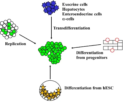
Specifically, significant attention has been focused to the formation of new fully
functional β-cells. Several methods of β-cell generation have been investigated including
the replication of pre-existing β-cells (15), transdifferentiation of endoderm-derived cell
differentiation from progenitors (controversially thought to reside in ducts or originating
during development and maintained into adulthood) (18, 19), and differentiation from
human embryonic stem cells (hESC) in vitro (20) (Figure 1.1). For type 1 diabetes, being
able to expand β-cell mass ex vivo, or in vivo after islet transplantation, could both
increase the number of patients that can be treated with a limited supply of donor islets
and improve the outcome after transplantation. For type 2 diabetes, the identification of
targets and pathways that mediate proliferation and/or apoptosis might lead to the
development of novel drugs that stimulate β-cell growth in the patient and thus allow for
Figure 1.1. The mechanisms by which new fully functional β-cells can be generated.
Approaches toward generating new β-cells for the treatment of type 2 diabetes or for cell
replacement therapy for type 1 diabetes include the use of instructive cues to induce the
replication of terminally differentiated β-cells in vitro or in vivo, the direct
reprogramming of endoderm-derived cell populations into β-cells by transdifferentiation
in vitro or in vivo, the differentiation of purified pancreatic progenitor cells in vitro, and
differentiation of human embryonic stem cells (hESC) in vitro. Green-filled circles
represent fully functional adult β-cells, blue stars represent terminally differentiated cells
of endoderm origin, red circles represent pancreatic progenitor cells (potentially residing
Homeostatic control of β-cell mass
The β-cell population is restricted to the endocrine pancreas or islets of
Langerhans, and is required for insulin production and secretion. The exocrine pancreas
is composed predominantly of acinar cells which secrete digestive enzymes, and duct
cells which transport these digestive enzymes out of the pancreas and into the duodenum.
The endocrine pancreas is highly innervated by blood vessels and apart from endothelial
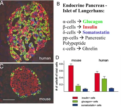
cells, nerves and fibroblasts, is primarily composed of five endocrine cell types: the
glucagon producing α-cell, insulin producing β-cell, somatostatin producing δ-cell,
pancreatic polypeptide producing pp-cell, and ghrelin producing ε-cell. Together, the
islets constitute about 1-2% of total pancreas volume, and range in size from just a few
cells to several thousand cells (21). It should be noted that the three dimensional islet
structure and relationship between endocrine cells differ between rodent and human
islets, however, the significance of this concerning the maintenance of β-cell mass is
unknown (Figure 1.2) (22, 23).
An organism‟s β-cell mass is determined by the product of the number and size of
its pancreatic β-cells. In adult mammals, β-cell mass is maintained by the balance
between cell renewal and growth (cell replication, hypertrophy, neogenesis), and cell loss
(cell death, atrophy, autophagy) (Figure 1.3A). It is well established that adult pancreatic
β-cells replicate very slowly after the establishment of β-cell mass during the neonatal
period (a period of high β-cell replication rates both in rodents and in humans) (24-26).
The absolute death rate of β-cells is extremely low, and counters the slow rate of β-cell
is a subject of controversy (28, 29), a recent study demonstrates that all β-cells of an islet
are similarly capable of numerous cell divisions (30), agreeing with gradual β-cell mass
expansion as rodents age (31, 32). In addition, it is now known that adult β-cells can
dynamically respond to systemic increases in insulin demand (here defined as an increase
in metabolic load) by dramatically expanding their functional mass, at least in rodents
and with limited certainty in humans, as seen during aging (32-34), pregnancy (35-37),
obesity (38, 39), genetic insulin resistance (40), and even during hyperglycemic
circumstances in models of β-cell loss (12, 41) and in both short- and long-term type 1
diabetic patients (an example of autoimmune β-cell loss in humans) (6, 42).
Current evidence suggests that dysregulation of the balance of β-cell gain
(proliferation) and loss (cell death) mechanisms is an essential feature in the pathogenesis
of diabetes mellitus. For example, a subset of obese individuals is unable to compensate
for insulin resistance, and thus develop type 2 diabetes mellitus. In an attempt to explain
why this occurs, gene expression studies identified a cell cycle regulatory module in islets
that distinguishes between diabetes-resistant and diabetes-susceptible strains of
leptin-deficient Ob/Ob mice, and successfully predicts their predisposition to diabetes onset
(43). In line with this, a recent study comparing obese non-diabetic and type 2 diabetic
human donors, correlates non-diabetic obesity with an increase in β-cell volume and
proliferation, and obese individuals with type 2 diabetes with heightened rate of β-cell
apoptosis without increases in cell volume and proliferation (44). Indeed, apoptotic
β-cells are often organized in pairs in pancreatic tissue sections from type 2 diabetics, a
finding that has been interpreted as β-cell apoptosis following mitosis as a mechanism of
overcome by β-cell loss, suggesting that diabetes onset is a result of a failure of β-cell
expansion rather than a decrease in existing β-cell mass only.
While the ability of the pancreas to modulate β-cell mass is certain, the source of
new endocrine cells in response to increased metabolic loads remains unclear and
controversial. The use of lineage tracing in recent studies has clearly demonstrated the
origin of newly derived β-cells in many physiological and pathological settings (Figure
1.3B). For example, evidence of neogenesis (the production of new β-cells arising from
the differentiation of progenitors) was limited for a long time to the detection of
insulin-positive cells within the ductal epithelium (46). However, a recent genetic lineage tracing
experiment, showing that cells expressing Cre recombinase under the control of the
carbonic anhydrase II promoter (a gene expressed at high levels in duct cells), strengthens
the argument that a portion of β-cells can arise from the ductal compartment after birth
and after pancreatic injury (18). Furthermore, the fetal differentiation program, as marked
by Cre recombinase under the control of the bHLH factor Ngn3, can be reactivated in the
adult mouse by the extreme injury stimulus of pancreatic duct ligation (47), though not in
response to the milder insult of partial pancreatectomy (48). Similarly, the
endoderm-derived α-cell, as marked by Yellow Fluorescent Protein under the control of glucagon
expression, can spontaneously transdifferentiate into insulin-producing cells after
extreme β-cell loss in mice (41). Nevertheless, genetic lineage tracing studies performed
in the young adult mouse indicate that the great majority of new β-cells throughout
adulthood and either after partial pancreatectomy or conditional ablation of β-cells are
derived through the homogenous replication of pre-existing ß-cells and few, if any, newly
importance of proliferation in the normal maintenance of β-cell mass, at least in rodents.
Of note, while the length of the post-replication quiescence period is prolonged with age,
it is shortened during times β-cell regeneration, for example, after conditional ablation of
β-cells (30).
There remains controversy regarding what β-cell gain mechanisms are dominant
physiologically in humans, and the ultimate decision of which mechanism is best utilized
to make new fully functional β-cells is still an unanswered question. In the next sections,
I will concentrate on the generation of new β-cells through the replication of pre-existing
β-cells using the pregnancy paradigm as an example, with the ultimate goal of deriving a
well-defined, step-by-step protocol to drive efficient non-oncogenic expansion of human
Figure 1.2: Anatomy of the mouse and human islet. Mouse islets exhibit a high degree
of cell segregation, favoring β- and α-cell homologous contacts (C). In striking contrast to
the „core-mantle‟ organization of mouse islets, human islets display β-cells intermingled
with α- and δ-cells, with frequent heterologous interactions between β- and α-cells (A).
Additionally, (D) adult human islets have fewer β-cells, but more α-cells, than mouse
Figure 1.3: Homeostatic control of β-cell mass in rodents and humans. (A) Control of
β-cell mass (the fulcrum of the balance) is based on the relative contribution of processes
that result in β-cell gain (replication, hypertrophy, neogenesis) and β-cell loss (death,
atrophy, autophagy). A net increase in β-cell mass occurs when mechanisms involved in
β-cell gain exceed those of β-cell loss. (B) This section depicts the experimental evidence
in rodents and humans of β-cell gain mechanisms during adaptive increases in β-cell
mass (neonatal period, pregnancy, obesity, and β-cell recovery after injury). This
highlights the plasticity of the β-cell‟s ability to increase its mass during different
physiological and pathophysiological (hyperglycemic) states and the relatively large
amount of knowledge that remains to be uncovered, especially with respect to human
β-cell biology. While the rodent evidence for neogenesis during the neonatal period and
after injury is from work by Dr. Susan Bonner-Weir and colleagues (31, 50), this data is
under much scrutiny based on recent lineage tracing showing that no β-cells originate
from the pancreatic ductal epithelium during both instances (51). Dark squares represent
evidence for β-cell gain mechanism only in rodent models; white squares represent
evidence found only in human autopsy pancreatic samples; striped squares represent
evidence both in rodents and humans; and question mark squares denote that there is no
Intracellular glucose metabolism controls β-cell proliferation
β-cells compensate for increased systemic demands for insulin by both an
increase in insulin secretory capacity, and an increase in β-cell mass. Recent studies
suggest that these two processes are inherently linked. While the exact mechanisms
underlying these processes are controversial, there is evidence that an unknown
circulating factor in insulin resistant animals induces β-cell proliferation in transplanted
islet grafts (52). Not excluding the importance of insulin signaling with respect to β-cell
proliferation, glucose is a possible candidate for this factor since it is well established that
glucose infusion, i.e. food intake, increases β-cell proliferation in mice (53). When
glucose enters the bloodstream, the workload imposed on a β-cell (the net insulin
secretion per β-cell as regulated by intracellular glucose metabolism) increases in order to
maintain euglycemia. Briefly, the catabolism of glucose principally through glucokinase
(Gck) and glycolysis raises the intracellular ATP concentration, which leads to the
closing of ATP-dependent potassium channels (KATP), depolarizing the plasma
membrane and opening voltage-gated calcium channels, allowing calcium to enter the
β-cell, and triggering insulin release by exocytosis. A negative feedback loop indirectly
restricts insulin release on the same β-cell, as insulin lowers blood glucose by stimulating
glucose uptake by peripheral tissues. The reduced blood glucose is detected by the β-cell
as a diminished glycolytic flux, slowing the release of insulin, and holding blood glucose
nearly constant despite large fluctuations in dietary intake (21, 54).
Recent studies have demonstrated that β-cell proliferation rates are also controlled
by the intracellular glucose metabolism (the rate of glycolysis of the β-cell itself) (55).
be made in response to metabolic load. First, increasing glycolytic flux in the β-cell
increases β-cell replication. For example, if systemic insulin demand is constant,
decreasing β-cell mass would increase the workload of the remaining β-cells, increasing
their compensatory proliferation rates. Conversely, an excess of functional β-cells would
lead to a reduced glucose metabolism per β-cell and a reduced β-cell proliferation rate. In
line with this, wildtype mice into which additional wildtype islets have been transplanted
show decreased endogenous β-cell proliferation (55). Second, if glucose metabolism is
blocked, as in diabetic patients exhibiting mutations in glucokinase and the KATP channel
(56, 57), extracellular glucose levels would uncouple from the intracellular metabolic
flux of the β-cell. Indeed, β-cells lacking glucokinase do not respond to a hyperglycemic
environment and instead behave as if exposed to hypoglycemia, exhibiting blunted
glycolytic flux and decreased β-cell proliferation (55). Conversely, a patient with an
activating mutation in glucokinase exhibits increased β-cell proliferation (57). These data
show that metabolic demand during adult life is a key determinant of cell cycle re-entry
of the β-cell. This suggests that adjusting β-cell number is equally as important as
increasing insulin secretion in successfully maintaining euglycemia during states of
Reversible β-cell mass expansion during pregnancy
It has been recognized for decades that increased β-cell mass is an adaptation to
the progressive insulin resistance related to increased fetal burden that develops during
pregnancy in women (35, 36, 58). The precise mechanism of β-cell mass expansion, i.e.
proliferation, neogenesis or increase in size, has been elucidated only in part (Figure 1.4)
(36, 59, 60). However, as with obese individuals, based on rodent studies, when
compensatory β-cell mass expansion fails during gestation, diabetes results (61).
Interestingly, long-term follow-up studies show that a significant percentage of women
who are diabetic during pregnancy develop type 2 diabetes later in life, emphasizing that
the ability of β-cells to successfully adapt to increases in metabolic load is a common
theme for preventing both gestational diabetes and type 2 diabetes (62).
In addition to the increased sensitivity of the β-cell to secrete insulin in response
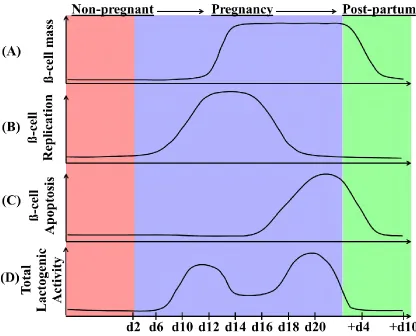
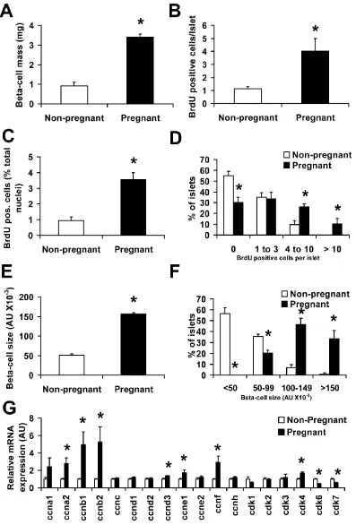
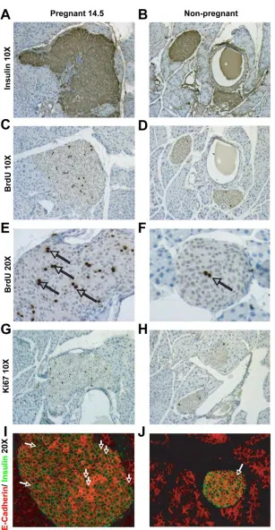
to glucose during gestation, studies in rodents found a 2-fold increase in β-cell mass and
demonstrated that β-cell proliferation also increases dramatically during pregnancy
(Figure 1.4A-B) (35, 63). The peak of bromodeoxyuridine (BrdU) incorporation, an
indicator of DNA synthesis during S-phase, occurs about two-thirds of the way through
the gestational period, with labeling returning to pre-pregnancy levels shortly before
parturition. Notably, this peak in DNA synthesis coincides with increased placental
lactogen (PL) levels, suggesting that lactogenic activity is vital for the ability of β-cells to
enhance proliferation and function in response to pregnancy (Figure 1.4D) (35, 58).
and DNA-to-protein ratio methods indicate β-cell hypertrophy in addition to β-cell
hyperplasia as a mechanism towards β-cell expansion during pregnancy in rodents (65,
66). Intriguingly, β-cell mass returns to normal levels within ten days after birth through
increased β-cell apoptosis, decreased proliferation and reduced β-cell size (Figure 1.4C)
(59, 60). In spite of the small case size considered, β-cell mass has also been shown in a
recent study to increase by 40% in pregnant women (37). Although the molecular
mechanisms underlying these processes are not yet known, the pregnancy paradigm is a
unique example of rapid and reversible β-cell mass expansion, with distinct bursts of both
β-cell proliferation and β-cell apoptosis occurring in a physiological setting.
As a proof of principle for direct regulation of β-cell proliferation by placental
lactogens, overexpression of PL in the β-cell caused a dramatic increase in β-cell
proliferation and β-cell mass, even resulting in hypoglycemia (67). Similarly, global
deletion of the prolactin receptor (Prlr), through which placental lactogens signal,
reduces β-cell mass and mildly impairs insulin secretion in non-pregnant mice (68). The
requirement of the prolactin receptor for β-cell adaptation during pregnancy was
demonstrated using pregnant mice heterozygous for the prolactin receptor null mutation.
These mice exhibited reduced β-cell proliferation, decreased β-cell size and mass, and
impaired glucose tolerance (66). Interestingly, the maternal genotype had a significant
effect on the phenotype of female offspring that became pregnant, as assessed by
heightened serum glucose levels (66), suggesting that in utero exposure to impaired
glucose homeostasis alters the epigenetic memory of β-cells (69).
These findings provide a link to the well-known phenomenon that the intrauterine
facing insulin resistance in the adult (69, 70). Indeed, individuals born to mothers with
gestational diabetes mellitus have a higher risk of obesity and type 2 diabetes (71).
Epidemiological studies in humans show very clearly how caloric intake by the mother
affects the future glycemic health of the child. Intrauterine growth retardation in rodents
is an experimental approach that has been used to investigate this phenomenon on the
molecular level. In this model, epigenetic marks at the promoter of the β-cell
transcription factor Pdx1 were found to be altered in the offspring of dams in which the
uterine arteries had been ligated, causing intrauterine growth retardation (72). Again, it is
clear that the metabolic state of the fetus determines the epigenetic fate of the β-cell, and
highlights the importance of understanding the physiological mechanisms underlying
maternal β-cell expansion during pregnancy.
Although activation of multiple signaling pathways (such as Stat5, Mapk and
classic insulin signaling mediators, such as phosphatidylinositol 3-kinase, Insulin
response substrate 1/2 and Akt) enhances β-cell compensation downstream of the
prolactin receptor in vitro (73-75), it is not clear whether placental lactogens stimulate
these pathways in vivo. In addition, Stat5- dependent downregulation of the tumor
suppressor gene menin (Men1) and subsequent inhibition of p18 and p27 are crucial
events in β-cell expansion during pregnancy (76). While substantial progress has been
made in elucidating the contribution of selected genes to β-cell compensation during
pregnancy, no systematic study investigating the global expression profile of islets in
response to pregnancy exists to consider which mechanisms specifically drive β-cell
Figure 1.4: β-cell dynamics during pregnancy in the mouse. (A) β-cell mass is
increased by (B) β-cell replication during the first two-thirds of gestation. After
parturition, maternal β-cell mass returns to non-pregnant levels by (C) β-cell apoptosis,
which increases through the end of pregnancy and is still detected 4-6 days after birth.
The graphs represent approximate changes in these processes before pregnancy (red,
non-pregnant), over the course of pregnancy (light purple) and post-partum (green), and show
what is believed to occur during rodent pregnancy based on previous studies. (D) Total
serum lactogenic hormone levels (such as placental lactogens) during pregnancy in the
mother are increased from gestational day 10 to 20, pointing to their key role in the
adaptation of the islet to pregnancy. Specifically, of the two identified rodent placental
lactogens, levels of PL-I peak at mid-gestation. As such, PL-I is considered to be the first
trigger to enhance β-cell proliferation and function, while PL-II is initially detectable on
day 12 of gestation and does not reach peak levels until closer to delivery (35, 77).
Additionally, it is thought that steroid hormones present at relative high levels at day 19
of pregnancy, such as progesterone, counteract the stimulatory effects of elevated
Regulation of β-cell proliferation by cell cycle genes
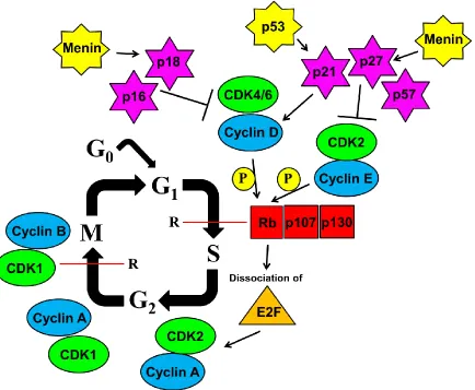
The cell cycle of the adult β-cell is comprised of four phases: (i) The first gap (G1)
growth phase, (ii) the DNA synthesis (S) phase, (iii) the second gap (G2) phase, and (iv)
the mitotic (M) phase (Figure 1.5). The expression of regulatory subunits called cyclins
fluctuates periodically throughout the cell cycle. The association of distinct cyclins with
specific cyclin-dependent kinases (cdks) activates the catalytic activity of their cdk
partners, and powers the cell through different phases of the cell cycle. Mitogenic
stimulation initiates exit from quiescence cells (G0), entry into G1-phase of the cell cycle
and up-regulation of D-type cyclins. Cyclin D complexes with its catalytic partner Cdk4
and/or Cdk6 to execute critical regulatory events in G1- phase. E-type cyclin expression is
up-regulated next, enabling the binding and activation of Cdk2. Together, these active
Cyclin/Cdk complexes inactivate the retinoblastoma protein (Rb) by
hyperphosphorylating it and/or both of its functional homologs (p107 and p130). This
facilitates progression through S-phase by the subsequent release of the E2F family of
transcription factors and the transcription of their target genes, including A-type cyclins,
allowing for the formation of the Cyclin A/Cdk2 complex. During G2- and M-phase, both
A-type and B-type cyclins associate with and activate the kinase activity of Cdk1 that is
required for progression through mitosis. Inactivation of Cdk1 in late mitosis, by the
decreasing availability of appropriate cyclins contributes to reset the cell to G1-phase (79,
80).
Surveillance mechanisms called checkpoints impose quality control in the cell
before it is allowed to proceed into the next phase (Figure 1.5). For instance, a cell will
not be permitted to enter into S-phase until all G1 processes have been properly
completed, for example the licensing of replication origins to form a pre-replicative
complex (81). Entry into G2-phase is blocked until a cell‟s entire chromosomal DNA has
been properly replicated. Furthermore, an activated checkpoint will not allow a cell to
enter into anaphase, when the paired chromatids are pulled apart, until all of its
chromosomes are properly assembled on the mitotic spindle during metaphase. In
addition, a cell is not allowed to advance into S- or M-phase if its DNA has been
damaged and not yet repaired. Cell cycle arrest is achieved during checkpoint activation
by two families of inhibitory kinases, the inhibitors of Cdk4 (INK4s) and the cyclin
inhibitory proteins (CIPs) or kinase inhibitory proteins (KIPs), which physically associate
with Cyclin/Cdk complexes to block their kinase activities. The INK4s include p15ink4b,
p16ink4a, p18ink4c and p19ink4d, and specifically inhibit the activity of the Cdk4/6-Cyclin D
complex present during G1/S-phase. The CIP/ KIP family which includes p21cip1, p27kip1
and p57kip2 inhibit the activity of Cyclin E/Cdk2, Cyclin A/Cdk2, Cyclin A/Cdk1 and
Cyclin B/Cdk1 activity present during later stages of the cell cycle. On the other hand,
sequestration of p21cip1 into Cyclin D/Cdk4 complexes alleviates p21cip1- mediated
inhibition of Cyclin E/Cdk2 activity and promotes G1/S phase transit. Of note, it is
thought that once cells have passed the restriction point in late G1-phase, they no longer
require extracellular mitogenic cues to complete the cell cycle (80).
There is overwhelming evidence that cell cycle genes controlling the G1/S-phase
transition in the cell cycle are likely central to the control of β-cell proliferation. While
the G2/M-phase transition cannot be understated, unfortunately there is a lack of
investigation into their predicted role in the β-cell. Indeed, the vast majority of cell cycle
genes governing G1- and S-phase are expressed in mouse and human islets (82, 83).
However, it is worth noting that there are important differences in the activity profile of
cell cycle genes between human and mouse islets. Fiaschi-Taesch and colleagues
catalogued the G1/S proteome of the human islet and found that Cdk4 and Cdk6 are
expressed at comparable levels, whereas only Cdk4 was found in the mouse (84).
Perturbation of G1/S-phase cell cycle regulators in the β-cell by the use of genetic
mouse models yields either deficiency or unconstrained proliferation (Figure 1.5). While
many tissues are unaffected by the global deletion of Cdk4 in mice, islets displayed β-cell
hypoplasia, leading to diabetes (85). This phenotype can be rescued by replacing the
endogenous Cdk4 locus with a constitutively active form of Cdk4 still under the control
of its endogenous promoter. Again, this mouse yielded a restricted phenotype, showing
marked β-cell hyperplasia leading to islet neoplasms (86). Additionally, global ablation
of both Cyclin D2 alleles in combination with a single allele of Cyclin D1 result in greatly
reduced β-cell mass and β-cell proliferation after birth, and severe diabetes by 3 months
of age (87). Conversely, overexpression of Cyclin D1 is frequently seen in human
pancreatic endocrine tumors (88). Mice with β-cell specific ablation of Rb and global
deletion of p130 leads to unrestrained cell cycle reentry as well as activation of apoptosis
(89). Together the aforementioned studies demonstrate that the D-type Cyclin/Cdk4/
phospho-Rb pathway is required for β-cell replication during adult growth in the mouse.
Interestingly, the decreased capacity with age of the β-cell to expand by
the Cdkn2a locus, which encodes the INK4 members p16ink4a and p19ink4d (34, 90, 91). In
fact, manipulation of p16ink4a expression in transgenic mice dramatically alters the
proliferative capacity of β-cells, precisely as would be expected if p16ink4a limits
proliferation in aging β-cells (92). Furthermore, recent genome-wide association studies
link both CDKN2A (p16ink4a) and CDKN2B (p15ink4b) to the risk of type 2 diabetes (93),
illustrating that G1/S-phase cell cycle regulators are not only important in rodent β-cell
proliferation, but also during the ontogeny of type 2 diabetes.
Finally, a complete picture encompassing all the signaling pathways that control
many of the mentioned cell cycle regulators in the β-cell is still not attained (Figure 1.5).
For example, while it is acknowledged that prolactin (PRL) signaling via the Jak2/Stat5
pathway increases expression of Cyclin D2, it is not known whether this is the only or
most important mechanism for cell cycle activation in the β-cell during pregnancy in vivo
(94). A known upstream regulator of cell cycle genes is the multiple endocrine neoplasia
(MEN) type 1 syndrome protein, menin. Mice deficient in menin specifically in β-cells
lead to the development of dramatic β-cell hyperplasia, and islet tumors (95). Increased
rates of β-cell proliferation are observed, and associated with reduced expression of
p18ink4c and p27kip1, both downstream targets of menin- dependent histone methylation
(96). Additionally, the transcription factor p53 is thought, among other functions, to be a
tumor suppressor, exemplified by its target gene p21Cip1. Unlike parental mice containing
either Rb-null or p53-null alleles, mice both heterozygous for Rb and null for p53,
develop frequent insulinomas similar to menin-deficiency (97, 98). These studies
demonstrate that p53 in addition to Rb is a critical upstream checkpoint of the cell cycle
β-cell whether extrinsic or intrinsic, the next challenge will be to identify the most feasible
and appropriate cell mitogens for driving cell cycle entry and progression in human
Figure 1.5: Regulation of β-cell proliferation by cell cycle genes. An overview of the
proteins that control β-cell cycle with emphasis on the G1/S-phase transition. The “P” in
the yellow circle indicates phosphorylation. The “R” next to the red line indicates the G1/
S- and G2/ M-phase checkpoints, respectively. All of the genes are discussed in the body
of the text. It should be noted, as indicated by the yet unexplained observation that
embryonic and adult cell division seem to be regulated independently (99, 100), that the
Attempts to expand functional β-cell mass by proliferation
in vitro
Initial efforts to expand functional β-cells by proliferation have not been
successful. Lineage tracing in purified mouse islets demonstrates that β-cells divide very
slowly, undergo gradual dedifferentiation to an insulin-, Pdx1-, and Glut2- negative state,
and are eventually replaced by cells of non-β-cell origin in vitro (101). In spite of this,
lineage tracing of primary adult human β-cells with a Cre-loxP system demonstrates that
dedifferentiated human β-cells, but not mouse, can be grown in culture for up to 16
passages (102). It is clear that spontaneous replication in primary human islets is
ultimately halted over time by cellular senescence, which is characterized by loss of
differentiated function (103). However, the redifferentiation of cells derived from adult
human β-cells expanded in culture could provide a sufficient number of β-cells needed
for islet transplantation at the current availability of human islet donors (104).
Understanding the molecular mechanisms involved in β-cell fate in vitro is critical for
attempts to redifferentiate these cells back into functional glucose-sensing, insulin
releasing β-cells after expansion (105).
Because primary β-cells do not proliferate in culture, attempts to bypass the
senescence program have been made by immortalizing pancreatic β-cells through the
forced expression of oncogenes such as the human homologue of the retrotranscriptase
subunit (hTERT), simian virus large T antigen (SV40T), and constitutively activated Ras
(106). While some attempts to do this still lead to dedifferentiation of human β-cells
(107), the use of reversible immortalization strategy for expansion of human islets has
pancreatic β-cells. Although many safeguards were used to eliminate the possibility of
developing tumorigenesis in this human β-cell line, the large scale cell growth needed to
meet the amount of cells required for islet transplantation therapy might result in somatic
mutations leading to increased cancer risk in the patient (108). Indeed, the cell cycle
regulators one might hope will be sufficient to drive cell cycle progression in the β-cell,
such as the G1/S-phase mediators described previously, are themselves members of
oncogenic pathways. Whether forced cell cycle entry by these molecules allows
non-oncogenic and safe progression of the cell cycle in the β-cell remains an important
unanswered question.
With the increased understanding of cell cycle regulation in the past five years, it
has become clear what single or a combination of potential human β-cell mitogens need
to accomplish to successfully expand functional human β-cell mass by proliferation in
vitro (Figure 1.6A). First, a β-cell cycle regulator must stimulate entrance into the cell
cycle (exit from G0- and entry into G1-phase), and ensure that precise duplication of the
genome is achieved to maintain genomic stability (progression from G1- into and through
S-phase). Without cell cycle entry, the β-cell would stay in G0-phase. Also, in addition to
the required high fidelity of DNA polymerases, the cell must be able to distinguish
between replicated and unreplicated DNA during G1-phase to ensure the precise
duplication of chromosomal DNA during S-phase of a single cell cycle. If the
chromosomal DNA is under-replicated as a result of too few active replication origins,
the chromosome is likely to be broken near the unreplicated region upon sister chromatid
separation during anaphase. The firing of a replication origin more than once would lead
sufficiently close together can undergo head-to-tail collision and lead to extruded double
stranded DNA fragments (109, 110). Of note, an additional level of yet unclear regulation
exists during S-phase called the „replication timing program‟ in which sequential patterns
of domains containing discrete foci or factories of DNA synthesis occur during the course
of S-phase (111).
The licensing of replication origins by the stable binding of the „pre-replicative
complex‟ (Pre-RC) only during G1-phase affords the cell the ability to distinguish
between active and inactive replication origins, defined here as a site on chromosomal
DNA where a bidirectional pair of replication forks initiate (81, 109). The major
component of the Pre-RC are the mini-chromosome maintenance 2-7 proteins (Mcm2-7),
whose binding onto origin DNA is essential for the replication origin to be able to initiate
a pair of replication forks. Mcm2-7 is thought to function as a DNA helicase traveling
ahead of the replication fork, as it is subsequently displaced from origin DNA as forks are
initiated. Therefore the Mcm2-7 complex is never associated with replicated DNA,
precluding the origin from being fired again during later phases of the cell cycle (81).
Furthermore, the down-regulation of other components of the Pre-RC complex at the
G1/S-phase transition such as chromatin licensing and DNA replication factor 1 (Cdt1),
thought to be the rate-limiting factor responsible for loading of the replicative
Mcm-helicase onto DNA, is critical to preventing re-licensing of replicated origins (110).
While there is no formal way of distinguishing early G1-phase from G0, the detection of
Mcm2-7 expression is a potentially powerful way of assessing cell cycle entry of β-cells
To date, experiments in primary human islets provide the proof of principle that
human β-cells can be stimulated to enter the cell cycle in vitro, confirming recent human
cadaver studies which determined that β-cell proliferation occurs neonatally (26) and in
increased frequency with greater proximity to pancreatic tumors (112). For example,
overexpression of a D-type Cyclin with its partner Cdk clearly stimulates β-cells to enter
the cell cycle, as assessed by BrdU incorporation, and Ki67 expression, a thymidine
analog marker of S-phase and a marker of late G1- to M-phase, respectively (83, 113). In
addition, overexpression of other regulators of the G1/S-phase transition, such as E2F1 in
primary rat islets also leads to activation of multiple phases of the cell cycle, as assessed
by BrdU incorporation, Ki67 expression and phosphorylation of histone H3 (pHH3), a
phosphorylation event that occurs in M-phase (114). Other examples of factors sufficient
for induction of multiple phases of the cell cycle in primary human β-cells include
overexpression of CYCLIN E in combination with CDK2, FOXM1, Nkx6.1, and
parathyroid hormone- related protein (PTHrP) (115-117). While it is clear that human
β-cells can be forced to enter the cell cycle in vitro, no assessment of genomic stability has
yet been demonstrated.
As implied in the previous section, progression through the remaining cell cycle
phases and subsequent cell cycle exit depend primarily on the successful completion of
activities during G1- and S-phase, namely the precise duplication of chromosomal DNA
during S-phase (Figure 1.6A). Conversely, genomic instability resulting from DNA
breaks generated during DNA replication (defined here as replication stress) can activate
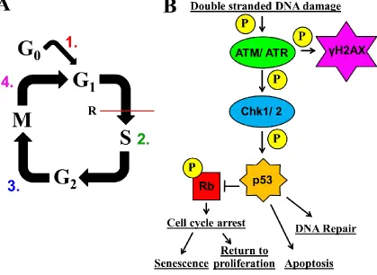
a G1/S-phase checkpoint, also referred to as the DNA damage response (Figure 1.6B). Of
ATM (ataxia telangiectasia mutated) and ATR (ataxia telangiectasia and Rad3-related
protein). These PI3K-like kinases in turn phosphorylate many adaptors and sensors,
including gamma-H2AX (γH2AX) on serine 139 which recognizes double stranded DNA
breaks and marks megabase lengths of DNA adjacent to break sites, and checkpoint-
transducer serine/threonine kinases, Chk1 and 2 (118). Originally thought to obey strict
phosphorylation dependence, Chk1 on ATR and Chk2 on ATM, crosstalk among these
kinases have now been documented (119). Activated ATM and ATR, directly
phosphorylate the p53 transcription factor within its amino-terminal transactivation
domain, specifically on serine 15. Also, Chk 1 and 2 converge on p53 phosphorylation,
particularly threonine 18 and serine 20. Serine 15 phosphorylation of p53 inhibits Mdm2
binding, an ubiquitin ligase that ensures rapid p53 turnover (118). Together these
modifications lead to the stabilization and subsequent accumulation of p53 protein in
response to DNA damage.
Once expressed, p53 functions as an integrator of diverse stress signals into
different cellular outcomes, including cell cycle arrest, senescence, DNA repair, and
apoptosis (120) (Figure 1.6B). For example, the transcriptional upregulation of its target
gene p21Cip1 silences the activation of the Cyclin E/Cdk2 kinase, blocking the
inactivation of Rb, leading to G1/S-phase cell cycle arrest in an attempt to give time for
DNA repair mechanisms to repair DNA damage (118). However, the exact response of
the cell to sustained p53 expression depends on the combination of, among others; cell-
type, tissue-type, type of stimulus, protein localization, and regulation of target gene
selection (120). Indeed, in support of the DNA damage response being utilized in the
hypomorphic mutant of p53, defective in apoptosis but not in cell cycle arrest, develops
diabetes. In these mice, β-cell mass is progressively depleted due to accumulated DNA
damage (sustained γH2AX expression), promoting a decrease in β-cell proliferation
through p53/p21-dependent cell cycle arrest (121). In addition, interventions that activate
the cell cycle in β-cells can also activate cell death pathways. For example, β-cell specific
overexpression of the oncogene c-Myc in transgenic mice not only increases β-cell
proliferation, but also increases apoptosis leading to the development of diabetes (122).
Furthermore, while sufficient to induce progression through one cell cycle, the ultimate
fate of primary rat β-cells overexpressing E2F1 is apoptosis (114). Multiple demonstrated
(dedifferentiation, apoptosis, senescence) and potential barriers (DNA damage response)
to attempted β-cell proliferation exist, and unfortunately, an in vitro protocol for efficient,
step-wise non-oncogenic stimulation of human β-cell proliferation to completion remains
Figure 1.6: Challenges that need to be overcome to successfully expand functional
β-cells by proliferation in vitro. (A)To successfully replicate, a sufficiency factor would
have to (1) enter the cell cycle, and license DNA at each replication origin, (2) properly
duplicate chromosomal DNA, (3) progress through G2/M- phases, and (4) exit the cell
cycle. We use this as a model and basis for characterization of potential β-cell mitogens.
(B) An oversimplified model of the DNA damage response pathway, showing the main
mediators of the double stranded DNA stimulus to cellular outcome. Briefly, ATM/ATR
are activated by double stranded DNA damage, and phosphorylate their targets, γH2AX,
and Chk1/2. Chk1/2 in turn with ATM/ATR stabilizes p53 expression. Depending on
many variables, p53 can activate diverse processes such as cell cycle arrest, senescence,
Maturity Onset Diabetes of The Young (MODY) transcription factors
regulate proliferation and survival in the adult β-cell
Maturity onset diabetes of the young (MODY) is a monogenic form of type 2
diabetes. The clinical criteria used to characterize MODY include (a) diagnosis before 25
years of age in at least one family member, (b) autosomal dominant inheritance pattern
and (c) defects in insulin secretion (123). Mutations in at least six identified genes define
the molecular genetic etiology of MODY. All encode various transcription factors
including hepatocyte nuclear factor 4α (HNF4α; MODY1), hepatocyte nuclear factor 1α
(HNF1α; MODY3), pancreatic and duodenal homeobox 1 (Pdx1; MODY4), hepatocyte
nuclear factor 1β (HNF1β; MODY5) and neurogenic differentiation factor 1 (NeuroD1;
MODY6), except for glucokinase (Gck; MODY2), a glycolytic enzyme important for
glucose sensing (124). The identification of glucokinase as a type 2 diabetes
susceptibility gene highlights MODY as an attractive model for studying the
multifactorial polygenic disorder of type 2 diabetes (125). It is now clear that mutations
in the different MODY genes result in clinical heterogeneity. While the frequency of
specific MODY gene mutations varies in MODY families from different countries, the
high percentage of the diabetic phenotype in carriers heterozygous for a MODY mutation
indicates the high penetrance of all known mutations (126, 127). Since the diabetic
phenotype of not all MODY families can be explained by the existing MODY genes,
additional MODY genes have been described such as kruppel-like factor 11 (Klf11;
MODY 7), carboxyl ester lipase (Cel; MODY8), and paired box 4 (Pax4; MODY9) (126,
MODY genes, well known for their role in glucose stimulated insulin secretion,
have been shown recently to be involved with β-cell mass homeostasis through the
regulation of proliferation and survival pathways. A recent study indicates that the rate of
β-cell proliferation is controlled by glycolysis or workload placed on an individual β-cell,
rather than blood glucose per se, linking together two previously assumed separate
cellular functions of the β-cell (55). Furthermore, it seems that the modulation of
glucokinase (MODY2) activity through genetic deletion or pharmacological activation, is
key to determining the rates of β-cell proliferation during adulthood, high fat diet and
following β-cell injury (55, 131). However, in line with the known toxicity of high
concentrations of glucose on β-cells (132), a human patient exhibiting an activating
mutation in glucokinase exhibits not only enhanced replication but also increased
apoptosis, although the reason for β-cell death is unknown (57). Indeed, mice
haploinsufficient for Pdx1 (MODY4) show blunted β-cell mass expansion in response to
high fat diet through increased endoplasmic reticulum (ER)-stress induced apoptosis
(133). Also, Pax4 (MODY9) protects adult β-cells from stress-induced apoptosis (134).
Together these studies of MODY genes demonstrate the importance of further identifying
molecular players leading to β-cell proliferation and/or β-cell survival during times of
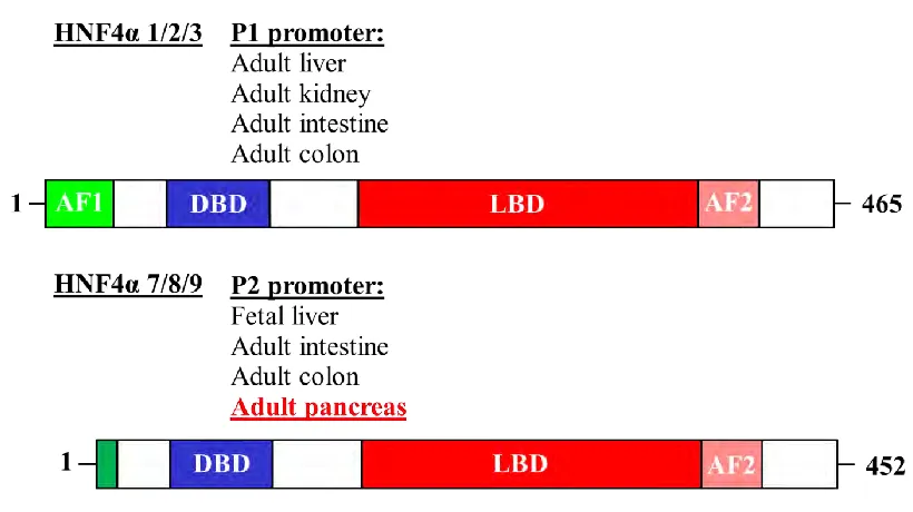
Hepatocyte Nuclear Factor- 4α: The MODY1 Gene, β-cell function and
β-cell proliferation
Hepatocyte nuclear factor 4α (HNF4α) is a highly conserved family member of
the nuclear receptor superfamily of transcription factors. A typical nuclear receptor is
characterized by a variable amino-terminal transactivation domain (AF-1; A/B region), a
conserved zinc-finger DNA binding domain (DBD) (C region), a hinge domain (D
region), a conserved ligand binding domain (LBD) containing a second (AF-2)
transactivation domain (E region), and an inhibitory carboxy-terminal domain (F region)
(135). The members of the nuclear hormone superfamily can be categorized based on the
following criteria: protein dimerization, structure of the cognate DNA binding site, and
intracellular localization. HNF4α is thought to define a distinctive subclass of nuclear
receptors, defined by stable homodimerization, predominantly nuclear localization, and a
binding preference for direct repeats of a hexamer half-site (DR+1 element) (136). While
the classical model of nuclear receptor transcriptional activation requires ligand binding
to induce a conformational change in the LBD that recruits co-activator complexes (137),
questions remain whether HNF4α transcriptional activity is always ligand- dependent.
The mechanism by which HNF4α is transcriptionally activated is unclear. The
crystal structure of the hydrophobic ligand binding domain of bacterially expressed
HNF4α has been solved, and demonstrates the ability of the binding pocket to
irreversibly hold various fatty acids (138). However, further structural studies on
bacterially expressed HNF4α show that co-activator binding and not the binding of a