Vol. 54,No. 2 JOURNALOF VIROLOGY, May 1985, p.317-328
0022-538X/85/050317-12$02.00/0
Copyright © 1985,American SocietyforMicrobiology
An
Unusual
Spliced
Herpes
Simplex Virus
Type 1
Transcript with
Sequence
Homology
to
Epstein-Barr
Virus DNA
R. H. COSTA,' K. G. DRAPER,' T.J. KELLY,2 AND E. K.
WAGNER'*
Department ofMolecular Biology and Biochemistry, University of California, Irvine, Irvine, California
92717,1
and Department of Molecular Biology and Genetics, Johns Hopkins University School of Medicine, Baltimore,Maryland 212052
Received 5 November 1984/Accepted 14 January 1985
High-resolution transcription mappinglocalizedaspliced 2.7-kilobase herpes simplex virus type 1 mRNA.
The4-kilobase intron ofthistranscriptencodes anested setof transcriptsontheoppositeDNAstrand. The nucleotide sequence of the DNA encoding the left-hand and right-hand exons of the spliced transcript was determined, and the salient featuresarepresentedhere.Ofmajorinterest isthatbothexonscontainedregions withinseveralhundred bases ofthesplicedonorandacceptorsiteswhich showedhomologytotworegions of the Epstein-Barr virus genome, which are themselves 3 kilobases apart. The spliced herpes simplex virus transcript encoded atranslational reading frame which could encode a protein with anapproximate sizeof 75,000 daltons. Thisvalue is in agreement with in vitro translation data. Thepredictedaminoacid sequence
of theherpes simplexvirusprotein hadsignificanthomologywithputative aminoacidsequencesencodedby the homologous Epstein-Barr virus DNA sequences.
Asrecentlv reviewed(24; E.Wagner, in B.Roizman, ed., Herpesviruses, vol. 3, in press), correlation between the
genetic maps and transcription maps for herpes simplex
virus type 1 (HSV-1) is generally excellent. Detailed
tran-scriptionmapsforthevirusindicatethatthe vastmajority of transcripts areunsplicedand havepromoter-control regions immediately 5' to the mRNA cap sites. Nested sets of
transcriptscanbe identified. Thesearepartially overlapping transcripts whichcansharepolyadenylation sitesyetencode unique polypeptides. Other partially overlapping clusters have been identified which have acommon 5' cap site but which differ in the polyadenylation site. Such a situation leads to mRNAs which are "redundant" in that they each encode the same protein. Variations on these patterns are common.
Splicing of mRNA is not common in HSV-1 mRNA metabolism. The reason for this is unclear, since other
herpesviruses encode spliced mRNAs. Furthermore, some
spliced mRNAs are expressed during HSV infection. In HSV-1 infection, most splices are short. It was therefore surprising to note the occurrence ofa late HSV-1 spliced
mRNA which is encoded bya DNA sequence containinga
4-kilobase (kb) intron. This transcript is located inaregion (0.185 to 0.225 map units
[m.u.])
with no reported geneticmarkers (25; P. Schaffer, personal communication). As described in this communication, analysis of the DNA sequence encodingthetranscript showed that it had
signifi-cant homology with an analogous region encoded by Ep-stein-Barr virus (EBV) DNA, the sequence of which has been reported by Baer et al. (3) Such homology iscompelling
evidence that this unusual (for HSV) spliced transcript encodes afunction important for the replication of herpes-viruses in general.
MATERIALS ANDMETHODS
Cells and virus. For RNA preparation, plaque-purified isolates of the KOS strain of HSV-1 were used to infect
* Correspondingauthor.
HeLa cells. Monolayer cultures of HeLa cells were grown at 37°C in Eagle minimal essential medium containing10% calf serum, penicillin, and streptomycin.
Enzymes. All restriction enzymes and bacterial alkaline phosphatase were obtained from Bethesda Research Labo-ratories. Digestions were carried out in buffers recom-mended by the supplier. Bacteriophage T4 polynucleotide kinase (Bethesda Research Laboratories) was used for 5' end labeling as described by Maxam and Gilbert (20). Escherichia coli DNA polymerase I (Klenow fragment;
Boehringer-Mannheim Biochemicals) was used to generate 3'-end-labeled DNA by the method of Maniatis etal. (19).
Isolation, labeling, and sizefractionation ofpolyribosomal
RNA. Monolayer cultures of HeLa cells (2 x 10⁷ cells per flask)wereinfected for30 minat amultiplicity of 10 PFU of virus per cell in phosphate-buffered saline containing 0.1% glucose and 1.0% fetal calf serum. Polyribosomes were
isolated from the cytoplasm ofHSV-1-infected cells by the magnesium precipitation method of Palmiter (22).
Polyad-enylic acid-containing [poly(A)] mRNA was isolated from total rRNAbyoligodeoxythymidylic acid-cellulose (Collab-orativeResearch, Inc.)chromatography. This isreferred to asHSVpoly(A)mRNA. Detailsof thisprocedurehave been presentedelsewhere(1, 2, 10-14, 16). RNAwasisolatedat6 h postinfection. RNA was size-fractionated by electropho-resison1.4% agarosegelscontaining10 mMmethylmercury hydroxide (4)aspreviously described (1, 2, 14, 16).
RecombinantDNA.AllrecombinantDNAclonesdescribed in this paper were derived from either BamHI-HindIII
fragment A-IO (0.151 to 0.182 m.u.), HindIll fragment J
(0.182to0.262m.u.), HindIII-BamHI fragmentJ-A(0.182to 0.223 m.u.),orBglII fragmentP(0.201 to0.233m.u.)of the KOS strain of HSV-1 cloned in pBR322. Several subclones were used: XhoI-HindIII fragment C'-IO (0.171 to 0.182 m.u.), HindIII-EcoRI fragment J-D (0.182 to 0.190 m.u.), EcoRI-SalIfragmentG-D (0.190to0.195m.u.),EcoRI-XhoI
fragmentG-U (0.190 to0.207 m.u.), Sallfragment I' (0.195 to 0.202 m.u.), Sall fragment X (0.202 to 0.219 m.u.), XhoI-BamHI fragment V-A (0.207 to 0.223 m.u.), Sall-BamHI fragment U-A (0.219 to 0.223 m.u.), BamHI-XhoI 317
on November 10, 2019 by guest
http://jvi.asm.org/
318 COSTA ET AL.
fragment A-V (0.223 to 0.226 m.u.), XhoI-BglII fragment L-P (0.226 to 0.233 m.u.), andSalI-XhoI fragment U-V (0.219 to 0.226 m.u.). Procedures for cloning HSV-1 DNA fragments in the pBR322 vector have been described previously (1, 7). Cloned DNA fragments were named as described previously and located by their map coordinates on the prototype arrangement of the HSV-1 genome (7).
Insitu Northern RNA blots. Unless notedotherwise, 10-,ug samples of HSV poly(A) mRNA were fractionated on meth-ylmercury gels and dried onto Whatman 3 MM paper under vacuum as previously described (10, 14, 16). The agarose film was floated off the paper in water and hybridized with appropriate nick-translated 35P-labeled DNA probes in50%
formamide containing 0.4 M Na+, 0.1 M HEPES
(N-2-hydroxyethylpiperazine-N'-2-ethanesulfonic acid), pH 8.0, 0.005 M EDTA, and Denhardt solution (9) at 50'C for 36 h. Blots were rinsed at 50°C. The first two rinses were in50%
formamide-2x SSC (lx SSC = 0.15 MNaCl plus 0.015 M
sodium citrate)-0.1%sodium dodecyl sulfate (SDS). The last
rinsewasin 0.lx SSC-0.1% SDS. Autoradiography was on Kodak XRP film with or without intensifying screens as
needed.
In vitro 32P-labeled DNA was made by nick-translating
appropriate DNA clones with DNA polymerase I, DNase I
(Boehringer-Mannheim), and 50
p.Ci
of[co-32P]dCTP
(3,000Ci/mmol; AmershamCorp.).
Isolation of restriction fragment-specific mRNA. Restric-tionfragment-specificmRNA wasisolatedfromHSVpoly(A) mRNAby preparativehybridizationtotheappropriateDNA covalently coupledtocellulose. Details ofcouplingDNA to
cellulose andpreparative hybridization have beendescribed previously (2, 7).
Nucleotide sequencing. As described previously (10, 14),
nucleotide sequenceanalysiswascarried out by the method
ofMaxamandGilbert(20).
Nuclease mapping of HSV-1 mRNA. S1 nuclease and
exonucleaseVIIanalysis of RNA was carried outessentially
as described by Berk and Sharp (5) and as described
previously (1, 6-8, 10-14, 16). Appropriate HSV-1 DNA
clones (10 ,ug) were cleaved at the desired site with the
appropriate restriction enzyme. The DNAthen was 5' end
labeled with
[_y-32P]ATP
(3,000 Ci/mmol;ICN)withpolynu-cleotide kinase(Bethesda Research Laboratories)to a
spe-cific activity of100,000cpm/,ug ofDNA. Alternatively, the DNA was 3' end labeled to the same specific activity by
using DNA polymerase I (Klenow fragment) (Boehringer-Mannheim).
The DNA fragments were then denatured and strand
separated on 5% acrylamide gels as described by Maxam andGilbert(20). The strand-separated DNA(from10
pg
of cloned DNA) was hybridized with 10jig
of infected-cell mRNAin 0.1 MNa+-0.1
MHerpes(pH8.0)-0.01MEDTA at 65°C for 6 to 16 h in a 30-,ul volume. Hybrids weresubjectedto S1 nuclease(Boehringer-Mannheim)or exonu-clease VII (Bethesda Research Laboratories) digestion as describedpreviously(10, 14). Materialwasfractionatedon a
denaturing 5% acrylamide gel, with 5'-end-labeled Hinfl-di-gested pBR322 DNA fragments used as size standards. Alternatively,
Si-protected
products were fractionated ona1.5% alkaline agarose gel, with 5'-end-labeled
HindIII-di-gested lambda DNA fragments used as size standards (14, 16).
In vitro translation. Translation of size-fractionated viral mRNAwascarried out in vitro withamicrococcal nuclease-treated rabbit reticulocyte system (New England Nuclear
Corp.), with
[35S]methionine
(>800 Ci/mmol) as theradio-active amino acid. Details of theprocedureandfractionation
of polypeptides in SDS-acrylamide gels by the method of
Laemmli (18) have been described in several previous pa-pers (8, 10,14, 16). Gelsweretreatedwith En3Hance (New England Nuclear Corp.) and dried under vacuum at 60°C,
and radioactive bands were localized by autoradiography with Kodak XRP film. Exposure was for 3 to 5 days at
-700C.
RESULTS
Location of transcripts between 0.15 and 0.27 m.u. The map (Fig. 1) illustrates pertinent restriction endonuclease
sitesand thelocation of HSV-1transcripts inthe 18-kilobase-pair regionbetween 0.151 and 0.272 m.u.Characterization of
the transcripts between 0.16 and 0.185 m.u. and between 0.225 and 0.27 m.u. is also shown (6, 8). The preliminary characterization ofthetranscripts between 0.185 and 0.225
m.u. isthe subject of this paper. Temporal classification of individual transcriptswasbased on themeasured abundance oftranscripts in the presenceand absence ofHSV-1 DNA
synthesisinhibitorsasdescribedpreviously (17).These data are not shown here.
Individual cloned HSV-1 DNA fragments which
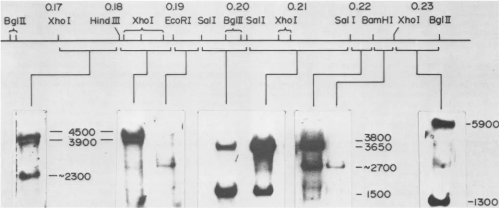
subdi-vided the region of interest were used as probes forin situ RNA hybridization (Fig. 2). The sizes oftranscripts were
calculated fromnucleotidesequencedata (seebelow; Draper
and Wagner, unpublished data). These do not include the length of poly(A) tails (ca. 200 bases [23]). Of particular interest is that a transcript of ca. 2.7 kb was seen when
probes mappingbetween 0.19 and 0.195 m.u. and between 0.22 and 0.23 m.u. were
used,
but not whenprobes
in the 4-kbregion, between 0.195 and 0.22 m.u., wereused. Each probethathybridizedtothe 2.7-kbtranscript extended onlyabout 1,200 to 1,300 base pairs on either side ofthe 4-kb segment of DNA. The 4-kb region ofDNA between 0.195
and 0.22 m.u. encoded a group of nested transcripts 1.5, 3.65, and 3.8 kb in length. The two larger transcripts were notreadily resolvable (Fig. 2), but were clearly resolved in other in situ RNA hybridization experimentsand exposures
(not shown). Data from
hybridization
experiments withsmallerprobes
spanning
the region between 0.195 and 0.22 m.u. indicated that the 1.5-kbtranscriptwasconfinedtotheleft-hand halfofthisregion. Thus, no evidencefor
noncon-tiguous portions ofthis mRNA was seen(datanot shown).
In vitrotranslationproductsof the1.5-, 2.7-,and 3.65-3.8-kb transcripts. The transcripts of interest all mapped with
BglII
fragment P (0.201 to 0.233 m.u.). This fragment wasused previously to isolate HSV-1 transcripts for in vitro translation (see Fig. 4a in reference 7). These earlier data
indicated that the 3.65-3.8-kb transcriptencodesan
80,000-dalton (Da)
polypeptide,
the 2.7-kbtranscript
encodes one ortwopolypeptides
between77,000and85,000Da, and the 1.5-kbtranscript uniquelyencodesa40,000-Dapolypeptide.
Theseresultswereconfirmedby usingvariousclonedHSV-1 DNA fragments that mapped between 0.19 and 0.225 m.u. Since the data fully confirm earlier
work, they
are not shown.Hybrid selectionwasusedtoisolatethetranscripts,
and this was followed by in vitro translation as describedabove. In anotherexperiment (not shown),the 2.7-kb mRNA wasisolatedby usingeitherEcoRI-SaIl fragmentG-D(0.190
to0.195m.u.)orBamHI-XhoIfragmentM-V(0.222to0.225
m.u.), and each sample was translated
separately.
Both isolatesyielded
the75,000-and85,000-Dapolypeptides.
The3.65-3.8-kb transcript encoded an
80,000-Da
polypeptide,
and the 1.5-kb
transcript unambiguously
encodeda40,000-Da
translation product.
J. VIROL.
on November 10, 2019 by guest
http://jvi.asm.org/
SPLICED HSV-1 mRNA 319
Map Units
0.15
1
0.164
0.182 0.190 0.202
0.222
0.241
0.258
0.272
BamHI
Major
Transcripts
HindM
EcoRI
HpaI
IHpaIi
Bgill
2.3
kb-82,000 d
a
exo
1.9
l
b
)((?)
60,000
d
45
kb-13y-
(?)
3y-
50,000d
(?acpsid- ICP
18.8)
BamHl
Hpal
I IHindlE Hpal
BamHl
1BamH J(BamHl
)mI>
Sall I
JSalI
SalI
BgiI
Sail
Sall
SaII
Bglll
Bgill
(85,OOOd
13y-2.7 kb
177,000d
3Y'-VP5-155,000 d
>
6kb
3.8kb/3,80,000d
J3r-
35,000d
1.3kb
I-1.5
kb-/3y-40,000d
FIG. 1. High-resolutiontranscription map of HSV-1 KOS in theregion between 0.151 and 0.272 m.u. Locations of selected restriction endonuclease sitesonthe prototypearrangement of the HSV-1 genome are shown. The position in map units is shown for several restriction sites (0.01 m.u. is equivalent to 1,500 base pairs). Locations of the mRNA transcripts within the region are illustrated with respect to restriction endonuclease sites. Arrowsrepresent the 3' ends, vertical lines represent the cap sites, and the caret delineates the position of the 2.7-kb spliced mRNA intron. The indicated mRNA sizes do not include the poly(A) tail. The molecular sizes of the in vitro translation products for each mRNA were determined by comigration with size standards on
SDS-9%o
acrylamide gels. Identification of transcripts mapping between 0.16 and 0.185 m.u. was described previously (8), as was characterization of the transcripts between 0.225 and 0.27 m.u. (6). Identification of transcripts between 0.185 and 0.225 m.u. isdescribedin the text.Nuclease mapping of the transcripts. The termini of the
2.7-, 1.5-, and3.65-3.8-kb transcriptswereprecisely located
by Si nuclease and exonuclease VII mappingwith hybrids between infected-cell polyribosomal poly(A) RNA (viral
mRNA) and strand-separated 5'- or 3'-end-labeled DNA
restriction fragments as described in detail previously (8, 14). Evidence that the 2.7-kb mRNAtranscript contains an
BgflE
...1 t
0.17
0.18
0.19Xho
I
Hindlm
XhoI
EcoRI SalI
l ~~~~~
1% l lintro of ca. 4 kb is based on analysis of hybrids between
HSV-1 viralmRNAandHindIII-BamHIfragmentJ-A DNA
(0.182to0.223 m.u.)5' endlabeled atthe BamHI site(Fig.
3). In this case, Si nucleasedigestion yielded a fragment 500 bases long, whereas exonuclease VIIdigestion resulted in a
fragmentca. 6.2kblong (Fig. 3A, tracks X and S). A major 5'end of the mRNA wasfound to lie 530 bases to the left (5')
0.20
Bg¶I
SailI
.A
I I
0.21
XhoI
0.22
0.23
Sal I BamHl Xhol
BglE
I I
_111 -5900
p-.
- 4500 -_
-
3900
-_3800
-3650
tl
--2300
A.
Is.IZ-..
4 -2700
-
1500
i
~-1300FIG. 2. InsituRNAhybridslocalizing mRNAs between 0.17 and 0.23 m.u. of the HSV-1 KOS genome. Samples (10 ,ug) of late polysomal poly(A)RNAisolated from infected cellswerefractionated onmethylmercury-containing agarose gels and immobilized by drying in vacuo. RNA wasdetectedby hybridization with specific nick-translated probes,as indicated by brackets below the restriction endonuclease map of thisregion. Sizes shown were determined by the position of HeLa cell rRNA (not shown). However, the length of the poly(A) tail (ca. 200 bases[23])wassubtractedto remainconsistent withthe sizes shown in the transcription map (Fig. 1). Noeffort was made to standardize the specific radioactivity of various probes. Therefore, relative mRNA abundance should not be compared between tracks.
VOL.54, 1985
i 4
I II L-- i __j
.... .. ... .. ... .... .... - ---
---I
on November 10, 2019 by guest
http://jvi.asm.org/
[image:3.612.60.559.66.272.2] [image:3.612.58.559.455.664.2]320 COSTA ET AL.
X S M
A.
0.182
-0,223*
- 2700n (X) A,_
0.223
0.182 0.190 0.195 0.202 0.207 0219 0226
HinDIl EcoRi SaI Sa1l XhoI SolI BamHI XhoI
._
I ...-...1I I . . 1_I....1 ..IB.
0.182-0.190*M s x
1000- _
t.p'
600- _
519/506- _ 398-
-345-
-298-
-221/220- OD
5'
C. 0.190*-0.207 M S
U
53600-- 1
519/506- 6
398- 345
-298-
_ 220/221-A
154-3'
D.
0.219-0.226*
M S
E.
0.207-0.223*
S M
1000- . -950
7---X600-
800
519/506-
*.'398-
... ...345-4,
298-'1
:;220/221
F.
0.226*-0.233
S M
-7 -1000
-1000 - 600
4- 519/506
-60 *-398
-519V506 345
*39-8298 _-398
- -22V220
3'
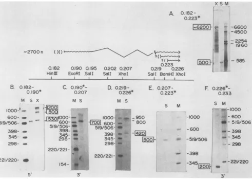
FIG. 3. Localization ofthe2.7-kbsplicedmRNAbyS1 nuclease and exonuclease VII mapping. The diagramsummarizesmapping data presented in thetextbypositioning the splicedmRNAwithrespect totherestriction endonuclease sites. The 4-kb intron is represented by awavyline, with possible minor spliceacceptorsites indicated byaquestionmark(?). Single-strandedDNA5' or3'end labeledatspecific restriction siteswashybridized with HSV-1 poly(A) mRNA, andthe DNAprotected fromnuclease digestionwassize-fractionated on an alkaline1.5%agarosegel(A) or ondenaturing5% acrylamide gels(BthroughF).Tracks: S,S1nuclease-digested material;X,exonuclease VII-digested material;M,sizemarkerofHinfl-EcoRI-digestedpBR322 DNA orHindlIl-digestedlambda DNA.Sizesareindicated in daltons. n, Nucleotides. Mappositions of the restriction fragments used inthe nuclease protection experimentsare shown. Asterisks denote the labeled restriction site.(A)HindIII-BamHIfragmentJ-ADNA;(B)HindIII-EcoRIfragmentJ-DDNA;(C)EcoRI-XhoIfragment G-U DNA; (D)SaIl-XhoIfragment U-V DNA; (E)XhoI-BamHI fragment V-A DNA; (F)XhoI-Bglllfragment L-P DNA.
of the EcoRI site at 0.190 m.u. by using HindIII-EcoRI
fragmentJ-D DNA(0.182 to 0.190m.u.)5'end labeledatthe EcoRIsite(Fig.3B). ExonucleaseVIIdigestionyieldedtwo otherbands,1,200 and 800basesinsize, whichareprobably artifacts since they were also observed inexonuclease VII
digestion experimentswithuninfectedHeLacell RNA(data
not shown). The 5' contiguous end of the 2.7-kb mRNA
(spliced donor portion)extendedonly about700basestothe 3' side of the EcoRI site at 0.190 m.u. (Fig. 3C), where mRNAhybridizedtoEcoRI-XhoIfragmentG-U DNA(0.190 to0.207 m.u.) 3' end labeled atthe EcoRI site.
Theright-hand portionof the 2.7-kbmRNA waslocated in a similar manner. S1 nucleasedigestionof hybrids between viral mRNA and SalI-XhoI fragment U-V DNA (0.219 to 0.226 m.u.) 5' end labeled at the XhoI site yielded fully protectedDNA(950 baseslong) and minor amounts of DNA
fragments 800 and 420 bases long (Fig. 3D). This result
suggested that a major splice acceptor is located near the SalI site at 0.219 m.u. and that other minor sites may be utilized. However, these minor bands were not seen when DNA 5' end labeled at the BamHI site at 0.223 m.u. was
used in such experiments. The majoracceptor was located 500 basestothe left (5')of theBamHI site at 0.223m.u. by
S1 nuclease digestion ofhybridsbetween viral mRNA and XhoI-BamHI fragment V-A DNA (0.207 to 0.223 m.u.) 5' end labeled at the BamHI site (Fig. 3E). The 3' end ofthe 2.7-kb mRNAwaslocated 200 bases to theright (3') ofthe XhoI siteat0.226m.u.bycarryingoutSi nucleasedigestion
ofhybrids between viral mRNA and XhoI-BglII fragment
L-P DNA(0.226to0.233m.u.)3' end labeledattheXhoIsite
(Fig. 3F).
The nested 1.5-kb and 3.65-3.8-kb mRNAs were tran-scribed from the oppositeDNA strand. These RNAs were
precisely mappedina mannersimilartothat shown inFig.4. The 3' endof the clustered
transcripts
wasmappedverynear the Sall site at 0.195 m.u. This was accomplished by Si nucleaseanalysisofhybrids between viral mRNA andSalIfragmentI'DNA(0.195to0.202m.u.)3' endlabeledat0.202 m.u. (Fig. 4A, track S), wherenearly full-length DNA was
protected from digestion.
Hybridization
between viral mRNA andEcoRI-SaiIfragment G-D DNA(0.190 to0.195 m.u.) 3' end labeledat theSallsite gave noSinuclease-re--6600
..i.
-4500 -22541 - 1960
W - 585
.I
s"
J. VIROL.
on November 10, 2019 by guest
http://jvi.asm.org/
[image:4.612.64.555.66.414.2]SPLICED HSV-1 mRNA 321
A
A
A
i500
0.195
0.202
Sal I
SaIl
I I
L
A.
(0.195-0.202S )
D
S M
V'-
Ioo.
_*
JL
d
3800
n
4
3650n
0222
0213
0.219
PvulE
SalI BamHI
I I..._
IL
lZ
B.
(0-202
0.219)
XC
X
S
D M
2i550
-10 0 0 '----''---''-'
-
600
-
506/519
-
398
450
-
345
-
298
-1000
-
600
>-519/
506
1-398
J
C.
(0.213*-0.222)
MM S
1000-
MO_i
600-i
-
506/-519
398-345-
.-
345
298--298
220/-'-' , ,I
a
Rf«^
-221
FIG. 4. Mapping ofmRNAs located between0.195 and 0.219 m.u. by S1 nuclease and exonuclease VII analysis. The diagram is a transcriptionmapsummarizing the data presentedinthetext.Details of the nucleasedigestionsaredescribed in thelegendtoFig. 3 and in thetext.Alltracksaredenaturing5%acrylamide gels. Bracketsspantherestrictionfragments utilized in the nucleaseprotection experiment. See the legendtoFig.3for details. Tracks: D,undigestedDNA;XC, exonucleaseVIIcontrol in whichuninfected cellmRNA wasusedfor hybridization. (A) Sall fragment I' DNA; (B)Sall fragmentXDNA; (C)PvuII-BamHIfragmentP-A DNA.
sistant material (data not shown). Therefore, the 3' end of the mRNA groupwasmappedtotheright(5')oftheSallsite at 0.195 m.u.
The 5' endofthe 1.5-kbmRNAwasmapped 450 basesto theright (5') oftheSall site at0.202 m.u.
Si
nuclease and exonuclease VII digestion of hybrids between viral mRNA and SalI fragment X DNA (0.202 to 0.219 m.u.) 5' end labeled at0.202m.u. bothyielded fragments450baseslong (Fig.4B,tracks Sand X). A smallerfragment(ca.250 bases) seenin the exonuclease VII-digested material wasprobablyan artifact ofdigestion since it was also seen in a control track of exonuclease VII-digested hybrids between this DNA and uninfectedcell mRNA (Fig. 4B, track XC).
The 5'endsofthe 3.8- and 3.65-kb mRNAs were shown to map 150 bases apart by
Si
nuclease digestion of hybrids between viralmRNAandPvuII-BamHI fragment P-A (0.213 to0.222m.u.) 5' end labeledatthePvuII site. Itwas found(Fig. 4C)thattwo Si nuclease-resistant fragments, 900 and 1,050 baseslong, wereproduced.
Correlation between transcript mapping and nucleotide sequencedata.Thenucleotide sequence of the DNA
encod-ing the left-hand and right-hand portions of the spliced
2.7-kb mRNA, aswellas the DNAcontaining the 5' end of the 1.5-kbmRNA,wasdetermined. These data are available
upon request. Specific features related to the transcripts
characterized hereinare shownin Fig. 5.
The precise 5' end ofthe 2.7-kb mRNAwaslocalized by using denaturing acrylamide gels tofractionate S1 nuclease
digests of hybrids betweenviral mRNA andanappropriate 5'-end-labeled 380-base AvaI restriction
fragment
found in HindIII-EcoRI fragment J-D DNA (0.182 to 0.190 m.u.). This fragment was electrophoresed next to a sequence ladderofDNAlabeledatthesamesite(Fig. 6A).The 5' endofthe 3.9-kb mRNAencodingtheputative 50,000-Da capsid protein characterized previously (8) is also shown. These dataindicate that cap site for thesetwotranscripts, encoded from opposite DNA strands, lie within 100 bases of each other. Note that the sequence ladder is of the DNA strand
complementaryto the mRNA sequence. The dataare sum-marized in Fig. 5A. There are reasonable TATA and CAT box homologies within 100 bases ofthe cap sites for both mRNAs, as is normally found for HSV-1 transcripts (24;
Wagner, in press).
Theputative splicedonorwaslocatedca.700 basestothe
right (3') ofthe EcoRI site at 0.190m.u. (Fig. 3C). It was
located (datanot shown) ca. 40bases to the left (5')of the SalI site at 0.195 m.u. by S1 nuclease
analysis
ofhybrids
between viral mRNA and a 3'-end-labeled 130-base Ava/I VOL. 54, 1985
I
_9 On/Do
on November 10, 2019 by guest
http://jvi.asm.org/
322 COSTA ET AL.
A
Cap-3.9kb-50,000d (capsid)MW
!0 20 30 40 50 60 70 80 90 100 110 120
CGCGAATr' lTAA GG-iCGCGCGCTGTCGATCA,vUCTAACGCGCGCGGGACMAGCCCGC6vTCCAATCCAAMTGG6CCGCCT GCAGCCATiAAGGCGCCAACAArCA
GCGC'7AAT677
CCAGCGCGCGAvCAGGATGAGATTBCGCGCGCGCCCTfAAA7CGGGCGGAGGTTAGGTTGCCrGCGGAACGTMCGGTAMCCGCGMMTCCap 2.7kb spliced mRNA
130 140 150 160 170 180 !90 200 210 220 230 240
GATMGGGTG7ACGCCCTCCrGGsGGCCAG6G6TMGCCMGTAAATCTCGGCTCGGAGGTGC6TCTC66Cm66CGTCGGCGCAGGGCC6CGsG66CGGCATCTCGGTCCA 7,GA
A(i&C;qT-GCGGGAGGGCoACGTCGACGTCCCAG°AACAGAAr"GAGCCG4TAiGCC6cTCmCACGAGACCGGTCUrGCAGCCGCGTCCC6GCGCACCCccCcGAA6CGAG6T
B
C
putative splice donor
Sa II
(0.195 mu)
10 20 30 40 50 60 170 80 90 100 110 120
'.CC7CTCGMCCGGACGGGTCGCGCATACCATC6TTTCCTCCAGCCACAACACACGTAAGTCCTCTMCGCAT6GGUTCCCAAGGGGCCCCGG6TCGACCCGACCA GPGAG6CG GCCAGC6CTCTGGABCACACGGCG6TTTCAGiGA GACC GAGAG6TCCCCG666CCCACT6GG G
poly(A)sig. (1.5-3.85kb
mRNA cluster)
130 140 150 160 170 180 190 200 210 220 230 240
ACCCACCCACCOCAGAAACACACAACiCAGC6C6GAG6GCTGCCCCrCMG69A76AT IMATTCGGGATCGC'TTGGGA6GCCC666CAACGGCCAGCCG6TGGG6CAAT GTOTG,TGGGT6GTATGTGTMGTUGTTG6TCTGCGCC.7C -CCAGACGGGGCACCrTGACTAAAATAAGCCCTAGCGAArTCCTCCGGGCCCGTTGCCG6TCG6CC'ACCCCMGAG
nominal cap
1.5 kb mRNA
10 20 30 40 50 60 70 80 90 100 110 120
,gTCACAGTGr7CCATC TGGACGATGTAACCAGTGTCCGMGTGCTGTAGTAOACGTATGCCGT C6C.AGCGGGCGGGGTC GCAgGTGG 6GTCGTiC6MU6 CGCACAGAG6CGACACGACAT,)TCAGlMIT GCATTACGGCAGCCTCAGCCGTiCGCCCGGCCC7CCAGCAG
130 r.TCGAG AAGAGCTC
D
possible
splice acceptor
10 24| 30 40 50 60 70 30 90 100 110 120
SGGGGSrGGGTGTGgGGTTGAGGCGA-TGCGGrjtCCC,iCGArGTCCGCGGGAAGG5CGTrGGG1MGGrvGCGCGTATGCGTATTCIGCCvACGGAGGCGTGCGTGCTTAtGCGCGGCGCG'MC CCrvCaCCAC4CACC4CACACTCCGCTACvGCnC4AGGGGGCTCAGGCGCCCTTCCC6CACCCAAiCCGCGvCGvCATACGCATPqAGCGGTTCC CCGCACGCACB*GG,ir'C6CCl;C6C4OA6
s
lice
acceptor
SalI
(0.219
mu)
130 SL 140
150
160 170 180 190 200 210 220 230 2407iCTGTCTC6AGAACCG'AGGCCAGACTi-ACCTGCCTG6TCGACGAGCCAAMC ATCGCCi,6GGTGC6GGCCAGCGATTAT6GGGMTCTCAACAGGCCAACTGCA6
AGAGACAGAGu-TCMA6G66UCCG6TrCTC"TTAGGACgG4WA-CAGCTGCTCCGGT
GAATAAGCGGGCCTACGCCAGGTCTGCTAATACCC6MGAGATTG6TCCG6TTG6AC6TTCNominal cap 3.6 kb mRNA
250 260 270 280 290 300 310 320 330 340 350 360
~~~~~~~~~~~~~~~~~""33,.rKG 7''A~--'-2 CCTATATATGCGAT TC TGCC6 A.7AT7 TTCGGACGTCGCACCAAACCGGGT,TrCAGTACGGC76G;CTTTCGTA.CCACTGTGC
:TAT
GTACGATATCAG1 6CCTAATAGPAAGACAGCrAGGT6Grr6TGGCCCmCCGGTCATG'CTCG0ACATG7GGAGGCGCCCCGGCGGCTGI
,'SGPAGAGTTrCACCACTGGATATATACGCTACTA6'TGACGGC
370 380 390 400 410 420
AGGGTGGTGACGCACACCGCCACGGCCTGTTCMGTTATATCCTCAACAAGCCCGTT
TCCCACCACTGCTGT6MGCG6TGCCGGCAACAATMAGGATTFC6G5A
Cap-
3.85
kb
mRNA
XhoI
(0.226
mu)
10 20 30 40 50 60 70 80 90 100 110 120
TCATGGCCTCCCAGGAGTCGTTCCGCGACGGTGCGCCTGCAGCCACC6CGGCGA6TTCT6GCCTCGAGCACT4TAACCTCACCGWCCGTCTCCCCCAAGACGTCC6TA
AGTRCCG6MG6tCCtCC6A6CAAGCG7CTGC6CACCOAGGGtcTGGCt6CAGCTCATA6ACGAGCtCGTCGAMATTG6GC'MGGCAGAGGGGGTGTACT6GCAMT
translation
terminator
130 140 150 160 170 180 190 200 210 220 230 240
CGtATlTCCGGAAAACGGPACGCGC6CTCGGATGACMATGtCGCC6TCATTATACCATCTACCTCGC6ACCCAGGCCGACCTCCGCACAvCATCGTCCCATCACAC6CGT.CTg6
6CATAAGGCC.I IGCMGCCGAGCC60c4TACGATB ACGATATac66mACT6T66TAAGG C6UTG
gGTGGCGGGG66TGAGCUGAGGCi6AGFG6C6c
6wpoly(A)sig.
(2.7kb
spliced
mRNA)
25 260 270 80 290 300 310 320
A6CGCCCCACACCTA6GTAT6CTAC6CACGCCACG6T6TCCGGCCT G66 66W666TGCCGGC
J. VIROL.
E
TEGEGGGTTATrTGTGTGGATECATAEGATGCGTGCTGGTGCCACAGCGGACGGTTCCCCCCCCCCTTCCCCCACGGCCG
on November 10, 2019 by guest
http://jvi.asm.org/
SPLICED HSV-1 mRNA 323
A
3.9kb
2.7kb
copj
I
cap
-~ ~ _ _
-~~ _m
AC
GA_OC
GT~
G
B
C
-S
0- cap
G
A
C
C
G
T
FIG. 6. Preciselocalization ofthe 5' ends of the 3.9-, 2.7-, and 3.8-kb mRNAs and ofaspliceacceptorsiteof the 2.7-kb mRNA. HSV-1 mRNAwashybridized with5'-end-labeled single-stranded DNA, followed by digestion with S1 nuclease. The nuclease-resistant materialwas
fractionated againstasequenceladder of the DNA fragment 5' end labeledatthesamerestrictionsite. (A) 5'-end-labeled 380-base-pair Aval
restriction fragment DNA located in HindIII-EcoRI fragment J-D (0.182to0.190 m.u.)localizing the mRNAcapsitesof the 2.7- and 3.9-kb mRNAsonthe DNAsequence.(B) XhoI-SaIIfragment V-X DNA (0.210to0.219 m.u.) 5' end labeledattheSalI sitetoposition the 2.7-kb splice acceptor(s)tothe DNAsequence.(C)SalI-BamHIfragment U-A DNA (0.219to0.222 m.u.) 5' end labeledattheSall sitetoprecisely localize the 5' endof the 3.85-kb mRNA (cap).
restriction fragment whoseright-hand (3') end corresponded to the SmaI site at nucleotides 102 through 107 (Fig. SB). This potential splicedonor sequence(Fig. 5B) shares nom-inal sequence similarity withother eucaryotic splicedonor sequences (21).
A potential polyadenylation signal, AATAAAAA, was
observed80 bases totherightof the Sail site at 0.195 m.u. (Fig. SB). Thelocation of this siteagreeswell withthe 3' end of the 1.5- and 3.65-3.8-kb mRNA nested group (Fig. 4A). This location was confirmed by Si nuclease analysis of hybrids between viral mRNA and a 110-base Sall-SmaI restrictionfragment extending tothe right of theSall siteat 0.195 m.u.and3' end labeledattheSmaIsite aftercleavage withXmaI togenerate a5' overhang (datanot shown).
The cap site for the 1.5-kb member of the cluster of mRNAs encoded by the strand opposite the 2.7-kb spliced mRNA intronwas located 450 bases tothe right (5') of the SalIsiteat0.203m.u. (Fig. 4B).Thenucleotidesequenceof
DNA inthisregion is showninFig.5C. Tentative location of thecapsite isca.25to30 basestotheleft(3')of thenominal
TATA box homology (bases 31 through 39, Fig. 5C). We suggest that this region is a normal late HSV-1 promoter region on the basis of comparative sequence analysis of HSV-1 promoters (reviewed inWagner, in press).
One splice acceptor for the 2.7-kb mRNA was precisely located 34 basestothe left(5')of theSallsiteat 0.219m.u.
bySi nucleaseanalysisofhybridsbetween viral mRNAand XhoI-SaIl fragment V-X DNA(0.210 to0.219 m.u.) 5' end labeled at the Sall site. An example of fractionated Si nuclease-resistant materialcomparedwithasequenceladder proceedingfromtheSallsiteat0.219m.u. is shown in Fig. 6B. Thecontiguous5' end of the RNA lies within bases 130 through 136 (Fig. SD), whose complement is shown in the ladder (Fig.6B).Theposition ofthisacceptoris960 basesto the left (5') of the XhoI site at 0.226 m.u., a result in excellentagreement with thedata shown in Fig. 3D.
Otherpotential splice acceptorswere suggested from the data (Fig. 3D). As discussed above, these correspond to regions ca. 150 and 530 bases to the right (3') ofthe major acceptorindicated here. Thereare potentialsitesat
nucleo-FIG. 5. Selected DNAsequencesin theregion between 0.19 and 0.25m.u.Thepositions of the mRNAcapsites, polyadenylation sites,
andputative splice donor andacceptorsitesweredeterminedbyS1nuclease analysis (6, 8, 10, 14)asdiscussed in thetext.(A) DNAsequence
around themRNAcapsites of both the3.9-kb50,000-Da (d) capsid protein (ICP 18.8) and the 2.7-kb spliced mRNA. (B) Nucleotidesequence
in thevicinity of the 2.7-kb mRNA splice donor site and the polyadenylation site [poly(A) sig.] of the nested 1.5-kb and 3.65-3.8-kb mRNAs. (C)DNAsequencedata 5'of the1.5-kb mRNAcapsite. (D) DNAsequencesintheregion of the spliceacceptorsite andapossiblealternate
acceptor site forthe 2.7-kbspliced mRNA. Sequences upstreamfromthe 5' ends of both the 3.65- and 3.8-kb mRNAsare included. (E) Nucleotidesequenceatthe 3'end ofthe 2.7-kbspliced mRNA; the potential translation terminator (bracket, toprow)and thepoly(A) site
areindicated.
G
A
CG
G T
C
VOL. 54, 1985
on November 10, 2019 by guest
http://jvi.asm.org/
[image:7.612.56.556.73.346.2]324 COSTA ET AL.
A.
940'
125,529
999'
125,587
CAC6TTCCTGCGTCTCGTGTTAGATCCCCCTGMAGCI4CGCG6CCG-T6CGCCACT
:::::: 4 ::: ::: : :: : :: :::: :
wAC-ATCMCAATG4^Gt-CMCGGATTGNGAAGATGCGCTGGAGAGCtr6CCA
.
ILCULALULbL.LRLL,
LLULUAUTULTCLUUA
U, X,LUIAWACCL
U I,TT
TLT
III*WULA*WbLL5bLb
C
ITT
I1IR I LbLjTULLCLjbLICA Ci6UITUA
GiT
HSV-1
EBV
HSV-1
EBV
1059'
TGCCCCTCATC6CGCTGTCGCTGGC-CTCCMTCGGGG"TC>AATC6GCTACACGGCG
125,647
TGGCCATCATCAGCCTCATCCTCTCGAATCICTCCMCGTGCA-ATAGGCTACGTGGCT
111 8'
CA-CATCCGCAAGCGACCGAGCC66TGMGGGAGATCGACGCCT6CCTGCGGGGCTG
125,706
CACCATCTCGCGTCCGCC-GTCCTGTTTTGCCC7GACCAAGAG
1177'
125,765
1237'
125,822
125,882
GT7CGGTTCGGCCCGATGACCACGTTA6GGGCCATCTCCTECTCGTTTCCGGA C7TCGACTC--CAGCGTGTA-GAGGTCACMGACCA6CACCATCACGMAGGCA
1(HSV-1
splice
donor-base
67of
fig.
6B)
U---uuu I W-UII"W9tIUI U,, Ltw, I "u,IWk
CATGIGGITCTCCAGCACCGTAATGTGTGCCACCTGMCATITAAGAATGT4AAGCC
HSV-1
SalI
site(O.195 mu)
T--CTM TTTCGCATGGCTCTCCCAG6G6CCCCG6GTCGACCCGACCCACACrCCACC
TGCGTCAGTACTGGCAATTGTAGAGCAT
AGCCCGGCTGTAAAGGTCGAAATCG
HSV-1
EBV
HSV-1
EBV
HSV-1
EBV
1287
HSV-1
EBV
HSV-1
EBV
rGACCCACATACACACACAACCAGCGC
GGGAG
G GTCGCCCCGGGCTMT
HSV- 1
125,941
cAGcAGG6TCC*GGTGTuGCTTAcATGG4CeCcTmCCCATuACoAAwCCCCCT6
E BVtides 172 and 203 (Fig. 5D). Both sites would change the
translational reading frame from that generated from the
major
acceptor. Another potential acceptor is located atnucleotide 22(Fig.5D)totheleft(5') ofthemajoracceptor.
This site does yield the sametranslationalreading frame as themajoracceptor. Itssignificancewill become apparent in
thenextsection, where homology between this HSV-1DNA sequence and that of EBV DNA is discussed. Finally,
several potential splice acceptor sequences are seenin the
region around the BamHI site at 0.223 m.u. (not shown). Thesecould function to generatetheputative spliceacceptor which was observed 420 bases 5' of theXhoI site at 0.226 m.u. (Fig.3D).
The capsite of the 3.8-kb mRNAwasdeterminedongels by using sequence ladders as size standards for the
Si
nuclease-resistant material generated by theSi
nucleasedigestionofhybrids between viral mRNA and SaIl-BamHI fragment U-A DNA (0.219 to 0.223 m.u.) 5' end labeled at the SalI site(Fig.6C). The sequence aroundthis cap site is shown in Fig. SD. A TATA box homology25 bases to the
right
(5')
of this cap site isclearly visible (nucleotides 337 through 343,Fig.SD).The sequence 155basestotheleft(3')ofthis site (nucleotide 165) is indicated asthe nominal cap
site forthe3.65-kb mRNA, sinceagoodTATAbox
homol-ogy is seen ca. 30 bases to the
right
(5') of this site (nucleotides 181 through 187, Fig. SD).The sequencein thevicinity oftheXhoI siteat0.226m.u. is shown in Fig. 5E. Ofparticular note are nucleotides 75
through 78, whichareatranslationterminator
signal (TGA)
that could close a longopen reading frame for the 2.75-kb spliced mRNA (seebelow), anda poly(A) signalat nucleo-tides 248through258, 185 basestotheright (3')of the XhoI site. The position of this signal sequence is in excellent agreementwith thepositiondeterminedforthe 3'end of the 2.7-kbsplicedmRNA(200 basestotheright [3']of the XhoI site at0.226 m.u.,Fig. 3F).
Homology between the 2.7-kb spliced HSV-1 mRNA se-quenceandsequences of EBVDNA.Because
spliced
mRNAs are sounusualin HSV-1transcripts,it seemedpossible
that the 2.7-kb mRNAmight beconserved among otherherpes-viruses. Therefore, the sequence of HSV-1 DNAencoding
thespliced 2.7-kb mRNA was
compared
with thecomplete
EBV DNA sequence (3). The computer search
algorithm
used was developed by D. J.
Lipman
and W. R. Pearson J. VIROL.on November 10, 2019 by guest
http://jvi.asm.org/
SPLICED HSV-1 mRNA 325
B.
129,001
129,061
1303'
129,121
1362'
129,170
1419'
129,230
1479'
129,290
1539'
129,350
1599'
129,410
1659'
129,470
GICCCGGCCGTG6CCIAGTCGTACTGGC
LC*AATbUILL
IWTTi IITTAlAIL AXLLCL bbAWTIb b'AbIjUTbL AlAI IL L(base
No.23 ,fig.
6D) HS\,-1 294
CTCGTTGGCC66GTGCGCGTTCATGGTTCG6666T6TGTG6GGTG6TAGGCGATGC66 AGGCGCTGCACCTCGAiGACCTCCTCTAAGAiG6ACCTCCCACCGtPGACGTAGACGCGG CTCCCCCAGTCCGCGGGAAG6GC-GTGGfXGGCGC6C6TATGCGTATTCGCCACGG
G-CCACCG--TCCG-GG&AGGTCAGTGGG6TCCCAGCTC---AGC-MTTCTCC4---(HSV-1
splice
acceptor-
bose
134-
Fig
6D)
'API
AGGCGTGCGTGCTTAT-GCGCGlGCCGT-TTMCTGTC-TCCAGGAAiTCCG6AuCCAG
AlILiLILILLLLAIAUIULLILULIILIIAILLIIWLI I ILHtIULMILLUbWbUUUb
HSV-1
Sail
(0.219
mu)
GACTTAACCTGCTCMTTGACIAC6GCcACMATTCGCCCGGATGCGGTCAGAIG
AATGA CCTC'riG67TGACGAGGUCTAMATCAAGAAGGAGCCCTGCCGGCG ATTATGGCTTTCTCMCCA66CCACA6CAArATTTCGTGTCGTCCACCACACC ATCCTGCTATGMCAGAAGTGCCAGTTATMCATCTCGTCTGAACTCG
GGAAGGCCAGTACGGCTTTXIGTACAACCTCCGCGGGCCGCCGACGAGCTTCAC
GCTGACCAGGCCACAGCCTT ATAGCTGAAGGTGCTAGGACGGCTGCTGAC
GGG6A6TiRATvr-.TGTAU66CATCGGCAGTTTGBACATGCAiCAGCATGGTC
GCCT6TTCGTTATATCCT-CACBCCCTlMCATCACGT6IC666GCG6TTCGC
TCAT6CCCCT6CMCGCCTGCACATCCCGTCCTACATCACCATGGACAG6CATCCG
I
HSV-1
EBV
HSV-1
EBV
HSV-1
EBV
HSV-1
EBV
HSV-1
EBV
HSV-1
EBV
HSV-1
EBV
HSV-1
EBV
HSV-1
EBV
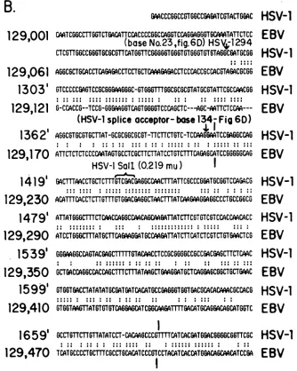
FIG. 7. Nucleotide sequence homology between HSV-1 KOS DNA encodingthe spliced 2.7-kbtranscriptand EBV B95-8DNA. The splice donor (A) andsplice acceptor (B) regions of HSV-1areshown,as arethe Sall sitesat0.195 and0.219m.u.Regionsofhomologyare designatedby:;-indicatesregions where gapswereintroducedbythecomputerhomology programtomaximize overall sequence fit.Regions of strongestcomputer-generated homologyarebracketedby verticallines.HSV-1DNAis numbered fromasite 120bases5'of the cap site of thespliced2.7-kbtranscript. EBV DNA is numbered asdescribedpreviously (3).
(Science, in press). Significant nucleic acid homology was foundto exist between the HSV-1 DNAsequence andtwo regions of the DNA of EBV B95-8. These regions are separated byca. 3,000bases in the EBVgenome(Fig. 7).
The 783 bases ofsequence to the left (5') ofthe HSV-1 splicedonorsiteneartheSallsiteat0.195m.u.wasfoundto be homologous to EBV DNA between bases 125,118 and 125,901. An optimized matchbetween375 of780 baseswas found, compared with a "random" expected value of less than 190 matches for nonhomologous EBV DNA. The
sequenceof EBV DNA is numberedasdescribed previously (3). Theregionofgreatesthomology(Fig. 7A) laywithin300 basesof thesplicedonorsequenceofHSV-1(182 matches). Homology was lost3' ofthesplice donorsite in the HSV-1
sequence(nucleotide 1287, Fig. 7A). Potential splice donor sites inthe EBV DNAsequence are seenatbase 125,875 as well asat several other places.
Considerable homology was seen between the HSV-1
DNAatthespliceacceptor regionand EBV DNAbeginning
at base 129,055. This region showeda match for 476 of900 bases. The homology began at a potential splice acceptor
site in HSV-1DNAlying145bases tothe left(5') oftheSalI
site at 0.219 m.u. This site
(nucleotide
23, Fig. SD) is numberedHSV-1base 1294 in Fig.7B. Homologyextended ca. 820 bases to the right (3') of this site, where it wasabruptlylost (notshown).The
position
wherehomologywas lostcorrespondstobase129,956 ofthe EBV DNA sequence. The 450-baseregionofhighesthomology neartheidentifiedspliceacceptorsis showninFig.7B (251 matches). Potential spliceacceptorsitesareevident in the EBV DNA sequence very nearthose identified in HSV-1 DNA.
The nucleic acid sequence homology seen between the HSV-1 DNAencoding thespliced2.7-kb mRNA and several
regions ofEBV DNA suggested that strongprotein homol-ogyshould also exist. The HSV-1 DNA sequence encoding
thesplicedmRNAcontainsatranslationstart
signal
(ACAA-VOL. 54, 1985
on November 10, 2019 by guest
http://jvi.asm.org/
[image:9.612.137.471.74.492.2]326 COSTA ET AL. J.VIROL.
A
201
211
221
231
241
251
261
H.
Y6TLELFW1MILNHATYFLAVLLGDHIEIQMFLRLVFEIPLFSDAVRHFRORATVFLVPRRHGKUWF
HSV-1
as ss a saa a a as iias
PARLEAFOKJWLHSFYFLISIKSLEITDTMFDIFOSAFGLEEMTLEKLHIFKOKASVFLIPRRHGKTI
EBV
169
179
189
199
209
219
229
271
281
291
301
311
321
331
LVPLIALSLASFRGIK!GYTAHIRKATEPVFEEIDACLRWFGSARVDHVKGE
TISFSFPDGSRSTIVF
H
SV-
1
WAIISLILSNLSWJQIGYHOKIHASAVFTEIIDTLTKSFDSKRVEYNKETSTITFRHSGKISST'MC
EBV
239
249
259
269
279
289
299
340
ASSHTW
splice donor site
ATCFNKW
309
putative
alternate
splice
acceptor
I
identified splice
acceptor
of fig.
6D
115
325
335
3454
355
365
375
B.
A6PPSPREGRGFGARMRIRORRRACLCARFFCLQGIRGODFNLLFVDEAFIRPDJGTIMGFLNi*C
H
SV-I
RGPPSG EGO
WGPSSAILOILSPOXCLASYPYFQSIRGOTFHLLFVDEANFIKKEALPAILGFMLQKDA
E
BV
42
52
62
72
82
92
102
385
395
405
415
425
435
445
K!IFVSSTNTGKASTSFLYNLRGAADELLNwVTYICDDHMPRWTHTNhTACSCYILNKPVFITMDGAVR
H
SV-I
KIIFISSVNSADOATSFLYKLKDAQERLLNVVSYVCQEHRQDFDMQDStMSCPCFRLHIPSYITMDSNIR
E B
V
110
120
130
140
150
160
170
455
465
475
485
495
505
515
RTADLFLAESCMQEIIG6GR
ETGDDRPVLTKSAGERFLLYRPST
TTN
SGLMAPDLYVVDPAFTAN
HSV-
1
ATMLFLDGAFSTELMDTSSLSOGSLSRTVRDDANaLELCRDTNPAGRLASSLYVYDPAYTNN
E
BV
180
190
200
210
220
230
240
522
532
542
552
562
572
582
TRASGTGVA6J6RYR
D
D
YI1FALEHFFLRALTGSAPADIARCWHSPTauLALHpGAFRGVRVAvEG
HSV-1
TSASGTGIAAVTHDRADPNRVIVLGLEHFFLKDLTGDALQOIATCWALVSSIVTLHP
HLEEVKVAVEG
E
BV
250
260
270
280
290
300
310
589
599
NSSODSAVAIATHV
HSV-1
NSSODSAVAIASII
EBV
319
329
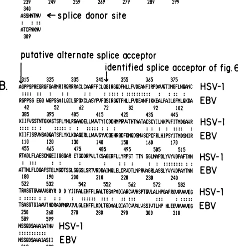
FIG. 8. Comparisonofpredicted amino acidhomology betweentheprotein encoded by the spliced HSV-1transcriptand theputative EBV polypeptide. Asdescribed inthe text, the HSV proteinisinitiatedatanATGtriplet331bases3' of the mRNA cap site and extends 646or 683 aminoacids, dependingonthe spliceacceptor used.(A) Amino acid sequence encoded around thesplice donorsiteofthe mRNA. The EBVpolypeptidehereisinitiatedatbase124,939. (B) Amino acid sequencenearthespliceacceptorsites.Thereading frameused for EBV herewasarbitrarily begunat base128,994. There isaterminator(X,number 62 in the amino acidsequence)atbase 129, 185. Thereading framethen stays open between bases 129,188 and 130,348. Both EBVreading frames werenotedpreviously (3).
on November 10, 2019 by guest
http://jvi.asm.org/
[image:10.612.67.544.198.690.2]SPLICED HSV-1 mRNA 327
TGG)331basestotheright(3')ofthe capsite(Fig.5A).This translational frame remains open through the splice donor
site (base 1287, Fig. 7A) and through boththe alternate and primary splice acceptor sites (nucleotides 23 and 134, Fig. 6D,corresponding toHSV-1 bases 1294 and 1405, Fig. 7B). Itterminates at the TGA codon near the XhoI site at 0.226 m.u. (base 76, Fig. SE). This translational reading frame
predicts proteins of 70,750 (646 amino acids)or 74,822 (683 aminoacids) Da or both, dependingon the splice acceptor used.The codonusefrequency oftheproteinswassimilarto
that seen forotherHSV-1 proteins predicted from
nucleot-ide sequencedata (reviewed in Wagner, in press). Further-more,thepredictedmolecularweight oftheproteinencoded by the spliced 2.7-kb mRNA was in good agreement with that predicted bythe in vitro translation data.
The EBV DNA sequence predicted a proteininitiated at base 124,938 (AACATGC) and continuing through the po-tentialsplicedonors noted inFig.7A. Aproteintranslational
readingframe is alsoseenin the EBV sequence
correspond-ingtothemajor spliceacceptorhomology shown in Fig.7B
(EBV DNAbase 129,114). There is, however, a translation terminator inthe EBV DNA sequence totheleft(5')of this spliceacceptorsite and to theright ofthesite homologousto theputative alternate HSV-1 acceptorsite 111 bases tothe
left(5') ofthemajorone(EBVbase 129,186, Fig. 7B).The
translation reading frame for the EBV DNA sequence
ter-minates at base 130,351. A residue molecularweightofca.
82,000 ispredicted fortheprotein encodedbythecombined translation frames if the chain terminator noted above is
ignored. If the major splice acceptor is used, then the
predicted protein would be ca. 79,000 Da.
The search algorithm of Lipman and Pearson (in press) was used to examine homology between the predicted
HSV-1protein encoded bythespliced2.7-kb mRNA and the
putative protein encoded by EBV (Fig. 8). Regions of striking homology were seen in the predicted amino acid
sequenceoftheproteins both on the N-terminal sideofthe
predicted splicedonor site (52 of146amino acidsmatched;
Fig.
8A) and on the C-terminal side of the twosplice
acceptorsites (135 of287amino acids matched; Fig. 8B). In
thislatter case,it isseenthathomologyextendstotheleft of theterminator signal in the EBV sequence (X, correspond-ingtoamino acid 62 ofEBV, Fig. 8B). Thepredicted amino acidhomology forthe proteins encoded between the poten-tial alternate splice acceptor sites of HSV-1 suggests that there may be alternate splicing patterns in the EBV
tran-script encodingtheprotein homologoustothatofHSV-1. At any rate,the very strikingaminoacidsequencehomologyis
compelling evidence for the conservation of this protein
between thesetwo ratherdistantly relatedherpesviruses.
DISCUSSION
Detailed location ofthetranscripts describedin this report revealedtheirgeneralsimilaritytoHSV-1transcripts mapped
in other regions. Thus, sequences recognizable as
promot-er-regulatory regions existjust5' tothetranscriptcapsites.
Polyadenylation signals are the nominal eucaryotic ones, andnested sets oftranscripts are present. Thecurrent data do show onestriking variationonthis theme: the existence ofatranscript containinga4-kbintron. Full characterization of thistranscriptwillrequirecDNAsequencing through the
potential splicedonor and acceptor sites. Availabilityof the DNA sequence within thisregionwillallow thesynthesis of
specific
DNA primers to carry outthe necessary synthesisandcloningof the cDNA required.
Although full characterization ofsplicing patterns for the
HSV transcript remains to be done, current data clearly reveal that this spliced transcript, which is so unusual for
HSV-1, encodes a protein conserved between HSV-1 and EBV.This suggests that theprotein may be important in the biology of herpesviruses as a group. The sequence homology is of the same order as that seen between the HSV-1 and EBV genes for ribonucleotide reductase (15; Wagner and
Draper, unpublished data). In the case of the spliced
tran-script, however, there was somewhat less homology be-tween theregions. There are, at this date, two other HSV
proteins whichhave been reported to have homology with EBV ones: DNApolymerase andthymidine kinase (3). For
thymidine kinase,thehomologyis minimal and isnot seenin the DNA sequence(15).
Although it cannot yet be said what the functions are of
the HSV genesencodingthesplicedmRNAand itsintron,it is clear that the combination ofprecise transcript mapping
andnucleotide sequenceanalysis has revealedapotentially interesting herpesvirus geneticmarker. Thepredictedamino acid sequence homology with a putative EBV protein is
striking. Furthermore, sequence data willallow the synthe-sis ofspecific synthetic polypeptides tobe used as immuno-gensforgenerating antibodies whichcanbe usedtoexamine
the properties andpotentialfunction of thisprotein. Finally, it is interesting that the arrangement of the
transcripts, vis-a-vis the nested set oftranscripts encoded from theopposite DNA strand of thespliced 2.7-kb mRNA
intron,mayalso begenerally conservedinthe EBV genome.
Analysis of potential translational reading frames of the EBV DNA indicates several longopenframes in theregion
between bases 126,000 and 129,000 (3). These translation
frames are on the opposite coding strand from the EBV
frames homologous to those of HSV-1 and could be
ex-pressedas anestedsetof
transcripts
analogoustothoseseen inHSV-1. Further sequenceanalysis
may revealhomologies
within such translational readingframes. ACKNOWLEDGMENTS
Thisworkwassupported by Public Health ServicegrantCA11861 fromtheNational CancerInstitute, bygrantMV159fromthe Amer-ican Cancer Society, and by a grant from the California Cancer ResearchCoordinatingCommitteetoE.K.W.SupporttoT.J.K.was
from Public HealthServicegrantCA16519 from the NationalCan.cer Institute. R.C. is a trainee supported by Molecular and Cellular BiologytraininggrantGM-07311.
We wishto thank M. Riceand G. Devi for their help. We also thank G. Hayward forlending us his copyofthe EBV sequence. Particular thanks goto R. Sandri-Goldin and K. Leary for critical discussionsof various aspectsof thispresentation.
LITERATURECITED
1. Anderson, K.,R.Frink,G. Devi,B.Gaylord, R.Costa, andE. Wagner. 1981.Detailed characterization ofthe mRNAmapping in the HindlIl fragment K regionofthe herpes simplex virus type genome.J. Virol.37:1011-1027.
2. Anderson, K., J. Stringer, L. Holland, and E. Wagner. 1979. Isolation and localizationofherpessimplexvirus type 1 mRNA. J. Virol.30:805-820.
3. Baer, R., A. Bankier, M. Biggin, P. Deininger, P. Farrell, T. Gibson, G. Hatfull, G. Hudson, S. Satchwell, C. Seguin, P. Tuffnell,and B.Barrell. 1984. DNA sequence andexpressionof the B95-8 Epstein-Barr virus genome. Nature (London) 310: 207-211.
4. Bailey, J. M., and N. Davidson. 1976. Methylmercury as a
reversible denaturing agent for agarose gel electrophoresis. Anal.Biochem. 70:75-85.
5. Berk,A.J.,and P. A.Sharp.1977. Sizingandmappingofearly adenovirusmRNAsby gelelectrophoresis ofS1 endonuclease-VOL.54, 1985
on November 10, 2019 by guest
http://jvi.asm.org/
328 COSTA ET AL.
digested hybrids. Cell 12:721-732.
6. Costa, R., G. Cohen, R. Eisenberg, D. Long, andE. Wagner. 1984. Directdemonstration that the abundant 6-kilobase herpes simplex virus type1mRNAmapping between 0.23 and 0.27 map units encodes the major capsid protein VP5. J. Virol. 49:287-292. 7. Costa, R., B.Devi, K. Anderson, B.Gaylord, and E.Wagner. 1981.Characterization of a major late herpes simplex virus type 1mRNA. J. Virol. 38:483-496.
8. Costa, R., K. Draper, L. Banks, K. Powell, G. Cohen, R. Eisenberg,and E. Wagner. 1983. High-resolution characteriza-tion of herpessimplex virus type 1 transcripts encoding alkaline exonuclease and a 50,000-dalton protein tentativelyidentified as acapsid protein. J. Virol. 48:591-603.
9. Denhardt, D. T. 1966. A membrane-filter technique for the detection of complementary DNA. Biochem. Biophys. Res. Commun. 23:641-646.
10. Draper, K., R. Costa, G. Lee, P. Spear, and E. Wagner. 1984. Molecular basis of the glycoprotein-C-negative phenotype of herpes simplex virus type 1 macroplaque strain. J. Virol. 51: 578- 585.
11. Draper, K., R.Frink, and E. Wagner. 1982.Detailed character-ization of an apparently unspliced beta herpes simplex virus type 1 gene mapping in the interior of another. J. Virol. 43: 1123-1128.
12. Frink, R., K.Anderson, andE.Wagner. 1981. Herpes simplex virus type 1 HindIll fragment Lencodes spliced and comple-mentary mRNA species. J.Virol. 39:559-572.
13. Frink, R., K. Draper, and E. Wagner. 1981. Uninfected cell polymerase efficiently transcribes early but not late herpes simplex virus type1mRNA. Proc.Natl. Acad. Sci. U.S.A.78: 6139-6143.
14. Frink, R., R. Eisenberg, G. Cohen, and E. Wagner. 1983. Detailed analysis of the portionof the herpes simplex virus type 1genomeencodingglycoprotein C. J. Virol. 45:634 647.
15. Gibson, T., P. Stockwell, M. Ginsburg, and B. Barrell. 1984. Homology between two EBVearly genes and HSV ribonucleo-tide reductase and 38K genes. Nucleic Acids Res. 12:5087-5099. 16. Hall, L., K.Draper,R.Frink, R. Costa,and E.Wagner. 1982. Herpes simplex virus mRNA species mapping in EcoRI frag-mentI. J. Virol. 43:594-607.
17. Holland, L., K. Anderson, C. Shipman, Jr., and E. Wagner. 1980.Viral DNA synthesis is requiredfor the efficient expres-sion of specific herpes simplex virus type 1 mRNA species. Virology101:10-24.
18. Laemmli, U.K.1970.Cleavage of structuralproteins during the assembly ofthe head ofbacteriophage T4. Nature (London) 227:680-685.
19. Maniatis, T., E. Fritsch, and J. Sambrook. 1982. Molecular cloning-a laboratory manual. Cold Spring Harbor Laborato-ries, ColdSpring Harbor, N.Y.
20. Maxam, A.,andW. Gilbert. 1980.Sequencing end-labeled DNA with base-specific chemical cleavages. MethodsEnzymol. 65: 499-559.
21. Mount, S. 1982. A catalogue of splice junction sequences. Nucleic Acids Res. 10:459-472.
22. Palmiter,R. D. 1974. Mg++precipitationof ribonucleo-protein complexes. Expedient techniques for the isolation of unde-graded polysome and messenger ribonucleic acid.Biochemistry 13:3606-3614.
23. Stringer, J.,L.Holland,R.Swanstrom,K.Pivo,and E.Wagner. 1977. Quantitation of herpes simplex virus type 1 RNA in infected HeLacells. J. Virol. 21:889-901.
24. Wagner, E. 1983. Transcription patterns in HSV infections. Adv.Viral Oncol. 3:239-270.
25. Weller, S., D. Aschman,W. Sacks, D. Coen, and P.Schaffer. 1983. Genetic analysis of temperature-sensitive mutants of HSV-1: the combined use of complementation and physical mapping for cistronassignment. Virology130:290-305.
J. VIROL.