0022-538X187/041092-06$02.00/0
Copyright© 1987, American Society forMicrobiology
Mutants
Defective in Herpes Simplex Virus Type 2 ICP4: Isolation
and
Preliminary Characterization
COLTON A. SMITHANDPRISCILLA A. SCHAFFER*
Laboratory of Tumor Virus Genetics, Dana-Farber Cancer Institute, and Department of Microbiology and Molecular Genetics, Harvard Medical School, Boston, Massachusetts 02115
Received 3 November1986/Accepted 2 December 1986
Vero cells were biochemically transformed with the geneencoding ICP4 of herpes simplex virus type 2 (HSV-2). These cellswereused as permissive hoststoisolate andpropagateHSV-2 mutantsdefective in this gene.Twomutants, designated hr259 andhr79, wereisolated. Neithermutantgrewinnontransformed Vero cells, but bothgrewtonearwild-type levels in HSV-2 ICP4-expressing cells. hr259 containsadeletion of about 0.6kilobases which eliminatesthe mRNAstartsite oftheICP4gene.hr79 containsamutationwhichmapsby markerrescue totheportion of theICP4 geneencodingthe carboxy-terminal half of the protein. Although hr259 failed to generate any detectable ICP4 mRNA in nontransformed Vero cells, h49 encoded an ICP4 mRNA which is wildtypewithrespecttosize. Innontransformed Verocellsinfected withhr259, only ICPO, ICP6, ICP22,andICP27werereadily detectable. Inthecaseofhr79,atruncated formof ICP4 appearedto be made in additionto ICPO, ICP6, ICP22, and ICP27. Both hr259 and hr79grew efficiently oncell lines transformed with the ICP4 geneof HSV-1 asevidenced by platingefficiencies and single-burst experiments. Similarly, cells transformed with the ICP4 geneof HSV-2 served as efficient hosts forthe growthofd120, HSV-1 ICP4 deletionmutant.
Herpes simplex virustype 1(HSV-1)andtype2(HSV-2) havemanysimilarities. Theirgenomes arecolinear(4), their transcriptional programs appear similar (7), and many of their proteins are functionally interchangeable (8). Despite these similarities, the two viruses exhibit significant differ-ences. Forexample, they exhibit hostrangedifferences(12), HSV-2 shuts off host protein synthesis more rapidly than HSV-1does (9), and HSV-2 reduces the cell surface expres-sion of class 1 H-2 antigens to a greater extent (13). It is reasonable to expect that further analysis will reveal addi-tionaldifferences.
In thisreport, wedescribeourinitial effortstodetermine whether the corresponding immediate-early proteins of HSV-1 and HSV-2 exhibit differences with respectto func-tion. The immediate-early proteins include ICP4, ICPO, ICP22, ICP27, and ICP47 and are operationally defined as products whosegenes aretranscribed in the absence ofprior viral protein synthesis. A variety of HSV-1 mutants with mutations in these geneshave been isolated and character-ized (6, 14, 20, 22; W. Sacks, personalcommunication). As yet, no analogous HSV-2 mutants have been reported. In this study, we describe the derivation and characterization oftwodistinctHSV-2 mutantsdefective in thegene encod-ing ICP4. We found that these mutantshave the samebasic phenotype exhibited by analogous HSV-1 mutants, indicat-ing that the ICP4s of thesetwoviruses perform similar roles during productive infection.
MATERIALSANDMETHODS
Cells and viruses. Vero and CV-1 cells were propagated andmaintainedaspreviously described(26). E5cells contain thegeneencoding HSV-1 ICP4 andexpresscomplementing levels of this protein upon infection with ICP4 deletion
mutants (N. DeLuca, manuscript in preparation). HSV-1 strain KOS and HSV-2 strain 186 were used as wild-type
*Correspondingauthor.
viruses in this study. d120 is an HSV-1 (KOS) mutant harboringadeletion inthegene encoding ICP4 (6).
Plasmids. The viral DNAsequences intheplasmids used in this study are illustrated in Fig. 1. pB6 contains the indicated HSV-2 BamHI-EcoRI fragment cloned into pBR325 (DeLuca, personal communication). pPst, a deriv-ative of pB6, contains the indicated PstI fragment cloned intopUC8 such that the PstI site locatedatabout mapunit 0.86isadjacenttotheunique HindIIl site of thepolylinker. pBal4was generated by cleaving pPstwithHindIII, digest-ing it with Bal 31 nuclease, and ligatdigest-ing it with HindIII linkers.pBal4 lacks
oris,
asdeterminedbyDNAsequencing (data not shown). p2-Bal3 was constructed by cleaving pBal4 with SphI, digesting it with Bal 31 nuclease, and ligating itwith EcoRIlinkers. The deletion in pdlBal4 was constructed by cleaving pBal4 at the NruI site located at +252 with respect to the ICP4 mRNA initiation site and digesting it with Bal 31 nuclease until about 0.6 kilobases (kb) had been removed.TheresultingDNAwasligatedwith BglII linkers. pdlBal4 lacks the mRNA initiation site, as demonstrated by restriction enzyme analysis (data not shown). Plasmids pR, pM,pg,pNB, and pNHaresubclones of p2-Bal3 inserted as HindIII fragments into pUC8. pSV2neo harbors the bacterial gene encoding G418 resist-anceunderthetranscriptional control of the simian virus 40 earlypromoter(24).Nucleic acid isolation. Cytoplasmic RNA and plasmid DNAwereisolatedaspreviously described (15). Infectious HSV-2 DNA was purified by previously described proce-dures(11).
Biochemical transformation of Vero cells with plasmid DNA. Vero cells were cotransfected with pSV2neo and pBal4, and G418-resistant colonies were isolated by the procedure described by DeLucaetal. (6). Theresulting cell lines were screened by plaque assay for the ability to complement d120. One cell line, designated n-33, was cho-senforuse asthepermissive host cell.
1092
on November 10, 2019 by guest
http://jvi.asm.org/
Eii
=
0.79 080 0.81 0.82i 0.83 084 0.85 0.86 ICPO ICP4 oris
P B S N BNBN8NrNrPBE
MapUnits
,f 11 -flil pB6
I pPst
pBal-4
<>- pdlBol-4
-> p2Ba1-3
pR
_ pM
I PM
I pg
pNB
- pNH
FIG. 1. Plasmids used in this study. Restrictionenzymecleavage
sites and subclones of the region of the HSV-2genomebetweenmap
units 0.773 and 0.865 are shownbeneath thearrowsindicating the
locations of the ICPO and ICP4 mRNAs. The black box designates oris. The precise location of HSV-2 ICPO mRNA has not been determined (dotsat the 3' and 5' ends of the ICPO transcript). The dashed linerepresentsthe L-Sjunction. Restriction sites shownare
BamHI (B),PstI (P), EcoRI (E), NcoI (N), NruI (Nr), and SphI (S). By virtue of their orientation in pUC8, the HSV DNA inserts in pBal4, pdlBal4, and p2-Bal3 terminateonthe rightataHindlIl site.
TheHSV DNAinserts inpPst,pBal4, and pdlBal4 terminateonthe
leftatthe PstIsite, whereas p2-Bal3 terminatesataninsertedEcoRI site.Plasmids pR, pM,pg,pNB, and pNHaresubclones of p2-Bal3 insertedasHindIll fragments into pUC8. See Materials and
Meth-ods fordetails ofthese constructions.
Markerrescue test. Marker rescue tests were performed essentially as described previously (18). The subclones of p2-Bal3 described in Fig. 1 were linearized with PstI and separatelycotransfected with the appropriate infectious
mu-tant virus DNAinto n-33 cells. Transfection progeny were assayed simultaneously on Vero and n-33 cells.
Southern andNorthern blot analyses. Southern and North-ern blot analyses were conducted as described previously (15).
Analysis of infected-cell polypeptides. Labeling with
[35S]methioninewascarried outasdescribedpreviously (5). Labeling with 32p, involved incubating cell monolayers in
phosphate-free Dulbecco modified Eagle medium for 6 h before infection and maintaining them in this medium until the timeof harvest. Sodiumdodecyl sulfate-polyacrylamide
gel electrophoresis (SDS-PAGE) analysis of labeled cell extractswas conductedby previously described procedures
(5).
RESULTS
It was our goal to generate HSV-2 mutants harboring deletions in the gene encoding ICP4. For isolation of such mutants, weassumed that HSV-2requires ICP4togrow, as in the case of HSV-1. Accordingly, we used the approach describedby DeLucaetal.for the derivation of HSV-1ICP4 mutants(6). This approach involved (i) generatingacell line expressing complementing levels of HSV-2 ICP4, (ii) con-structingan appropriate deletion in the plasmid-borne copy of theHSV-2 ICP4gene,(iii)cotransfectingthe complement-ing cell line with this deletion plasmid and infectious wild-type HSV-2 DNA, (iv) screening the transfection progeny
forplaque isolatesunabletogrowonnontransformed Vero cells, and (v) determining whether these isolates had ac-quired theengineered deletion.
Generation of cell lines expressing HSV-2 ICP4. To gener-atecell lines able to express complementing levels ofHSV-2
ICP4, we cotransfected Vero cells with pBal4 (Fig. 1) and
pSV2neo. pSV2neo contains the gene specifying G418 re-sistance (24). G418-resistant cell lines were derived and screened for the ability to support the growth of d120, an HSV-1 ICP4 deletion mutant (6). This screening procedure was chosenbecauseprevious studies indicated that HSV-2is able tocomplement HSV-1 mutants defective in ICP4 (8). Of the 19G148-resistant cell lines tested, 4 complementedd120.
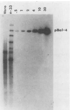
Of these four, cell line 33 (referred to hereafter as n-33)
complemented d120 most efficiently and was therefore fur-ther characterized by Southern blotting (Fig. 2). n-33 cells harbor approximately one intact copy of the HSV-2 ICP4 gene per haploid equivalent. ICP4 sequences smaller and larger than the intact gene werealso evident, suggestingthat deletions and rearrangements of ICP4 sequences had oc-curred. Hybridization of pBal4 to Vero cell DNA (lane 1)
was not surprising in view of the extremely high G+C content of the HSV-1 ICP4gene (16, 21).
Derivation of HSV-2 ICP4 mutants. pdlBal4 contains an engineered 0.6-kb deletion that eliminates thetranscriptional initiation site of the HSV-2 ICP4 gene (Fig. 1). As antici-pated, pdlBal4 did not stimulate the expression of chloram-phenicol acetyltransferase (CAT) when cotransfected into CV-1 cells with a plasmid containing the gene encoding CAT under the transcriptional control of the HSV-1 thymidine kinase promoter, a promoter of the early kinetic class responsive to theICP4 products of HSV-1 and HSV-2 (data not shown). Likewise, pdlBal4 failed to complement the growth of d120 in a transient assay (data not shown). Therefore, it was expected that HSV-2 mutants containing this constructed deletion in both copies of the ICP4 gene would not be viable. Accordingly, n-33 cells were cotrans-fected with pdlBal4 and wild-type HSV-2 DNA, and the progeny were plated on n-33 cells. Of500 plaque isolates
tested fortheabilitytogrow on n-33 cells versus Vero cells,
pBaI-4
[image:2.612.89.252.75.237.2]It
soFIG. 2. Southern blot analysis of HSV-2 DNA in n-33 cells. DNA from n-33 cells and Vero cells was cleaved with PstI and HindIII andsubjectedtoSouthern blotanalysiswith,asprobe,the HSV-2DNAinsert ofpBal4(Fig. 1). pBal4cleaved withPstI and HindIlI wasincludedtovisualize0.5(31pg),1(62pg),2(124 pg),4 (248pg), 10(620 pg),and 30(1.86 ng)copiesofviral DNAper 3 x
109basepairsofcellularDNA. A10-,ugportionof cellularDNAwas
analyzed.
-I -L--i T
on November 10, 2019 by guest
http://jvi.asm.org/
[image:2.612.368.492.464.656.2]f3g II1: EcoRI BglI1 E.o
00 O o 10
-4 K - C4 - C4 K - C4
00w
_mm
[image:3.612.115.256.76.271.2]A
FIG. 3. Restriction analysis of hr259, hr79, and wild-type DNAs. Ontheleft, k andmrefertofragments in the BgII digest of HSV-2 (186) DNA. On the right, k andmrefertofragments in the EcoRI digest of strain 186 DNA. Dots designate detectable fragments resulting from the incorporation of the deletion contained in pdlBal4. (A) BglII and EcoRI digests stained with ethidium bromide. (B)Same DNA digestsasshown in panel A, but blotted and probed
withplasmidpg(Fig. 1).
3, designated hr259, hr79, and hrl2, exhibited the desired host range.hrl2 exhibitedaplating efficiency of1.1 x 10-2 (calculated as PFU per milliliter on Vero cells divided by PFU per milliliter on n-33 cells). Upon reassay onthe two cell types, hrl2 plaques arising on Vero cells exhibited a plating efficiency of unity. Because hrl2 exhibited a high frequency of reversion, it was notanalyzed further. hr259 exhibited a plating efficiency ofless than 5 x 10⁻⁶. hr259 harbors thedeletion engineered in pdlBal4 inbothcopies of the ICP4 gene (Fig. 3). It is evident that hr259 DNA lacks BglII fragments k and m and contains three smaller
frag-ments instead (Fig. 3A), only one of which hybridizes to plasmid pg(Fig. 3B). Since k andmeach containonecopy of the ICP4gene (25) and since pdlBal4 contains aunique BglII site, it is reasonabletoconclude that hr259 contains the deletion constructed in pdlBal4 in both copies of the ICP4
gene.Toconfirmthis, EcoRI digestswereconducted. These
tests demonstrate thathr259 possesses shortened forms of EcoRIfragments mand k(Fig.3A), each of which contains acopyof theICP4gene(25). As expected, pghybridizesto bothshortenedforms of EcoRIfragmentsmand k(Fig. 3B). hr79exhibitedaplating efficiency of 3 x
10-4,
and, like hrl2,plaques arisingonVero cells exhibitedaplating efficiency of unityupon reassay on the two cellstypes. The location of the mutation responsible for the host range phenotype of hr79 was determined by marker rescue with the cloned restrictionfragments listed in Table1.Themutationmapsto
theportion of the ICP4geneencoding the carboxy-terminal halfof theprotein (pNB;Table 1). Rescue with pNB yielded similar results in three independent tests. hr79 does not
appeartocontainanyobvious deletions inthe ICP4gene, as
evidencedby the analysis in Fig.3 and by the factthat pNB hybridizes to the same-sized fragment in NcoI-BamHI-digested hr79 DNAasitdoes in similarly digested wild-type 186DNA(datanotshown).
Phenotypic analysis of hr259and hr79. Cytoplasmic RNA
[image:3.612.325.564.88.179.2]was extracted at5 h postinfection from anisomycin-treated
TABLE 1. Marker rescue of the host-range mutation inhr79a
Plasmid Percent
rescue
pUC8 ... <0.04%
p2-Bal3... 2.60%
pR ... <0.04%
pM... <0.07%
pg... <0.04%
pNB... 0.30%
pNH ...<0.09%
a n-33 cellswerecotransfectedwithhr79DNAand thedesignatedplasmids
linearized with PstI. Titers of the progeny of the cotransfection were
determined simultaneously on n-33 and Vero cells. Percent rescue was
calculated as:(PFU per milliliter on Verocells/PFU per milliliter on n-33cells)
x 100.
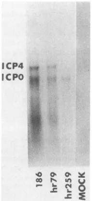
Vero cells infected with hr259, hr79, or HSV-2 (186) and
subjectedtoNorthern blotanalysiswithpBal4astheprobe
(Fig.4). pBal4 is able to detect both ICP4 and ICPO mRNAs
(Fig. 1). Both the 4.7-kb ICP4 mRNA and the 3.4-kb ICPO mRNA were produced during infection with hr79 and HSV-2 (186). As expected, the pBal4 probe detected only ICPO mRNAin extracts of hr259-infected cells.
SDS-PAGEanalysisof extracts of cells infected withhr79, hr259,orHSV-2(186)andlabeled with [35S]methionine from 3 to 15 h postinfection revealed that the mutant viruses exhibited identical polypeptide profiles in nonpermissive Verocells(Fig. 5). ICP6, ICPO, and ICP27 weresynthesized
in relatively small amounts, and the shutoff of host-specific translation was efficient but notcomplete. The status of the three other immediate-early proteins, ICP4, ICP22, and
ICP47, was notclear in thisexperiment. SDS-PAGE analy-sis of extracts of infected n-33 cells revealed that these cells complemented hr259 and hr79 veryefficiently and that hr79 apparently does not specify a full-length thymidine kinase
ICP4 ,
1.C
PO*S.
-.C so ob og
FIG. 4. Northernblotanalysis of cytoplasmicRNAextracted at 5 h postinfection from anisomycin-treatedVerocells infectedat a multiplicity ofinfection of 20 PFU per cell with hr259, hr79, or HSV-2(186).pBal4 (Fig. 1)wasusedasprobe.Verocell monolay-erswereincubated in 100F.Manisomycin for 1 hbefore infection and weremaintainedin thisconcentration of drug untilthetime of harvest.
on November 10, 2019 by guest
http://jvi.asm.org/
[image:3.612.396.491.456.663.2]MOC K hr259 hr 79 186
...-i i.-.1
_w
_~.II'
_
_ ~ ~~~~~~~~~I
a. g
11
15
27
36
41 43 44
V 33 V 33 V 33 V 33
FIG. 5. SDS-PAGE analysis of extracts of Vero or n-33 cells infectedat amultiplicity of infection of 20PFU percell withhr259, hr79,orHSV-2(186) and labeledwith[35S]methioninefrom3 to 15 hpostinfection. VdesignatesVerocells;33designatesn-33 cells.
(ICP36). Whether or not hr79 specified thymidine kinase activity hasnot been determined.
Togain further insight into the mutant phenotype exhib-ited by these viruses, SDS-PAGE analysis was conducted with extracts of Vero cells infected with hr79, hr259, or
HSV-2 (186)andlabeledwith
32p,
from 1.5 to 5 h postinfec-tion (Fig. 6). In this experiment, ICP4 was detectable in extractsof HSV-2 (186)-infected cellsbut nothr259-infectedcells. In the case of hr79, a set of novel proteins was
detectablein the 110- to120-kilodaltonrange. Marker rescue
data(Table 1)areconsistent withtheimplicationthatthese
proteins may represent truncated forms ofICP4. It is also
evident from this experiment that both hr259 and hr79 synthesized ICPO, ICP6, ICP22, and ICP27 under
noncomplementing conditions. The status of ICP47 was not
evident in thisexperiment. It isunclear why neither ICP22
nor ICP0 was detectable in the wild-type extract, but this
may reflect the fact that
immediate-early
transcription isdown-regulated
by
ICP8,aproteinnotspecifiedby
hr259or hr79in Vero cells (Fig. 5) (10).Functional interchangeability of the HSV-1 and HSV-2 ICP4 products. Although earlier studies demonstrated that HSV-2 ICP4 can substitute for HSV-1 ICP4 to support the
growth of HSV-1, it has not been determined whether the converseistrue(8).Toaddress this
question,
weperformed
the
plating-efficiency
assays andsingle-burst
studies showninTables 2 and 3. Theplating efficiencies of hr79 and hr259 were similar to that of the HSV-1 ICP4 mutant d120 on HSV-1
ICP4-expressing
E5 cells(Table 2).
Likewise,
d120plated as well as the HSV-2 ICP4 mutants on HSV-2
ICP4-expressing
n-33 cells. Moreover,complementation
yields
of all three mutantsin both E5 and n-33 cells wasat least 3 orders ofmagnitude
moreefficientthan in Vero cells asassessedby single-burst
experiments (Table 3).Hencewe conclude that thetwoICP4sarefunctionally
interchangeable
during productive
infection in cell culture.DISCUSSION
In this report, we describe two distinct HSV-2 mutants
defective in the gene
encoding
ICP4. Ourprincipal
motiva-tionforgeneratingthesemutants wastodetermine whether HSV-2 ICP4 mutants behave differently from their HSV-1counterpartsundernonpermissive conditions.Wefound that theirphenotypes arebasicallythe same. Under
nonpermis-sive
conditions,
hr259 induced detectable amounts ofonly
ICPO, ICP6, ICP22,
andICP27,
the same spectrum ofproteins
inducedby
d120,
an HSV-1 mutant whose ICP4gene is almost completely deleted (6). ICPO, ICP22, and ICP27areproductsofimmediate-earlygenes,and, as
such,
their
synthesis
does notrequire
ICP4 or any other viralprotein. Hence,that hr259 and hr79 induced the synthesis of these proteins under nonpermissive conditions was not unexpected. ICP6 is formally classified as an early, or 1,
protein (17). 1B
proteins are the products of genes whichrequire prior viral protein synthesis but not viral DNA
synthesisfor maximum transcription. As evidencedby the
phenotypes
of the mutants described in this report, thesynthesisofICP6 is apparently not as dependent on ICP4as is thesynthesisofother 13 proteinssuch asICP36.This isnot unexpected,since,unlike many other 13 genes, the ICP6 gene is transcribed to some extent evenwhen cycloheximide or concavanine is presentthroughout infection (7,
19).
In addition to describing mutants with mutations in the
I
0 >
I
'4-r
0b 4wF
IW
aft~~
i6
O
22
27
[image:4.612.108.246.74.367.2]__4_
FIG. 6. SDS-PAGEanalysis ofVerocellsinfectedata multiplic-ityof infection of 20 PFUpercell withhr259,hr79,orHSV-2 (186) and labeled with 32p, from 1.5 to 5 h postinfection. The dot designates novelpolypeptides in thehr79-infectedextract.
on November 10, 2019 by guest
http://jvi.asm.org/
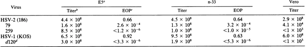
[image:4.612.395.466.419.693.2]TABLE 2. Titersandplating efficiencies ofhr79, hr259, d120, HSV-2 (186), and HSV-1 (KOS) on E5, n-33, and Vero cells
E5a n-33 Vero
virus
Titerb EOPC Titer EOP Titer
HSV-2 (186) 4.4 x 10⁸ 0.66 4.5 x 10⁸ 0.64 2.9 x 10⁸
79 1.6 x 10⁸ 2.6 x 10⁻⁴ 1.3 x 10⁸ 3.2 x 10⁻⁴ 4.1 x 10⁴
259 8.5 x 10⁸ <1.2 x 10⁻⁶ 1.0 x 10⁸ <1.0x 10⁻⁵ <1 X 10³
HSV-1(KOS) 6.5 x 10⁸ 0.92 9.5 x 10⁸ 0.63 6.0 x 10⁸
d120d 3.0 x 10⁸ <3.3 x 10⁻⁶ 1.9 x 10⁸ <5.3 x 10⁻⁶ <1 x 10³
aE5cells aretransformed with an HSV-1DNAfragment encoding ICP4.
bTiters are expressed as PFU per milliliter.
cEOP, Efficiency of plating, calculated asPFUper milliliteronVerocellsdividedbyPFUpermilliliteronICP4-transformed cells.
[image:5.612.64.562.92.169.2]dd120is an HSV-1ICP4 deletion mutant.
TABLE 3. Yieldsof d120, HSV-1(KOS), hr259, hr79, and HSV-2 (186)onVero, n-33, and E5 cells
Yield(PFU/ml)oncelltypea Ratio
VirusRai
Vero E5 n-33 (E5/n33)
HSV-1 (KOS) 4.0 x 10⁷ 1.2 x 10⁸ 8.0 x 10⁷ 1.5 d120 <1.0 x 10⁴ 5.4 x 10⁶ 1.7 x 10⁴ 3.2 HSV-2 (186) 1.9 x 10⁷ 1.3 x 10⁷ 3.3 x 10⁷ 0.4 hr259 <1.0 x 10⁴ 1.2 x 10⁷ 2.4 x 10⁷ 0.5 hr79 <1.0 x 10⁴ 5.7 x 10⁷ 2.4 x 10⁷ 2.4
aCellswereinfectedat amultiplicity of infection of0.1 PFU percell and
harvested 18 hpostinfection.
HSV-2 genefor ICP4, thisreport establishesthat the ICP4
products encoded by HSV-1 and HSV-2 are functionally interchangeable during productive infection. Although we were the first to demonstrate complementation of HSV-1
ICP4temperature-sensitivemutantsby HSV-2 ICP4 (8), this
reportisthefirsttodemonstratethe reverse. Thiscomplete interchangeability is significant, since the two proteins ap-pear to bebiochemically distinct. Not onlydothey differin molecular mass by about 10kilodaltons (19), but they also
exhibit type-specific epitopes (2, 23), although one
cross-reactivemonoclonal antibody hasbeenidentified(1). More-over, the first 47 amino acids ofHSV-2 ICP4 have been
identifiedand exhibit no homology with the aminoterminus of the HSV-1 ICP4(27). However, thetwoproteinsshare at least some nucleic acid homology and exhibit considerable
functional colinearity as evidenced by the existence ofan arrayofintertypic ICP4genes whose chimericproductsare
fully functional (3; C. A. Smith andP. A. Schaffer,
manu-script in preparation). Further study of the HSV-2 ICP4 molecule andits structural andfunctional homologies with
its HSV-1 counterpart shouldhelpto reveal themechanism by whichtheseproteins function.
ACKNOWLEDGMENTS
We thank N. DeLuca, L. McMahan, and W. Sacks for helpful comments onthemanuscriptand M. Datz for manuscript prepara-tion.
Thisinvestigationwassupported by Public Health Service grant CA20260 from the National Cancer Institute.C.A.S. wassupported by National Science Foundation Graduate Fellowship RCF-84-50074.
LITERATURE CITED
1. Braun, D. K., L. Pereira, B. Norrild, and B. Roizman. 1983. Application of denatured, electrophoretically separated, and immobilized lysates of herpes simplex virus-infected cells for detection of monoclonal antibodies and for studies of the properties of viral proteins.J.Virol.46:103-112.
2. Courtney, R. J., and M. Benyesh-Melnick. 1974. Isolation and
characterization of a large molecular-weight polypeptide of herpessimplex virustype 1. Virology 62:539-551.
3. Davison, A. J., H. S. Marsden, and N. M. Wilkie. 1981. One functionalcopyof the long terminalrepeatgenespecifying the immediate-early polypeptide IE110 suffices for a productive infectionof human foetallung cells by herpes simplexvirus.J. Gen. Virol. 55:179-191.
4. Davison, A. J., and N. M. Wilkie.1983.Location andorientation ofhomologoussequencesin thegenomesof fiveherpesviruses. J.Gen. Virol. 64:1927-1942.
5. DeLuca, N. A., M. A. Courtney, and P. A. Schaffer. 1984. Temperature-sensitive mutants in herpes simplex virus type 1 ICP4permissive for earlygeneexpression.J.Virol. 52:767-776. 6. DeLuca, N.A., A. McCarthy, and P. A.Schaffer. 1985.Isolation andcharacterization of deletionmutantsofherpessimplexvirus type 1 in the gene encoding the immediate-early regulatory protein, ICP4.J. Virol.56:558-570.
7. Easton, A. J., and J. B. Clements. 1980.Temporalregulationof herpessimplex virustype2transcription and characterization of virus immediate early mRNA's. Nucleic Acids Res. 8:2627-2645.
8. Esparza, J., M. Benyesh-Melnick, and P. A. Schaffer. 1976. Intertypic complementation and recombination between tem-perature-sensitivemutants ofherpessimplex virustypes 1 and 2. Virology 70:372-384.
9. Fenwick, M.L.,L.S.Morse,andB.Roizman.1979.Anatomy of herpessimplex virusDNA. XI.Apparentclustering of functions effectingrapid inhibition ofhost DNA andproteinsynthesis.J. Virol. 29:825-827.
10. Godowski, P.,and D. M.Knipe. 1986.Transcriptionalcontrolof herpesvirusgene expression: genefunctionsrequiredfor posi-tive and negative regulation. Proc. Natl. Acad. Sci. USA 83: 256-260.
11. Goldin, A. L., R. M. Sandri-Goldin, M. Levine, and J. C. Glorioso. 1981. Cloning of herpes simplex virus type 1
se-quencesrepresenting the wholegenome.J. Virol. 38:50-58. 12. Goodman, J. L., and J. G. Stevens. 1986. Passage ofherpes
simplex virustype 1 onchickembryo fibroblasts confers viru-lence for chickembryos. VirusRes.5:191-200.
13. Jennings,S.R., P. L.Rice,E. D.Klosteweski, R. W. Anderson, D. L. Thompson, and S. S. Tevethia. 1985. Effect of herpes simplex virus types 1 and 2 on surface expression of class I major histocompatibility complexantigens on infected cells. J. Virol. 56:757-766.
14. Longnecker, R., and B. Roizman. 1986.Generation ofan invert-ing herpes simplex virus 1 mutant lacking the L-S junctiona sequences, an origin of DNA synthesis, and several genes including those specifying glycoprotein Eand the a47 gene. J. Virol. 58:583-591.
15. Maniatas, T., E. F. Fritsch, and J. Sambrook. 1982. Molecular cloning:alaboratory manual. ColdSpring HarborLaboratory, Cold Spring Harbor,N.Y.
16. McGeoch, D. J., A. Dolan, S. Donald, and D. H. K. Brauer. 1986.CompleteDNAsequence ofthe shortrepeatregionin the genome ofherpes simplex virus type 1. Nucleic Acids Res. 14:1727-1745.
17. Morse, L.S.,L.Pereira, B. Roizman, and P. A. Schaffer. 1978.
on November 10, 2019 by guest
http://jvi.asm.org/
Anatomyofherpessimplexvirus (HSV)DNA. XI.Mapping of viralgenesby analysis of polypeptides andfunctions specified by HSV-1 x HSV-2 recombinants.J. Virol. 26:389-410.
18. Parris, D.S.,R. A. F.Dixon,and P. A. Schaffer.1980.Physical
mapping of herpes simplex virus type 1 ts mutants by marker
rescue: correlation ofthephysical and genetic maps. Virology
100:275-287.
19. Pereira, L., M. H. Wolff, M. Fenwick, and B. Roizman. 1977. Regulationofherpesvirus macromolecular synthesis. V. Prop-erties ofapolypeptides made in HSV-1 and HSV-2 infected
cells. Virology 77:733-749.
20. Post, L. E., and B. Rolzman. 1981.Ageneralized technique for deletion of specificgenesinlargegenomes: agene22ofherpes simplex virus 1isnotessentialfor growth. Cell25:227-232. 21. Puga, A., J. Gomez-Marquez, P. R. Brayton, E. M. Cantin,
L. K.Long, M. Barbacid, and A. L. Notkins. 1985.The imme-diate-early enhancerelementofherpes simplex virustype1can
replacearegulatory region of thec-Ha-rasl oncogenerequired fortransformation. J. Virol. 54:879-881.
22. Sacks,W.R.,C. C. Greene,D. P. Aschman, and P. A.Schaffer. 1985.Herpes simplex virustype 1ICP27 isanessential
regula-toryprotein. J. Virol. 55:796-805.
23. Showalter, S. D.,M.Zweig, and B. Hampar. 1981. Monoclonal
antibodiestoherpes simplex virustype1proteins, including the immediate-early protein ICP4. Infect. Immun. 34:684-692. 24. Southern, P. J., and P.Berg.1982. Transformationof
mamma-lian cells to antibiotic resistance with a bacterial gene under
control oftheSV40early regionpromoter.J.Mol.Appl. Genet. 1:327-341.
25. Spear, P. G., and B. RQizman.1981. Herpessimplex viruses,p.
626-628. In J. Tooze (ed.), DNA tumor viruses. Cold Spring Harbor Laboratory, Cold Spring Harbor, N.Y.
26. Weller, S. K., K. J. Lee, D. J. Sabourin, and P. A. Schaffer. 1983. Genetic analysisoftemperature-sensitive mutantswhich define thegenefor the major herpes simplexvirustype1 DNA binding protein. J.Virol.45:354-366.
27. Whitton, L. J., and B. Clements. 1984Replication origins anda sequence involved in coordinate induction of the
immediate-earlygenefamilyareconserved inanintergenic regionofherpes
simplexvirus. NucleicAcids Res.12:2061-2079.
28. Wilcox, K. W., A.Kohn, E.Skylynskaya,and B.Roizman.1980. Herpes simplexvirusphosphoproteins. I.Phosphate cycleson
and offsome viralpolypeptides andcan alter theiraffinityfor DNA. J. Virol. 33:167-182.
![FIG. 5.infectedhhr79, postinfection. SDS-PAGE analysis of extracts of Vero or n-33 cells at a multiplicity of infection of 20 PFU per cell with hr259, or HSV-2 (186) and labeled with [35S]methionine from 3 to 15 V designates Vero cells; 33 designates n-33 cells.](https://thumb-us.123doks.com/thumbv2/123dok_us/1362860.89704/4.612.395.466.419.693/infectedhhr-postinfection-analysis-multiplicity-infection-methionine-designates-designates.webp)