!
"
#"#
$ % %#
&
''' (
Segmentation on Brain Image-A Method
Prof. Samir Kumar Bandyopadhyay Professor, Dept of Computer Science & Engineering,
University of Calcutta, 92 A.P.C. Road, Kolkata – 700009, India
Abstract:Magnetic Resonance Imaging (MRI) is a medical imaging technique. MRI, invented in 1970, is a popular method in medical imaging. MRI scanning is relatively safe and unlike other medical imaging modalities, can be used as often as necessary. Moreover, it can be adapted to image brain. Clinical MRI is based on the hydrogen nucleus due to their abundance in the human body and their magnetic resonance sensitivity.
Radiologist used it for the visualization of the internal structure of the body. MRI provides rich information about human soft tissues anatomy. MRI helps for diagnosis of the brain tumor. Images obtained by the MRI are used for analysing and studying the behaviour of the brain. Image segmentation is a key task in many image processes and computer vision applications.
In this paper we presented a method for brain segmentation. It is based on the image segmentation method, which refers to the major step in image processing, the inputs are images and, outputs are the attributes extracted from those images. It covers imaging modalities, magnetic resonance imaging and methods for noise reduction, inhomogeneity correction and segmentation.
Keywords: Brain, MRI, Image analysis, Segmentation, Tumor detection
I. INTRODUCTION
Brain is the kernel part of the body. Brain has a very complex structure. Brain is hidden from direct view by the protective skull. This skull gives brain protection from injuries as well as it hinders the study of its function in both health and disease. But brain can be affected by a problem which cause change in its normal structure and its normal behaviour .This problem is known as brain tumor. Brain tumor causes the abnormal growth of the cells in the brain. The cells which supplies the brain in the arteries are tightly bound together thereby routine laboratory test are inadequate to analyse the chemistry of brain. Computed tomography and magnetic resonance imaging are two imaging modalities that allow the doctors and researchers to study the brain by looking at the brain non-invasively [1].
The success of an image analysis system depends on the quality of segmentation. In the analysis of medical images for computer-aided diagnosis and therapy, segmentation is often required as a preliminary processing task. Medical image segmentation is a complex and challenging task due to the intrinsically imprecise nature of the images. The accurate segmentation of MR images into different tissue classes, especially gray matter (GM), white matter (WM) and cerebrospinal fluid (CSF), is an important task. Moreover, regional volume calculations may bring even more useful diagnostic information. Among them, the quantization of gray and white matter volumes may be of major interest in neurodegenerative disorders such as Alzheimer disease, in movement disorders such as Parkinson or Parkinson related syndrome, in white matter metabolic or inflammatory disease, in congenital brain malformations or perinatal brain damage, or in post-traumatic syndrome.
The accurate identification of the brain tumor boundary and its components is crucial for their effective treatment, but is rendered challenging due to the large variations in tumor size, shape and location, and the inherent inhomogeneity, presence of edema, and infiltration into surrounding tissue. Most of the existing tumor segmentation methods use supervised or unsupervised tissue classification based on the conventional T1 and/or T2 enhanced images and show promising results in differentiating tumor and normal tissues [1-3].
Images obtained by the MRI are used for analysing and studying the behaviour of the brain. Image intensity in MRI depends upon four parameters. One is proton density (PD) which is determined by the relative concentration of water molecules. Other three parameters are T1, T2, and T2* relaxation, which reflect different features of the local environment of individual protons.
One of such is brain image segmentation which is quite complicated and challenging but its accurate segmentation is very important for detecting tumors, edema, and necrotic tissues. Accurate detection of these tissues is very important in diagnostic systems. Also, magnetic resonance imaging (MRI) is an important imaging technique for detecting abnormal changes in different parts of the brain in early stage. MRI imaging is a popular way to obtain an image of brain with high contrast. MRI acquisition parameters can be adjusted to give different grey levels for different tissues and various types of neuropathology.
The human body consists primarily of water and bones. Moreover, trace elements exist in different parts of human body, such as iodine in the thyroid, tellurium in the liver and iron in blood. Medical imaging techniques use different properties of these elements.
single-photon emission computed tomography (SPECT), ultrasound and magnetic resonance imaging (MRI). The x-ray, invented by Wilhelm in 1895, is based on the measurement of the transmission of x-ray through the body. However, a disadvantage of x-ray is the high level of radiation emitted which can cause diseases such as cancer and eye cataract.
In x-ray computer assistance tomography (CT), image is reconstructed from a large number of x-rays. In PET, radio nuclides are injected into patient’s body which attach to a specific organ. SPECT is a nuclear medicine tomographic imaging techniques which able to produce true 3D image. It uses gamma rays. Ultrasound measures the reflection of ultrasonic waves transmitted through the body and is the best modality for investigation of soft tissues.
Researchers have been able to achieve good segmentation. There are lots of methods for automatic and semi-automatic image segmentation, though, most of them fail because of unknown noise, poor image contrast, and weak boundaries that are usual in medical images.
II. RELATED WORKS
Existing methods are classically divided into region based and contour based methods. In the First class, Clark et al. [1] have proposed a method for tumor segmentation using knowledge based and fuzzy classification, where a learning process prior to segmenting a set of images is necessary. Other methods are based on statistical pattern recognition techniques such as [2-4]. These methods fail in the case of large deformations in the brain. Existing contour based methods are not fully automatic and need some manual operation for initialization. Lefohn et al. [5] have proposed a semiautomatic method using level sets. Another segmentation method based on level sets was introduced by Ho et al. [6] that uses T1-weighted images both with and without contrast agent for tumor detection. A method by deformable model and neural network was introduced by Zhu and Yang [7] that processes the image slice by slice and is not a real 3D method.
For classification of brain images as normal and abnormal El-Syed et al [7] proposed a hybrid technique. In the technique features of the brain MR image are extracted using Discrete Wavelet Transform (DWT) and then reduced using PCA. Classification is based on these features. Two types of classifiers are used for classification one is feed forward fack propagation neural network and second is K-nearest neighbours. Maximum 98.6% accuracy is achieved in the method.
For MRI brain image segmentation Sripama [8] proposed fuzzy symmetry based genetic clustering technique. In this method clusters are evolved by variable length genetic fuzzy clustering technique. For measuring quality of cluster fuzzy point symmetry based cluster validity index is proposed in this paper. Experiments are performed on different T1, T2 and PD brain images. This technique performs better then FCM and Expectation Maximization algorithm. But this technique does not consider spatial information and sometime does not segment brain image correctly. This technique also does not work properly for the data sets which have same point as a centre for different clusters.
Zhang proposed a Hidden Markov Random Field Model and the Expectation-Maximization algorithm for segmentation of brain MR images [9]. This is a fully automatic technique for brain MR images segmentation. This method is based on estimation of threshold that is heuristic in nature. Thus most of the time, this method does not produce accurate results. It is also computationally very expensive.
Fuzzy c-means (FCM) clustering [10] is an unsupervised system that has been effectively applied in fields such as astronomy, geology, medical imaging, target recognition, and image segmentation for clustering, classifier designs and feature analysis.
III. PROPOSED METHOD
Brain image segmentation is one of the most important parts of clinical diagnostic tools. Brain images mostly contain noise, inhomogeneity and sometimes deviation. Therefore, accurate segmentation of brain images is a very
difficult task. However, the process of accurate
segmentation of these images is very important and crucial for a correct diagnosis by clinical tools.
Medical image segmentation is a key task in many medical applications such as surgical planning, post-surgical assessment, abnormality detection, and so on.
Ultrasound images contain speckle noise which degrades the quality of the images. Eliminating such noise is an important pre-processing task. Ultrasound imaging is
widely used in the field of medicine. It is used for imaging soft tissues in organs like liver, kidney, spleen, uterus, heart, brain etc. The speed, low cost of imaging and the portability of scanning machine makes it very popular. The common problem in Ultrasound image is speckle noise which is caused by the imaging technique used that may be based on coherent waves such as acoustic to laser imaging [11][12].
This paper [13] describes and analyses an algorithm for reducing such speckle noise. This algorithm is based on mathematical morphology. It is a modified form of MIC and it is called as MMIC. It differs from MIC by not using the histogram for calculating the threshold of the image. It is also using a different technique for reconstructing the features that are of speckle’s size. Moreover it uses structuring elements which are having arbitrary structures which resemble the shapes of the speckles. This algorithm produces better result when compared to the original MIC in time complexity as well as output quality.
A morphological algorithm known as Morphological Image Cleaning algorithm is good in reducing noise in different types of images including scanner images [14]. This algorithm finds the residual image which is the difference between the original image and the smoothed image. It separates the features from the residual image and puts it back into the original image so that features are preserved. This algorithm is an iterative procedure which works with disk shaped structuring elements with different radius.
existence of a static magnetic field, they line up with the field and the net magnetization of protons tends toward the direction of the field. In existence of enough energy, it is possible to make the net magnetization zero. In the relaxation process an induced electronic signal is recorded. The strength and duration of the signal depend on three quantities:
A. (proton density)
B. spin-lattice relaxation time: the time which describes how fast the net magnetization takes to relax back to its equilibrium (T1).
C. spin-spin relaxation time: with this time, magnetization components decrease to zero (T2).
In scanning of a person’s body, by using different parameters setting, it is possible to
obtain three different images of the same body: T1-weighted, T2-T1-weighted, and -weighted.
Segmentation is the partitioning of an image to several segments. The main difficulties in Segmentation are: [a] Noise
[b] The bias field (the presence of smoothly varying intensities inside tissues)
[c] The partial-volume effect (a voxel contributes in multiple tissue types)
The paper is based on the image segmentation method, which refers to the major step in image processing, the inputs are images and, outputs are the attributes extracted from those images. Segmentation divides image into its constituent regions or objects.
Segmentation plays an important role in image analysis. The goal of segmentation is to isolate the regions of interest (ROI) depending on the problem and its characters. Many applications of image analysis need to obtain the regions of interest before the analysis can start. Therefore, the need of an efficient segmentation method has always been there.
A gray level image consists of two main features, namely region and edge. Segmentation algorithms for gray images are generally based on two basic properties of image intensity values, discontinuity and similarity. In the first category, the approach is to partition an image based on abrupt changes in intensity, such as edges in an image. The principle approaches in the second category are based on partitioning image into regions that are similar according to a set of predefined criteria.
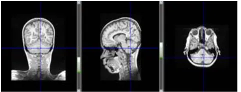
A MRI of brain tumor contains two distinctive regions, the exposed region and the unexposed region. The principal feature of the image is contour, otherwise known as the boundary. The contour can be obtained by partitioning the image into two regions-tumor and non-tumor. Figure 1 shows different Isometric View of Original Brain Volume.
[image:3.612.59.300.615.707.2]
Figure 1 Different Isometric View of Original Brain Volume
Algorithm:
Input: Raw MRI (orgM)
Output: Segmentation of Tumor Begin:
Step1. Open orgM Step2. Create Stack
Step3. Loop row=0 to orgM.Height col=0
Stack.push=row,col,orgM.Width Call Procedure hCheck() row=row+1
End Loop
Step4. Loop col=0 to horM.Width row=0
Stack.push=col,row,horM.Height Call Procedure vCheck() col=col+1
End Loop Step5. End
Procedure: hCheck() Begin:
Step1. If Stack=Empty Return Else
row=Stack.Pop Start=Stack.Pop Last=Stack.Pop If Start=Last
Call Procedure hCalc(row,Start,Last) Call Procedure hCheck()
Else j= Start
gval= orgM.Pixel[row,j] Loop k=j+1to Last
kval= orgM.Pixel[row,k] If kval < gval-MDT OR kval > gval+MDT
midval= (Start + Last) / 2 Stack.push=row,Start,midval Stack.push=row,midval+1,Last Break
EndIf k=k+1 End Loop If k > Last
Call Procedure hCalc(row,Start,Last) Call Procedure hCheck()
Else
Call Procedure hCheck() EndIf
EndIf EndIf Step2. Return
Procedure: hCalc(row,Start,Last) Begin:
Step1. If Start <> Last Len=0
Loop l=Start to Last
Array[len]=(orgM.Pixel[row,l]/16)*16 len=len+1
End Loop
Loop l=Start to Last
horM.Pixel[row,l]=MODE(Array) l=l+1
End Loop Else
horM.Pixel[row,l]=(orgM.Pixel[row,l]/16)*16
EndIf Step2. Return
Procedure: vCheck() Begin:
Step1. If Stack=Empty Return Else
col=Stack.Pop Start=Stack.Pop Last=Stack.Pop If Start=Last
Call Procedure vCalc(col,Start,Last) Call Procedure vCheck()
Else j= Start
gval= horM.Pixel[j,col] Loop k=j+1to Last
kval= horM.Pixel[k,col] If kval < gval-MDT OR kval > gval+MDT
midval= (Start + Last) / 2 Stack.push=col,Start,midval Stack.push= col,midval+1,Last Break
EndIf k=k+1 End Loop If k > Last
Call Procedure vCalc(col,Start,Last) Call Procedure vCheck()
Else
Call Procedure vCheck() EndIf
EndIf EndIf Step2. Return
Procedure: vCalc(col,Start,Last) Begin:
Step1. If Start <> Last Len=0
Loop l=Start to Last
Array[len]=(horM.Pixel[l,col]/16)*16 len=len+1
l=l+1 End Loop
Loop l=Start,Last
resM.Pixel[l,col]=MODE(Array) l=l+1
End Loop Else
resM.Pixel[l,col]=(horM.Pixel[l,col]/16)*16 EndIf
Step2. Return
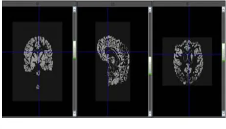
[image:4.612.321.552.96.228.2]Figure 2 shows the result.
Figure 2 Output after Segmentation
IV. CONCLUSIONS
Brain tumor diagnosis is a very crucial task. Segmentation consists of skull removal and tumor extraction phases. It is based on the image segmentation method, which refers to the major step in image processing, the inputs are images and, outputs are the attributes extracted from those images. Quantitative results show that our proposed system performed very efficiently and accurately.
V. REFERENCES
[1] M. Clark, L. Lawrence, D. Golgof, R. Velthuizen, F. Murtagh and M. Silbiger, IEEE Transactions on Medical Imaging 17(April 1998)
[2] M. Kaus, S. Wareld, A. Nabavi, E. Chatzidakis, P. Black, F. Jolesz and R. Kikinis, Segmentation of meningiomas and low grade gliomas in MRI, in MICCAI , (Cambridge UK, 1999)
[3] N. Moon, E. Bullitt, K. Leemput and G. Gerig, Model-based brain and tumor segmentation, in ICPR, (Quebec, 2002)
[4] M. Prastawa, E. Bullitt, S. Ho and G. Gerig, Medical Image Analysis 18, 217 (2004)
[5] A. Lefohn, J. Cates and R. Whitaker, Interactive, GPU-Based Level Sets for 3D Brain Tumor Segmentation, tech. rep., University of Utah (April 2003)
[6] S. Ho, E. Bullitt and G. Gerig, Level set evolution with region competition: Automatic 3D segmentation, 2006 [7] El-Sayed A,El-Dahshan, Abdel-Badeeh M.Salem and
Tamer H.Younis, A hybrid technique for automatic MRI brain images classification, Studia Univ, Babes Bolyai,Informatica, Vol LIV,2009
[8] Sriparna Saha and Sanghamitra Bandyopadhyay, MRI Brain Image Segmentation by Fuzzy Symmetry Based Genetic Clustering Technique, Evolutionary Computation, 2007, pp .4417-4424
[9] Yongyue Zhang, Michael Brady, and Stephen Smith, Segmentation of Brain MR Images Through a Hidden Markov Random Field Model and the Expectation Maximization Algorithm, IEEE Transaction on Medical Imaging, vol.20, No. 1,2001, pp. 45-57
[10] J. C. Bezdek, Pattern Recognition with Fuzzy Objective FunctionAlgorithms New York: Plenum Press, 1981 [11] L.Gagnon and A. Jouan, “ Speckle filtering of SAR
complex-wavelet-based and standard filters”, SPIE proc. #3169, 1997
[12] B.Aiazzi, L. Alparone and S.Baronti, “Multiresolution local statistics speckle filtering based on ratio laplacian pyramid”, IEEE Transaction on Geoscience and Remote Sensing Vol. 36, No. 5, 1998
[13] T.Ratha Jeyalakshmi and K.Ramar, “A Modified Method for Speckle Noise Removal in Ultrasound
Medical Images”, International Journal of Computer and Electrical Engineering, Vol. 2, No. 1, February, 2010