University of South Carolina
Scholar Commons
Theses and Dissertations
2015
Illuminating the Interactions and Functions of
Glutaredoxins, BolA Proteins, and Erv1 in Iron
Homeostasis
Adrian Colleen Dlouhy
University of South Carolina - Columbia
Follow this and additional works at:https://scholarcommons.sc.edu/etd Part of theChemistry Commons
This Open Access Dissertation is brought to you by Scholar Commons. It has been accepted for inclusion in Theses and Dissertations by an authorized administrator of Scholar Commons. For more information, please [email protected].
Recommended Citation
I
LLUMINATING THEI
NTERACTIONS ANDF
UNCTIONS OFG
LUTAREDOXINS,
B
OLA
P
ROTEINS,
ANDE
RV1
INI
RONH
OMEOSTASISby
Adrienne Colleen Dlouhy
Bachelor of Science
North Carolina State University, 2009
Submitted in Partial Fulfillment of the Requirements
For the Degree of Doctor of Philosophy in
Chemistry
College of Arts and Sciences
University of South Carolina
2015
Accepted by:
Caryn Outten, Major Professor
John Dawson, Committee Member
Andrew Greytak, Committee Member
Erin Connolly, Committee Member
ii
iii
A
CKNOWLEDGEMENTSFirst, I would like to acknowledge my advisor, Dr. Caryn Outten. She has been
helpful and encouraging throughout my time in her lab, even when it seemed like none of
my experiments were working. She’s given me many great opportunities to learn and grow
as a scientist. I would also like to thank my committee members, Dr. John Dawson, Dr.
Andrew Greytak, and Dr. Erin Connolly for all of their support and advice.
Additionally, I would like to thank all of my Coutten (and Woutten) labmates, past
and present: Max Darch, Khaleh Thomas, Sam Bouldin, Angela-Nadia Albeţel, John
Hepburn, Haoran Li, Kirsten Collins, Hatice Ozer, Crystal Conway, Malini Gupta, Rabi
Behera, Jingjing Hu, Nin Dingra, and Zuqin Xue. In particular, Henry for getting me started
in the lab and setting the bar high. Sam for taking me under her wing and being there for
me, even after moving to Tennessee. Angela for trying to teach me EPR and to be nicer.
John for all the snacks and talks while I was running AA. And especially Max and Khaleh
for keeping me sane and sticking it out together for six years. I would also like to thank all
the other graduate students and postdocs who helped along the way when they could,
listening to talks, reading abstracts, and showing me the ropes.
Finally, I would like to thank my family and all of my non-graduate school friends
for being encouraging, even when they had no idea what I was talking about. They are
unwavering in their support and always have my back. And I love them even though they
iv
A
BSTRACTIron is a redox-active protein cofactor required for essential cellular functions such
as respiration, however excess intracellular iron can generate damaging reactive oxygen
species. Understanding how cells regulate iron levels is critical for treatment of human
diseases that span from anemia to iron overload disorders. Glutaredoxins (Grxs) with a
CGFS active site are highly conserved proteins shown to have roles in iron homeostasis
and iron-sulfur cluster assembly, thus earning them the title “the Iron Whores”. They can
exist either as a [2Fe-2S] cluster-bound dimer or an apo monomer, suggesting conservation
of structure and function. In addition, Grxs interact with the BolA family of proteins, which
also have genetic connections to metal and sulfur metabolism. In the model eukaryote
Saccharomyces cerevisiae, CGFS-type Grxs and the BolA-like protein Fra2 were shown to transfer an Fe-S cluster to the transcriptional activators Aft1 and Aft2, inhibiting their
DNA binding activity.
E. coli express one CGFS glutaredoxin, Grx4, and two BolA-like proteins, BolA and YrbA. Grx4 forms [2Fe-2S]-bridged homodimers alone, while co-expression of Grx4
with BolA or YrbA yields [2Fe-2S]-bridged heterodimers. In vitro studies indicate
differences in Fe-S cluster binding between these two heterodimers. These results reinforce
the idea that Grx4 acts as Fe-S transport and delivery proteins, while interaction with
v
In the fission yeast Schizosaccharomyces pombe, the CGFS glutaredoxin Grx4
interacts with and regulates the iron-dependent transcriptional repressor Php4. Similar to
its homologues, Grx4 forms a [2Fe-2S]-bridged homodimer alone, and a [2Fe-2S]-bridged
heterocomplex when co-expressed with Php4. Comparison of these complexes indicates
differing cluster coordination environments. These results suggest that when iron is
sufficient, Grx4 interacts with Php4 to form a [2Fe-2S] cluster-bound complex,
communicating cellular iron status and inhibiting Php4 activity.
Erv1 is a sulfhydryl oxidase involved in importing proteins into the mitochondria.
In S. cerevisiae, Erv1 was implicated in cytosolic Fe-S cluster proteins maturation and iron
regulation. However, these studies were performed on a single erv1 mutant strain, erv1-1,
that we discovered has additional defects in glutathione metabolism. To investigate the
Erv1-dependent connection between GSH metabolism and iron homeostasis, we measured
GSH levels and Fe-S protein activity in a variety of erv1 mutants. Only the erv1-1 strain
has significantly reduced GSH levels, due to a mutation in a glutathione biosynthesis gene.
This mutation causes dysfunctional iron regulation and iron accumulation. Together, these
results suggest that the Fe-S cluster maturation and iron regulation defects reported in the
vi
T
ABLE OFC
ONTENTSACKNOWLEDGEMENTS ... iii
ABSTRACT ... iv
LIST OF TABLES ... ix
LIST OF FIGURES ...x
LIST OF ABBREVIATIONS ... xiii
CHAPTER 1:INTRODUCTION AND SCOPE OF THESIS ...1
INTRODUCTION AND SIGNIFICANCE ...1
PROPERTIES OF THE IRON METALLOME ...2
IRON METALLOPROTEINS ...12
IRON UPTAKE,TRAFFICKING, AND STORAGE ...29
REGULATION OF IRON ...42
CONCLUSIONS ...58
SCOPE OF THESIS ...59
COPYRIGHT RELEASE ...62
CHAPTER 2:BOLA AND YRBAFORM DISTINCT FE-SCLUSTER COMPLEXES WITH GRX4 IN E. COLI ...67
ABSTRACT ...67
INTRODUCTION ...68
MATERIALS AND METHODS ...71
vii
CONCLUSIONS ...110
CHAPTER 3:GRX4REGULATES PHP4FUNCTION VIA FE-SCLUSTER BINDING IN S. POMBE ...113
ABSTRACT ...113
INTRODUCTION ...114
MATERIALS AND METHODS ...118
RESULTS ...122
DISCUSSION ...143
CHAPTER 4:IDENTIFYING FUNCTIONS OF BOLA-LIKE PROTEINS IN S. CEREVISIAE ...147
ABSTRACT ...147
INTRODUCTION ...148
MATERIALS AND METHODS ...149
RESULTS AND DISCUSSION ...157
CONCLUSIONS ...171
COPYRIGHT RELEASE ...174
CHAPTER 5:THE SULFHYDRYL OXIDASE ERV1DOES NOT HAVE A DIRECT ROLE IN CYTOSOLIC FE-SCLUSTER PROTEIN MATURATION AND IRON REGULATION ...175
ABSTRACT ...175
INTRODUCTION ...176
MATERIALS AND METHODS ...179
RESULTS AND DISCUSSION ...183
CONCLUSIONS ...196
ACKNOWLEDGEMENTS ...198
viii
INTRODUCTION ...199
AFT1/2GEL SHIFT PROTOCOL ...200
BOLAGEL SHIFT PROTOCOL ...202
β-GALACTOSIDASE ASSAYS IN YEAST ...203
IN-GEL ACONITASE ASSAY ...205
MEASURING IRON LEVELS IN YEAST USING ATOMIC ABSORPTION ...208
ix
L
IST OFT
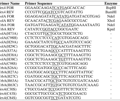
ABLESTable 2.1 Primers used in Grx4-BolA/YrbA study ...72
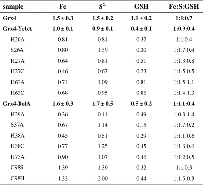
Table 2.2 Fe, S, and GSH content of Grx4-BolA/YrbA complexes ...80
Table 2.3 Molecular weight determination of proteins and complexes ...94
Table 2.4 Thermodynamic parameters for binding of BolA/YrbA to Grx4 ...98
Table 2.5 Fe-S cluster-loading in YrbA and BolA mutants...103
Table 2.6 SPR data of BolA DNA-binding affinity experiments ...107
Table 3.1 Fe-S cluster content in Grx4 and Php4-Grx4 complexes ...126
Table 3.2 Gel filtration data of Grx4 and Php4 complexes ...131
Table 4.1 Strains used in Chapter 4 ...154
Table 4.2 Aft2 EMSA titration results ...161
Table 5.1 Strains used Chapter 5 ...180
Table 6.1 IRDye oligos used in Aft1/2 gel shift assays ...200
Table 6.2 Set-up of Aft1/2 binding reactions ...201
Table 6.3 In-gel assay lysis buffer ...205
x
L
IST OFF
IGURESFigure 1.1 Comparison of Fe-binding siderophore moieties ...8
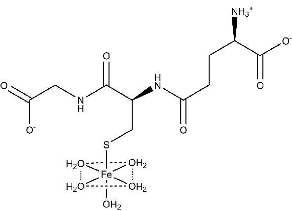
Figure 1.2 Structure of glutathione-coordinating complex with Fe (II) ...10
Figure 1.3 Heme cofactor ...14
Figure 1.4 Heme biosynthesis in eukaryotes ...16
Figure 1.5 Common forms of iron-sulfur cluster cofactors ...20
Figure 1.6 Iron-sulfur cluster biogenesis in E. coli ...21
Figure 1.7 Mitochondrial and cytosolic Fe-S cluster assembly in yeast ...25
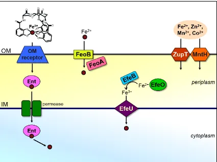
Figure 1.8 Iron uptake systems in E. coli...31
Figure 1.9 Iron uptake systems in S. cerevisiae ...33
Figure 1.10 Iron import and export in mammalian cells ...36
Figure 2.1 Comparison of UV-vis/CD spectra of Grx4, Grx4-BolA, and Grx4-YrbA ...79
Figure 2.2 GRX-HED assay of Grx4, Grx4-YrbA, and Grx4-BolA ...82
Figure 2.3 Comparison of EPR spectra of Grx4, Grx4-BolA, and Grx4-YrbA ...83
Figure 2.4 Comparison of resonance Raman of Grx4, Grx4-BolA, and Grx4-YrbA ...85
Figure 2.5 CD-monitored pH titration of Grx4-YrbA ...86
Figure 2.6 CD-monitored pH titration of Grx4 and Grx4-BolA ...88
Figure 2.7 CD-monitored titration studies of Grx4 with YrbA ...89
Figure 2.8 CD-monitored titration studies of Grx4 with BolA ...90
Figure 2.9 GSH-mediated destabilization of [2Fe-2S] clusters ...92
xi
Figure 2.11 Isothermal titration calorimetry data ...97
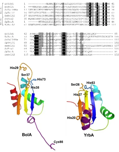
Figure 2.12 Sequence alignment and structural models of E. coli BolA and YrbA ...99
Figure 2.13 Comparison of UV-vis/CD spectra of BolA mutants ...101
Figure 2.14 Comparison of resonance Raman spectra of BolA and YrbA mutants ...104
Figure 2.15 Comparison of UV-vis/CD spectra of YrbA mutants ...105
Figure 2.16 EMSAs of BolA and YrbA complexes ...109
Figure 3.1 Comparison of UV-vis/CD spectra of Grxs ...123
Figure 3.2 UV-vis/CD spectra of Php4-Grx4 ...125
Figure 3.3 EPR spectra of Grx4 and Php4-Grx4 ...127
Figure 3.4 SDS-PAGE of purified Grx4 and Php4 proteins ...129
Figure 3.5 Gel filtration of purified Grx4 and Php4 ...130
Figure 3.6 CD-monitored titration of Php4 into [2Fe-2S] Grx4 ...133
Figure 3.7 CD-monitored titration of apo-Grx4 into [2Fe-2S] Php4-Grx4 ...134
Figure 3.8 CD comparison of purified and reconstituted [2Fe-2S] Php4-Grx4 ...135
Figure 3.9 CD-monitored titration of apo-Php4-Grx4 into [2Fe-2S] Grx4 ...136
Figure 3.10 CD-monitored titration of apo-Php4 into [2Fe-2S] Grx4 with Fra2 ...138
Figure 3.11 CD-monitored titration of Php4 and Grx4 in S. pombe extract ...139
Figure 3.12 UV-vis/CD spectra of Php4 (C221/227A)-Grx4 ...142
Figure 3.13 SPR of Grx4-Php4 interactions ...144
Figure 4.1 Domain structures of S. cerevisiae Grx5 and BolA proteins ...150
Figure 4.2 UV-vis/CD spectra of reconstituted Grx5 ...158
Figure 4.3 UV-vis/CD spectra of reconstituted Grx5-Aim1/Yal044w ...159
xii
Figure 4.5 EMSAs of [2Fe-2S] Aft2 with FET3 ...163
Figure 4.6 EMSAs of Aft2 with MRS4 ...165
Figure 4.7 EMSA of Aft1 with FET3 ...166
Figure 4.8 Spot tests of BolA knockout strains ...167
Figure 4.9 Spot tests of BolA knockouts on YPD compared to YPG ...168
Figure 4.10 SDH, aconitase, and β-galactosidase assays of BolA knockouts ...170
Figure 4.11 GSH assays on BolA knockouts ...172
Figure 5.1 Total GSH levels in erv1 and mia40 mutants ...185
Figure 5.2 In-gel aconitase assays ...188
Figure 5.3 β-galactosidase assays of JRY675 strains ...190
Figure 5.4 β-galactosidase assays of YPH499 strains ...192
Figure 5.5 β-galactosidase assays of W303 strains ...193
xiii
L
IST OFA
BBREVIATIONSAA ... atomic absorption
ABC ... ATP-binding cassette
AFT ... activator of ferrous transport
ALA ... δ-aminolevulinic acid
ALAD ... aminolevulinate dehydratase
ALAS ...ALA synthase
ARE... AU-rich elements
β-ME ... β-mercaptoethanol
BMP ... bone morphogenic protein
BPS ... bathophenanthroline disulfonate
BSA ... bovine serum albumin
CD ... circular dichroism
C/EBPα ...CCAAT enhancer-binding protein α
CIA ... cytosolic Fe-S protein assembly
Cp ... ceruloplasmin
CPgenIII ... coproporphyrinogen III
CPOX ... coproporphyrinogen oxidase
DEAE ... diethylaminoethanol
DHBA ... dihydroxybenzoic acid
dNDP... deoxynucleoside diphosphate
xiv
DTMB ... 5,5’-dithiobis-(2-nitrobenzoic acid)
DTT ... dithiothreitol
EDTA ... ethylenediaminetetraacetic acid
EMSA ... electrophoretic mobility shift assay
EPR ... electron paramagnetic resonance
ER ... endoplasmic reticulum
ERV1... essential for respiration and viability
EXAFS ... extended X-ray absorption fine structure
FECH ... ferrochelatase
FeRE ... iron-responsive element
Fe-S ... iron-sulfur
FET3 ... ferrous transport
FIH1 ... factor inhibiting HIF
FIT3... facilitator of iron transport
FPN ... ferroportin
FRA ... Fe repressor of activation
Grx ... glutaredoxin
GSH... reduced glutathione
GSH1... γ-glutamylcysteine synthetase
GSSG ... oxidized glutathione
GST ... glutathione S-transferase
HCP1 ... Heme carrier protein 1
HED ... 2-hydroxyethyl disulfide
HH ... hereditary hemochromatosis
xv
HMB ... hydroxymethylbilane
HRG ... heme responsive gene
IMS ... intermembrane space
IPTG ... isopropyl β-D-1-thiogalactopyranoside
IRE ... iron regulatory element
IRP ... iron regulatory protein
ISC ... iron-sulfur cluster
ITC ... isothermal titration calorimetry
LIP... labile iron pool
MALDI-TOF... matrix-assisted laser desorption/ionization-time of flight
Mfrn ... mitoferrin
MIA40 ... mitochondrial intermembrane space import and assembly
MRS4 ... mitochondrial RNA splicing
MTT ... 3-(4,5-dimethylthiazol-2-yl)-2,5-diphenyltetrazolium bromide
NADPH ... nicotinamide adenine dinucleotide phosphate
NDP... nucleoside diphosphate
NES ... nuclear export signal
NRAMP ... natural resistance-associated macrophage protein
NTP ... nucleoside triphosphate
OD ... optical density
ONPG ... ortho-nitrophenyl-β-galactoside
PBGD ... porphobilinogen deaminase
PCBP1 ... poly (rC) binding protein 1
PCR ...polymerase chain reaction
xvi
PIXE ... particle-induced X-ray emission
PLP ... pyridoxal 5-phosphate
PMS... post-mitochondrial supernatant
PMSF ... phenylmethanesulfonyl fluoride
PPgenIX ... protoporphyrinogen IX
PPIX ... protoporphyrin IX
PPOX ... protoporphyrinogen oxidase
qRT-PCR... quantitative real-time PCR
RNR ... ribonucleotide reductase
ROS ...reactive oxygen species
RNS ... reactive nitrogen species
RT ... room temperature
SC ... synthetic complete
SCF ... SKP1-CUL1-F-box
SDH... succinate dehydrogenase
SDS-PAGE ... sodium dodecyl sulfate polyacrylamide gel electrophoresis
SOD... superoxide dismutase
SPA tag ... sequential peptide affinity tag
SPR ... surface plasmon resonance
ssDNA ...salmon sperm DNA
STEAP ... six-transmembrane epithelial antigen of the prostate
SUF ... sulfur utilization factor
TAP tag ... tandem affinity purification tag
TB ... Tris-borate
xvii
TCA... tricarboxylic acid
TE ... Tris-EDTA
Tf ... transferrin
TfR ... Tf receptor
TOM ... translocase of the outer membrane
TRX... thioredoxin
TTP ... tristetraprolin
TZF ...tandem zinc finger
UPgenIII ... uroporphyrinogen III
URO3S ... uroporphyrinogen III synthase
UROD ... uroporphyrinogen decarboxylase
UTR... untranslated region
VHL ... von Hippel-Lindau
WT ... wild-type
XANES ... X-ray absorption near edge structure
XAS... X-ray absorption spectroscopy
XRFM ... X-ray fluorescence microscopy
1
CHAPTER
1
I
NTRODUCTION ANDS
COPE OFT
HESIS1INTRODUCTION AND SIGNIFICANCE
Organisms use a variety of transition metals as catalytic centers in proteins,
including iron, copper, manganese, and zinc. Iron is well suited to redox reactions due to
its capability to act as both an electron donor and acceptor. In cells, iron is a cofactor for a
wide variety of metalloproteins involved in energy metabolism, oxygen binding, DNA
biosynthesis and repair, synthesis of biopolymers, cofactors, and vitamins, drug
metabolism, antioxidant function, and many others. Because iron is so important for
survival, organisms utilize several techniques to optimize uptake and storage to ensure
maintenance of sufficient levels for cellular requirements. However, the redox properties
of iron also make it extremely toxic if cells have excessive amounts. Free iron can catalyze
the formation of reactive oxygen species such as the hydroxyl radical, which in turn can
damage proteins, lipids, membranes, and DNA. Cells must maintain a delicate balance
between iron deficiency and iron overload that involves coordinated control at the
transcriptional, post-transcriptional, and post-translational levels to help fine tune iron
utilization and iron trafficking.
1Dlouhy, A. C.; Outten, C. E., The iron metallome in eukaryotic organisms. Met Ions Life
2 PROPERTIES OF THE IRON METALLOME
Intracellular Concentration, Oxidation State, and Speciation. Iron is the most abundant metal on Earth, thus it is not surprising that almost all organisms have
evolved to exploit the unique chemical properties of this ubiquitous transition metal. Iron
primarily exists in either the ferrous (Fe2+) or ferric (Fe3+) oxidation state in biological
systems. Due to its critical role in cell metabolism, iron constitutes a significant portion of
the cellular metallome (Eide et al, 2005). Intracellular iron concentrations vary with cell
type, environmental conditions, and disease state. The iron concentration of human
erythroid cells was measured at 300-400 µM (Epsztejn et al, 1999), while isolated rat
hepatocytes maintain iron concentrations close to 1 mM (Petrat et al, 1999). Iron overload
diseases caused by mutations in iron handling proteins can lead to 10- to 20-fold increases
in these intracellular iron levels in specific tissues (Ceccarelli et al, 1995; Gao et al, 2010;
Petrak et al, 2006). The local bioavailability of iron also strongly influences intracellular
concentrations. For example, analysis of the single-celled model eukaryote S. cerevisiae
demonstrated intracellular iron concentrations ranging from 250 µM to 600 µM depending
on the iron content of the growth medium (Eide et al, 2005; Miao et al, 2011).
E. coli grown in rich media are 0.014% iron by weight in exponential phase, where
0.02% is approximately 106 atoms of iron per cell (Abdul-Tehrani et al, 1999). Iron content
almost doubles to 0.026% in stationary phase, when cells begin iron storage. Cells grown
under iron starvation conditions can be as low as 0.002% iron by weight. In stationary
phase, almost 75% of cellular iron is in the ferric state. While the distribution of this iron
is unclear, it is thought that the majority is bound to proteins as heme, Fe-S clusters, and
3
metalloproteins are non-heme iron, with 40% of those containing an Fe-S cluster (Andreini
et al, 2009). Up to half of the cellular iron can be contained within iron storage proteins
under iron-replete growth conditions (Abdul-Tehrani et al, 1999).
To better study the iron metallome in eukaryotes, biophysical probes such as
Mössbauer and electron paramagnetic resonance (EPR) have been recently employed to
measure not only the absolute iron concentration, but also the types of iron and how this
varies within specific organelles (Lindahl and Holmes-Hampton, 2011). Lindahl and
colleagues have used an integrated biophysical approach to characterize the iron speciation
in S. cerevisiae whole cells and organelles under several growth conditions (Miao et al,
2011; Cockrell et al, 2011; Holmes-Hampton et al, 2010; Miao et al, 2009). These studies
clearly demonstrate that the mitochondria and vacuole are the two central hubs of iron
metabolism in this organism. In general, yeast mitochondria contain 700-800 µM Fe. In
respiring cells, most of this mitochondrial iron is present as the prosthetic groups of the
respiratory complexes (~70% [4Fe-4S]2+ clusters and heme centers), with the remaining
iron present as [2Fe-2S]1+ clusters in enzymes and as non-heme, high spin Fe2+ ions.
Conversely, in fermenting cells the iron from respiratory complexes is reduced to ~30% of
the total iron, there is an increase in non-heme iron, and the appearance of ferric phosphate
nanoparticles. Mutations in Fe-S cluster assembly and trafficking proteins leads to
increased concentration of these nanoparticles with a concomitant rise in reactive oxygen
species (Miao et al, 2011; Miao et al, 2009). The other major iron repository in yeast is the
vacuole. Vacuoles isolated from fermenting yeast contain an average of 220 µM Fe in the
ferric state, which is expected given the acidic environment of this organelle (pH ~ 5).
magnetically-4
interacting Fe(III) nanoparticles, which interconvert based on changes in pH (Cockrell et
al, 2011).
Characterization of iron speciation in other organelles and organisms is still in the
initial stages as most published studies are based on a single technique, instead of
verification by several methods. In addition, various techniques have been used to study
different organisms, making comparison of results challenging. Iron can be found in a
variety of different forms based on location (and thus pH and redox potential), available
ligands, and cellular need. The integrated approach described above is one of the most
promising for studying the iron metallome: by combining Mössbauer, EPR, X-ray
absorption spectroscopy (XAS), electronic absorption spectroscopy, and electron
microscopy, one can resolve different groups of iron species (such as Fe-S clusters, hemes,
and nanoparticles) at a relatively low concentration. In particular, XAS techniques such as
X-ray absorption near edge structure (XANES) and extended X-ray absorption fine
structure (EXAFS) give information about oxidation state, geometry, and ligation. All
forms of iron can be detected and quantified, so changing levels of species can also be
monitored (Ortega et al, 2009).
Another promising method for future studies is X-ray fluorescence microscopy
(XRFM), which provides information about metal distribution, oxidation state, and
coordination. XRFM offers high spatial resolution of biological samples by detection of
emitted X-rays from the sample after irradiation. Pairing this with XANES can provide
more information about iron speciation and subcellular distribution. A combination of
XRFM and XAS studies on brain tissue from Alzheimer’s disease patients showed an
5
Fluorescence intensity from XRFM studies is directly proportional to the element
concentration, providing some quantitative analysis of samples. Quantification of
transition metals in cells and organelles can also be accomplished using particle-induced
X-ray emission (PIXE). Like XRFM, PIXE analysis detects X-rays that are emitted which
are characteristic of elements in the sample. This technique is capable of detecting and
quantifying trace elements (including Fe, Mn, Zn, and Cu) in the µg/g range (Ortega et al,
2009).
Subcellular Distribution. As mentioned above, the majority of cellular iron in eukaryotes is found in the mitochondria and the cytosol for utilization in iron-dependent
proteins. While yeast store excess iron in the vacuole, mammals express iron storage
proteins such as ferritin and mitochondrial ferritin for this purpose. In addition, iron is
recycled in lysosomes after iron-containing proteins are degraded. For example, human
liver and spleen cells from patients with hemochromatosis (an iron overload disease) were
found to contain iron-loaded lysosomes (siderosomes), hemosiderin (a degradation product
of ferritin), and ferritin (Iancu et al, 1997). There has not been a significant amount of research focused on the concentration and chemical nature of iron in the endoplasmic
reticulum (ER). However, the existence of iron pools in this organelle is likely since a
number of heme and non-heme iron proteins are located in the ER. More information on
iron-containing organelles and iron storage proteins is covered in more detail in a later
section.
Iron Bioavailability. Although iron is one of the most abundant elements on Earth, the environment is usually oxygenated, non-acidic, and aqueous. Under these
6
state. One way that organisms improve iron bioavailability is by acidifying the local
environment. The solubility of ferric iron is pH-dependent, changing from 10-18 M at pH 7
to 10-3 M at pH 2. By lowering the pH of the surrounding environment, organisms facilitate
solubilization and uptake of iron. ATP-driven proton transporters move H+ ions from the
cytosol across the plasma membrane to control the pH at the cell surface (Kaplan and
Kaplan, 2009). Humans also use an acidic environment to facilitate uptake of dietary iron.
Uptake mainly occurs though enterocytes in the duodenum, which receives the acidic
contents of the stomach. Iron can then be absorbed for storage in intestinal cells or delivery
to other cells (Kaplan and Kaplan, 2009; Hentze et al, 2004).
Many microorganisms, including E. coli and some fungi, also secrete low
molecular weight compounds known as siderophores into their surroundings, which form
high-affinity (~10-33 M) complexes with ferric iron to make it bioavailable for uptake.
Transporters on the cell surface then recapture the Fe3+-siderophores complexes. For
infectious microorganisms, these molecules help the invading pathogen acquire iron from
the host for survival. Interestingly, two reports suggest that mammalian cells may also
synthesize their own siderophores (Bao et al, 2010; Petrat et al, 2002; Devireddy et al, 2010). In both cases, the siderophore-like compound was isolated by screening for
molecules that bound to siderocalins, a class of lipocalins that specifically bind exogenous
siderophores. Siderocalins are weapons in the immune system arsenal, designed to prevent
the invading organisms from acquiring iron by sequestering Fe3+-bound siderophores
(Correnti and Strong, 2012). However, these new studies suggest that siderocalins may also
bind Fe3+ complexed with endogenous siderophores to facilitate iron trafficking. The
catechol-7
like compounds (Bao et al, 2010), as well as a molecule with a 2,5-dihydroxybenzoic acid
(2,5-DHBA) iron-binding moiety, which is isomeric to 2,3-DHBA found in the bacterial
siderophore enterobactin (Figure 1.1) (Devireddy et al, 2010). BDH2, a homologue of
bacterial EntA (which catalyzes 2,3-DHBA production), was found to be responsible for
2,5-DHBA production. Knockdowns of BDH2 suggested that the 2,5-DHBA-containing
mammalian siderophore is involved in regulating both cytosolic and mitochondrial iron
levels (Devireddy et al, 2010).
Intracellular Labile Iron Pools. The vast majority of iron is bound to proteins and enzymes for use as a cofactor or stored in ferritin, vacuoles, and lysosomes. The
remaining iron in the cell is proposed to be part of a labile iron pool (LIP), also known as
chelatable or free iron, which is most likely present as ferrous complexes given the neutral
pH and reducing conditions inside the cell. The LIP is thought to constitute only 0.1-3% of
total cellular iron (Petrat et al, 2001). EPR studies on E. coli cells suggest this to be around
10 μM (Keyer and Imlay, 1996). The LIP is thought to act as a crossroads in iron
trafficking, providing iron for incorporation into metalloenzymes, feeding pathways for
heme and Fe-S biosynthesis, and directing excess iron towards storage or export proteins
(Hentze et al, 2010). The LIP is also assumed to be dynamic in nature, shrinking and
growing in response to the needs of the cell. Only recently has research focused on
uncovering the chemical nature of iron in the cytosolic LIP (Hider and Kong, 2011). At
physiological conditions, small molecular weight ligands such as phosphates, citrate,
cysteine, and glutathione (GSH) are available to bind Fe(II) (Petrat et al, 2011). While the
predominant ligand for the LIP remains an open question, potentiometric and binding
8
________________________________________________________________________
____________________________
9
sufficient binding affinity to buffer the labile Fe(II) pool, forming the proposed
pentaaquo-Fe(II)-GSH complex shown in Figure 1.2. This iron-GSH complex is suggested to be a
way for cells to distinguish between Fe(II) and Mn(II), which have similar intracellular
concentrations. In addition, the Fe(II)-GSH complex may play a role in iron trafficking
based on the interaction of GSH with monothiol glutaredoxins, which are essential for iron
regulation and trafficking (Muhlenhoff et al, 2010).
There is some evidence that a labile iron pool exists in individual organelles as well
as the cytosol. Using fluorescent indicators, the LIP in mammalian mitochondria was
measured between 1 and 16 µM depending on the cell type, which constitutes <0.4% of
total mitochondrial iron (Petrat et al, 2002; Rauen et al, 2007; Sturm et al, 2005). Studies
on mitochondrial iron speciation in yeast suggest that a somewhat larger pool of non-heme,
high spin Fe2+ is used in assembly of hemes and Fe-S clusters. The exact nature of this iron
is still unknown, although it is presumably loosely bound by low molecular weight ligands
similar to the cytosol. In actively respiring mitochondria, this pool constitutes ~2% of total
mitochondrial iron, but grows to 20% during fermentation when the rate of Fe-S cluster
and heme biosynthesis decreases. Since the total mitochondrial iron in yeast is nearly
identical in respiring vs. fermenting cells, these results demonstrate dynamic shifts in
subcellular iron speciation rather than mitochondrial iron import in response to changes in
energy metabolism (Holmes-Hampton et al, 2010).
Iron Toxicity: Oxidative Stress and Formation of ROS. Although iron is required for many cellular processes, excess iron levels can be toxic to cells. Iron has a central role
in the production of one of the most reactive oxygen species (ROS) found in the cell, the
non-10
________________________________________________________________________
__
11
enzymatically by reacting with the superoxide anion (O2•−) (Eq. 1.1) and hydrogen
peroxide (H2O2) (Eq. 1.2) (Kehrer, 2000).
Fe3+ + O2•− → Fe2+ + O2 (1.1)
Fe2+ + H2O2 → Fe3+ + HO− + HO• (Fenton reaction) (1.2)
Net reaction (1) + (2):
O2•− + H2O2 → O2 + HO− + HO• (Haber-Weiss reaction) (1.3)
ROS such as H2O2 and O2•− are produced naturally in vivo through enzymatic
reactions and auto-oxidation from endogenous compounds and have well-documented
roles in signal transduction pathways and immune cell response (Rovira et al, 2007).
However, when left unchecked, these molecules together with HO• have the ability to
initiate oxidative damage to DNA, lipids, and proteins, all of which contribute to cell death,
aging, and various diseases. Thus iron overload diseases are often characterized by elevated
levels of biomarkers for oxidative stress, including protein carbonyls, DNA oxidation
products, lipid peroxidation, advanced glycation end products, and malondialdehyde
formation. Accumulation of iron in the brain coupled with oxidative stress is also a
common feature of neurodegenerative diseases such as Alzheimer’s and Parkinson’s
disease (Jomova and Valko, 2011).
Iron Toxicity: Iron Interference in Other Metal Trafficking Pathways. Recent studies also suggest that iron toxicity may not be solely due to iron-catalyzed ROS
formation. Kaplan and coworkers demonstrated that toxicity is not dependent on the
presence of oxygen, since iron is toxic to yeast even under anaerobic growth conditions
(Lin et al, 2011). Alternatively, iron toxicity may stem from the interference of excess iron
12
excess iron on manganese trafficking to the antioxidant enzyme superoxide dismutase
(SOD). Eukaryotic cells express an SOD in the mitochondria, SOD2, which preferentially
binds manganese over iron under normal conditions. SOD2 is an essential antioxidant
enzyme since deletion of the SOD2 gene leads to neonatal lethality in mouse models
(Lebovitz et al, 1996; Y. Li et al, 1995). SOD2 activity requires the correct insertion of
manganese into the enzyme, while misincorporation of iron renders it inactive
(Naranuntarat et al, 2009; Yang et al, 2006). Studies of SOD2 mismetallation in yeast
revealed the presence of two distinct iron pools in the mitochondria, one being
“SOD2-inert” and the other “SOD2-reactive”. Disruption of iron homeostasis increases the reactive
pool (without significantly affecting total mitochondrial iron), allowing for iron
incorporation into SOD2. In particular, disruptions in the late stages of mitochondrial
Fe-S cluster biogenesis led to diversion of iron to Fe-SOD2. A somewhat similar situation was
observed in a mouse model of the iron overload disease hereditary hemochromatosis, albeit
via a different mechanism. In this case, cytosolic iron overaccumulation was found to
disrupt trafficking of copper, zinc, and manganese to mitochondria, leading to deficiencies
of these essential metals in this organelle. Consequently, Mn-SOD2 activity was
significantly reduced leading to lower respiratory activity and increased lipid peroxidation
(Jouihan et al, 2008).
IRON METALLOPROTEINS
Mono- and Dinuclear Non-Heme Iron Proteins. While heme iron and Fe-S clusters are two of the most common ways that proteins use iron as a cofactor, it is found
in other forms. Non-heme iron cofactors can be bound directly to proteins as mononuclear
13
for specific roles. Many non-heme iron centers catalyze similar reactions to heme enzymes.
For example, both heme and non-heme diiron enzymes can act as monoxygenases that
insert oxygen atoms into substrate molecules. Non-heme iron proteins catalyze a wide array
of reactions, such as converting nucleoside diphosphates (NDP) to deoxyNDPs
(ribonucleotide reductase), catalyzing biomineralization of iron for intracellular storage
(ferritin), sensing oxygen (prolyl hydroxylases), synthesizing eicosanoids (lipoxygenases),
and modifying histones (lysine demethylases).
Due to the multitude of different non-heme iron centers, there is not a singular
system for assembly and insertion of these cofactors. In most cases, a specific set of
proteins are required: a chaperone for iron delivery, redox proteins to maintain the
oxidation state of iron, and enzymes involved in protein folding that allow for proper
insertion of the metal. For metalloproteins that merely need their cofactor inserted (such as
mononuclear iron), cells can minimize metal misincorporation by compartmentalizing
proteins and using metal chaperones. The metal concentrations in different subcellular
compartments can vary, and a metalloprotein’s metal affinity is usually tailored to these
specific ranges.
Heme-Containing Proteins. Organisms utilize heme-containing proteins for a variety of processes, including sensing and transport of oxygen, energy metabolism,
transcriptional regulation, and protein stability. Heme consists of iron bound to a porphyrin
ring (shown in Figure 1.3), where the iron can act as an electron source or sink for redox
and electron transfer processes. In mammals, heme is one of the most important iron
cofactors. It is best known as an oxygen carrier when bound to hemoglobin in red blood
14
15
cytochrome c transfers electrons between Complexes III and IV in the electron transport
chain of the mitochondria. The cytochrome P450 enzyme family catalyzes the oxidation of
many organic compounds, including lipids, hormones, and xenobiotics. Catalases and
peroxidases are both hemoprotein families that protect against peroxide damage. Catalase
prevents H2O2 damage by catalyzing its decomposition to H2O and O2. Peroxidases use
heme to convert peroxides into alcohols using electron donors and protons, again to prevent
damage caused by reactive peroxides. Recently, it was shown that the nuclear receptor
Rev-erbα binds heme and regulates circadian rhythmicity as well as other metabolic
pathways (Yin et al, 2007).
The steps involved in the synthesis of heme are well conserved from prokaryotes
to eukaryotes (Figure 1.4). As mentioned previously, free iron is toxic to cells due to
generation of ROS. Both porphyrin and heme are also toxic, generating oxygen radicals
and peroxidase activity, respectively. To reduce the risk of these potentially toxic
molecules, heme biogenesis is linked to intracellular iron concentrations and synthesis of
hemoprotein precursors. The heme synthesis machinery is distributed in both the cytosol
and the mitochondria in eukaryotes, requiring intermediates in this pathway to be shuttled
across membranes. Transport of these porphyrin intermediates must be tightly regulated,
again to reduce the risk of toxic components accumulating in the cell (Hamza, 2006).
The first phase in heme biosynthesis is formation of the pyrrole. Initially, ALA
synthase (ALAS) catalyzes this condensation reaction between succinyl-CoA and glycine
to form 5-aminolevulinic acid (ALA) in the mitochondrial matrix. ALA is then transported
to the cytosol, possibly via exchange for glycine by the mitochondrial carrier protein
16
________________________________________________________________________
_
17
two ALA molecules to form monopyrrole porphobilinogen. After formation of the
monopyrrole is complete, porphobilinogen deaminase (PBGD) catalyzes assembly of the
unstable tetrapyrrole hydroxymethylbilane (HMB) from four molecules of
porphobilinogen. Formation of the tetrapyrrole macrocycle is completed by
uroporphyrinogen III synthase (URO3S), which catalyzes the ring inversion and closure of
HMB to make uroporphyrinogen III (UPgenIII) (Ajioka et al, 2006; Shoolingin-Jordan et
al, 2003). Once the tetrapyrrole is formed, the side chains need to be modified to form the
correct porphyrin before insertion of iron. Uroporphyrinogen decarboxylase (UROD)
catalyzes the removal of carboxyl groups from the acetic acid side chains of UPgenIII to
form coproporphyrinogen III (CPgenIII). Coproporphyrinogen oxidase (CPOX) catalyzes
the conversion of CPgenIII to protoporphyrinogen IX (PPgenIX) via oxidative
decarboxylation of the pyrrole ring propionate groups to vinyl groups. CPOX is cytosolic
in yeast, and located in the mitochondrial intermembrane space (IMS) in higher eukaryotes.
Several studies suggest that the ATP-binding cassette transporter protein ABCB6 is either
the CPgenIII transporter, or is somehow involved in the transport of CPgenIII to the IMS
in mammals (Krishnamurthy et al, 2006). Protoporphyrinogen oxidase (PPOX) located on
the outer surface of the mitochondrial inner membrane catalyzes oxidation of PPgenIX to
protoporphyrin IX (PPIX) in the IMS (Ajioka et al, 2006).
The final step in forming heme is the insertion of ferrous iron into PPIX by
ferrochelatase (FECH) in the mitochondrial matrix. There is some evidence that FECH
physically interacts with PPOX across the mitochondrial inner membrane to allow
substrate channeling of PPIX between these two enzymes (Schultz et al, 2010). Human
18
oxide, while S. cerevisiae and bacterial ferrochelatases lack the Fe-S cluster (Dailey, 2002).
Studies indicate that ferrous iron may be imported into the matrix by Mrs3/4 importers
(Mfrn1/2 or mitoferrin in mammalian cells). The yeast homologue of human frataxin,
Yfh1, is proposed to act as an iron chaperone and donate Fe(II) to ferrochelatase for heme
biosynthesis (Park et al, 2003). However, human frataxin does not seem to be involved in
heme biosynthesis, although it may have a role in Fe-S protein assembly (Sheftel et al,
2010; Rouault, 2012).
Once heme is fully assembled, it must be transported from FECH in the matrix
across one or more membranes to target hemoproteins found in various organelles, such as
the IMS, cytosol, nucleus, ER, and lysosomes (Severance and Hamza, 2009). FECH may
act as a heme shuttle for proteins in the matrix that are in the same vicinity, such as
cytochrome P450. For proteins outside the matrix, there is no known heme chaperone in
mammals, although heme chaperones for cytochrome c have been identified in plants and
bacteria (Spielewoy et al, 2001). Some cytosolic heme-binding proteins have also been
suggested to have a role in heme transport, including glutathione S-transferases (GSTs)
from liver and red blood cells (Harvey and Beutler, 1982). Hemoproteins found in the
secretory pathway may obtain heme in the ER, indicating a role for the ER in heme
delivery. The ER and mitochondria have been shown to physically interact via a tethering
complex that may provide a path for heme transport from the mitochondria (Schultz et al,
2010).
Iron-Sulfur Cluster-Containing Proteins. Similar to the heme cofactor, organisms employ iron in the iron-sulfur cluster cofactor for its versatility in electron
19
form as complexes of iron (Fe2+ or Fe3+) and inorganic sulfide (S2-) in various
arrangements, with two of the most common being [2Fe-2S] and [4Fe-4S] clusters (Figure
1.5). Certain prokaryotes and cyanobacteria also utilize larger, more complex clusters that
incorporate additional metals such as molybdenum, nickel, and vanadium. For example,
the nitrogenase enzyme contains a FeMoco cofactor cluster (MoFe7S9) and a P cluster
(Fe8S7) in addition to a [4Fe-4S] cluster in the heterodimer form (Johnson, 1998). Evidence
for these types of complex clusters in higher eukaryotes is lacking. Fe- S proteins can also
interconvert between cluster forms. The enzyme aconitase is active in the [4Fe-4S] cluster
form, while partial disassembly to a [3Fe-4S] cluster renders it reversibly inactive. Fe-S
clusters are usually bound to proteins via coordination of the iron to sulfur from cysteine
and nitrogen from histidine, although serine and arginine have also been shown to ligate
Fe-S clusters. In addition to binding to protein residues, the iron can bind other small
molecules such as glutathione (GSH), homocitrate, CO, and CN−. Recent studies also
demonstrate that a carbon atom coordinates all 6 Fe at the core of the FeMoco cluster in
nitrogenase from nitrogen-fixing bacteria (Lancaster et al, 2011). In addition to their
well-known redox function in electron transfer reactions, Fe-S clusters are also involved in heme
biosynthesis, DNA synthesis and repair, ribosome assembly, tRNA modification,
nucleotide and amino acid metabolism, and biogenesis of Fe-S proteins (Sheftel et al,
2010).
Assembly and Insertion of Iron-Sulfur Clusters in E. coli. E. coli utilize two pathways for iron-sulfur cluster biogenesis, depending on growth conditions (Figure 1.6).
The primary machinery is the ISC (iron-sulfur cluster) system, while the SUF (sulfur
20
________________________________________________________________________
_
21
Figure 1.6. Iron-sulfur cluster biosynthesis pathways in E. coli. (a) The ISC pathway uses the cysteine desulfurase IscS to obtain sulfur from cysteine. Ferrous iron enters the pathway, and the Fe-S cluster is built in the scaffold protein IscU. Ferredoxin and the ATP-dependent chaperones HscA and HscB aid in transfer of the assembled cluster to target apo proteins. The A-type carrier protein IscA may also be involved in cluster transfer. (b) The SUF pathway uses a complex of SufS and SufE as the cysteine desulfurase. The cluster is
assembled on a complex of SufB, SufC, and SufD, which may use the FADH2 cofactor and
22
Homologues of the ISC system are found in the mitochondria of eukaryotes, while
homologues of the SUF system are found in chloroplasts (Lill et al, 2006; Balk and Pilon,
2011; Lill et al, 2012). All Fe-S cluster assembly systems follow the same basic strategy:
a cysteine desulfurase produces sulfur, a scaffold protein accommodates cluster building,
and a carrier delivers the cluster to the target proteins (Py and Barras, 2010; Saini et al,
2012). While iron donation certainly needs to occur as well, this step is not
well-characterized and sources of iron remain unclear.
The ISC system is composed of five proteins that are expressed from the isc operon
(Figure 1.6a). IscS is a pyridoxal-5’-phosphate-dependent enzyme that acts as the
desulfurase, obtaining sulfur from L-cysteine (Schwartz et al, 2000). This sulfur is bound
to the enzyme as a persulfide before being transferred to the scaffold, IscU (Smith et al,
2001; Urbina et al, 2001). As the scaffold protein, IscU accepts the Fe and S, promotes
cluster assembly, and transfer to targets (Agar et al, 2000; Smith et al, 2001; Urbina et al,
2001; Chandramouli et al, 2007). IscS forms a dimer and each subunit interacts with one
IscU, forming a 2:2 stoichiometric complex. NMR studies suggest that IscU can exist in
either a disordered or structured conformational state (Kim et al, 2009). IscS binding causes
IscU to convert to the structured form, which also stabilizes the cluster-bound form (Kim
et al, 2012). Ferredoxin may also function in the ISC system, assisting in coupling two
[2Fe-2S] clusters to form one [4Fe-4S] on IscU (Chandramouli et al, 2007). In order to
transfer the Fe-S cluster to target proteins, IscU interacts with the chaperones HscA and
HscB (Hoff et al, 2000). These chaperones enhance the rate of cluster transfer in an
23
chaperones may bind to IscU and stabilize a conformer with low affinity for the [2Fe-2S]
cluster, thus facilitating cluster release (Bonomi et al, 2011).
The SUF system functions with two protein complexes: SufBCD and SufSE
(Figure 1.6b). SufSE is a heterodimer and acts as the sulfur donor for cluster assembly.
SufS is a homologue of IscS whose activity is improved by interaction with SufE (Mihara
et al, 2000; Outten et al, 2003). Sulfur is bound as a persulfide on SufS before being
transferred to SufE for donation (Loiseau et al, 2003). The structure of SufE is similar to
that of IscU, although it does not have the elements needed for cluster binding or interaction
with HscA/B (Goldsmith-Fischman et al, 2004). SufBCD is the scaffold complex, and can
bind and transfer a [4Fe-4S] cluster (Chahal et al, 2009; Wollers et al, 2010). SufB binds
the cluster and acts as the actual scaffold while interacting with SufC and SufD. SufC has
ATPase activity that is essential for cluster assembly, although its function remains unclear
(Saini et al, 2010). SufD is a SufB paralog and seems to be involved in the entry of iron to
the complex (Nachin et al, 2003; Outten et al, 2003). SufBCD stimulates the desulfurase
activity of SufSE, with SufC being necessary for the interaction between SufB and SufSE
(Outten et al, 2003; Layer et al, 2007). While SufBCD is mainly found in the BC2D form,
it can also exist with other ratios of B:C:D (Wollers et al, 2010). ATPase activity is
facilitated by the C2D2 complex (Petrovic et al, 2008). The B2C2 complex may be involved
in Fe-S cluster assembly for [2Fe-2S] ferredoxin, and it may act as the last scaffold in
assembly (Chahal and Outten, 2012). It has been shown that one BC2D complex binds one
FADH2 molecule, which may act as an electron donor or mobilization of Fe3+ from
ferritins, ferric citrate, or the frataxin homologue CyaY-Fe3+ (Pandolfo and Pastore, 2009;
24
The ISC and SUF systems also contain homologous A-type proteins (IscA and
SufA, respectively) that were originally thought to be alternative scaffolds since clusters
can be reconstituted and transferred from them (Ollagnier-de Choudens et al, 2001, 2003,
2004). It was then shown that clusters can be transferred from IscU to IscA or SufBCD to
SufA, but not in the reverse direction (Chahal et al, 2009; Ollagnier-de Choudens et al,
2004). These results and further experiments indicate that IscA and SufA are involved in
transfer of clusters from the scaffold to target proteins, consequently they were designated
A-type carriers (Loiseau et al, 2007; Gupta et al, 2009; Vinella et al, 2009). E. coli also
contain another A-type carrier, ErpA, which seems to be involved in specialized cluster
delivery. In addition, the monothiol glutaredoxin family has been shown to bind and
transfer a [2Fe-2S] cluster, thus they may also act as carriers of this type of cluster (Iwema
et al, 2009; Yeung et al, 2011).
Assembly and Insertion of Iron-Sulfur Clusters in Eukaryotes. There are two identified systems in eukaryotes for assembly of iron-sulfur clusters: the mitochondrial
iron-sulfur cluster (ISC) assembly machinery and the cytosolic Fe-S protein assembly
(CIA) machinery (Figure 1.7). In the mitochondria, Fe-S protein maturation occurs via two
distinct stages. First, Fe(II) and sulfur are combined on a homodimeric scaffold protein
(Isu) to form a labile Fe-S cluster. Once formed, the nascent Fe-S cluster is then transferred
to its ultimate target protein via additional accessory proteins. In the initial stage, sulfur is
obtained from free cysteine via the pyridoxal phosphate-dependent cysteine desulfurase
Nfs1. Both yeast and human Nfs1 are mainly mitochondrial, although a portion of Nfs1
localizes to the nucleus and cytosol in both systems. In yeast, the mitochondrial form is
25
________________________________________________________________________
_
Figure 1.7. Mitochondrial and cytosolic Fe-S cluster assembly in yeast. (a) In the mitochondria, sulfur is obtained from the cysteine desulfurase Nfs1, interacting with Isd11. Nfs1 transfers sulfur as a persulfide to Isu1/2. (b) Iron is imported to the mitochondria by the transporters Mrs3/4 and possibly donated to Isu1/2 through frataxin (Yfh1 in yeast). (c) Electrons are donated by NADH through the ferredoxin-ferredoxin reductase pair
Yah1-Arh1 to reduce S0 to S2-. (d) The assembled Fe-S cluster is transferred to target proteins by
26
may act as a sulfur donor for other pathways requiring mobilized sulfur, such as cytosolic
tRNA thio-modification (Nakai et al, 2004). Nfs1 function is dependent on formation of a
stable complex with a partner protein named Isd11, although the specific role of Isd11 is
not clear. The Nfs1-Isd11 complex produces sulfane sulfur (S0) via persulfide formation,
thus a source of electrons is required to reduce this molecule to sulfide (S2-) for Fe-S cluster
synthesis. This electron transfer is most likely performed by the ferredoxin-ferredoxin
reductase pair Yah1 and Arh1 in yeast (FDX1/2 and FDXR in humans) using electrons
from NADH (Rouault, 2012; Lill and Muhlenhoff, 2008).
It is clear that the Nfs1-Isd11 complex transfers sulfur to the Isu scaffold proteins
(Isu1 and Isu2 in yeast, ISCU in humans). Similar to Nfs1, a small amount of human ISCU
exhibits cytosolic localization where it may function as a scaffold for extramitochondrial
Fe-S cluster biogenesis (Rouault, 2012; Lill and Muhlenhoff, 2008). The mechanisms of
cluster assembly on scaffold proteins, including the order of Fe and S binding and the
sources of iron, remain open questions. As far as iron delivery, one proposal is that Yfh1,
the yeast homologue of human frataxin, delivers iron to the Isu proteins. Yfh1 was shown
to bind iron in vitro, and to bind Isu1/Nfs1 in an iron- dependent manner in the
mitochondria, possibly enabling iron transfer (Subramanian et al, 2011). More recently,
frataxin was proposed to stabilize the active form of the Nfs1-Isd11-Isu complex thereby
promoting sulfur transfer and enhancing Fe-S cluster formation (Bridwell-Rabb et al,
2012).
Once the cluster is assembled, it must be transferred from the scaffold to the target
apoprotein. Bacterial U-type scaffolds (IscU, NifU) are capable of making and transferring
27
assemble both types of clusters. The cluster transfer system is formed by the chaperone
Ssq1 (an Hsp70 chaperone), a J-type accessory chaperone, Jac1, and the
chaperone/nucleotide release factor, Mge1. While not required for cluster assembly on Isu
proteins, depletion of these chaperones results in cluster accumulation on Isu1. This system
most likely takes the transiently bound cluster from the Isu scaffold and inserts it into the
target protein. The monothiol glutaredoxin Grx5 also may play a role in cluster transfer, as
Grx5 depletion results in cluster accumulation on Isu1, although a specific role has not yet
been determined. Studies on zebrafish and human forms of Grx5 show that it is important
for cytosolic Fe-S assembly, and thus may regulate heme synthesis by facilitating Fe-S
cluster assembly on IRP1 (Rouault, 2012; Lill and Muhlenhoff, 2008). Additional
mitochondrial Fe-S biogenesis assembly factors (e.g. Isa1, Isa2, Iba57, BolA3, Nfu1, Ind)
are proposed to function as intermediate scaffold/delivery proteins between ISCU and
specific subsets of target proteins (Sheftel et al, 2010).
In addition to the mitochondrial ISC system, yeast and mammalian cells have
cytosolic iron-sulfur assembly (CIA) components that build clusters for cytosolic and
nuclear proteins. Since the mitochondrial form of yeast Nfs1 is essential for both
mitochondrial and cytosolic Fe-S cluster assembly, one theory is that the mitochondria
exports a sulfur-containing substrate produced by the ISC machinery that is used by the
CIA machinery to build and/or insert Fe-S clusters (Sheftel et al, 2010). In yeast, the ABC
transporter Atm1 (ABCB7 in humans) is proposed to export the unidentified
sulfur-containing substrate (compound X) from the mitochondria to the cytosol since depletion of
Atm1 inhibits cytosolic Fe-S assembly. A recent study also implicates the mammalian
28
biogenesis (Ichikawa et al, 2012). The mitochondrial IMS-localized sulfhydryl oxidase
Erv1 and the ubiquitous tripeptide GSH are also implicated in export of the
sulfur-containing substrate from the mitochondria for cytosolic Fe-S cluster assembly, (Lange et
al, 2001; Sheftel et al, 2010). However, a recent study reported that the cytosolic Fe-S
assembly defect reported for Atm1-depleted strains was an artifact of the strain background
used in the initial report, indicating that Atm1 activity is not required for cytosolic Fe-S
cluster assembly in yeast (Bedekovics et al, 2011). More recently, the identity of compound
X was demonstrated to possibly involve some form of glutatihione. Atm1 and its
homologue in plants, ATM3, were shown to be stimulated by and preferentially transport
GSSG over GSH (Schaedler et al, 2014). Since an activated persulfide is required for
Fe-S assembly (Fe-S0 versus S2-), it was suggested that GSSG helps transport this intermediate.
GS-S0H would be too reactive to export from the mitochondria. Instead, the persulfide may
be transported more stably as GS-S-SG. Thus, GSH plays an intricate role in Fe-S cluster
assembly and iron homeostasis. In addition, studies in human cells suggest that the
cytosolic Fe-S cluster assembly machinery is independent of the mitochondrial system
(Rouault, 2012).
Regardless of the specific details regarding initial Fe-S cluster assembly in the
cytosol and the requirement of the mitochondrial ISC system, it is clear that a number of
additional proteins are essential for assembling Fe-S clusters for cytosolic/nuclear proteins.
Two potential scaffold proteins are the P-loop NTPases Cfd1 and Nbp35 that form a
[4Fe-4S]-bridged heterotetramer. Yeast Nar1 (IOP1 and IOP2 in humans) is similar to bacterial
iron hydrogenases, although it has no hydrogenase activity. Nar1 has two Fe-S clusters
29
Depletion of mammalian IOP1 disrupts cytosolic Fe-S cluster assembly, and homology of
Nar1/IOP1/2 to other hydrogenases has led to the suggestion that these proteins may act as
electron donors in the CIA system, either in assembly or transfer of a cluster. A fourth
member of the CIA machinery is Cia1, which might play a role late in biogenesis after
Nbp35 and Nar1, possibly in cluster transfer to target proteins. In addition, Dre2, which is
localized to both the cytosol and the mitochondrial intermembrane space, was recently
found to be a possible link between the mitochondrial ISC and cytosolic CIA systems. Dre2
can be purified with both a [4Fe-4S] and a [2Fe-2S] cluster, which seem to play catalytic
or structural roles based on cluster stability. Since depletion of Dre2 impairs cytosolic
Fe-S cluster assembly, it may work early in the CIA pathway and deliver the IFe-SC system
substrate necessary for cytosolic cluster assembly. The human homologue of Dre2,
anamorsin (or CIAPIN1) is proposed to function in electron transfer in the CIA system,
similar to Dre2. While the function appears conserved between yeast and humans,
anamorsin only binds a [2Fe-2S] cluster (Banci et al, 2011). Currently, the only proteins
known to require the CIA system bind [4Fe-4S] clusters (Sheftel et al, 2010; Sharma et al,
2010), thus there may exist a parallel pathway for assembly of cytosolic [2Fe-2S] clusters
such as those found on the Grx3/4 proteins (H Li et al, 2011).
IRON UPTAKE, TRAFFICKING, AND STORAGE
Iron Uptake and Transport in E. coli. Iron uptake in E. coli is divided into
two systems that target either more soluble Fe2+ in the free form, or the poorly soluble Fe3+
bound as a complex. Direct uptake pathways generally target ferrous iron as it is more
bioavailable, although some systems can transport ferric iron (Andrews et al, 2003). The
30
FeoA and FeoB proteins (Kammler et al, 1993). Other proteins such as ZupT and MntH
function as more general divalent metal transporters for zinc, manganese, cobalt, and iron.
ZupT is a ZIP-like protein that mainly acts as a zinc transporter, while the Nramp-like
transporter MntH was originally identified in manganese uptake (Grass et al, 2002; Grass
et al, 2005; Makui et al, 2000; Kehres et al, 2000). The EfeUOB system is involved in uptake of ferrous iron under aerobic and low pH growth conditions and is homologous to
the yeast Ftr1-Fet3 system (Cao et al, 2007). Studies suggest that EfeO binds Fe2+ which
is oxidized before transfer to the ferric permease EfeU at the inner membrane (Rajasekaran
et al, 2010). EfeB likely accepts the electrons from iron before EfeU transporting it across the inner membrane (Figure 1.8).
E. coli use siderophores for uptake of ferric iron through outer membrane receptors (Koster, 2001). Siderophore complexes are transported by the TonB-ExbB-ExbD system
in the periplasm and the inner membrane and an ABC transporter facilitates transfer to the
cytoplasm (Chu et al, 2010). Siderophores, such as the commonly studied ferrichrome and
enterobactin, are synthesized in the cell then secreted to the environment with transport
proteins across the inner and outer membranes (Furrer et al, 2002). Siderophore-iron
complexes are taken up by receptors in the outer membrane using the TonB system (Koster
et al, 2001). There is a range of siderophore receptors with different specificities which can
also take up siderophores produced by other species (Andrews et al, 2003).
Siderophore-iron complexes are then transported across the periplasm to the cytoplasm by periplasmic
binding proteins and inner membrane transporters. Iron is released from the siderophore
via reduction by ferric reductases, or by hydrolysis of the siderophore backbone (Fontecave
31
32
Iron Uptake and Transport in Yeast. The budding yeast S. cerevisiae expresses an extensive system of membrane transporters for uptake of environmental iron (Figure 1.9).
As previously mentioned, yeast have the ability to scavenge iron-loaded siderophores from
their surroundings. Arn1, Taf1, Sit1, and Enb1 (also known as Arn1-4, respectively) are
transporters specific for Fe3+-siderophore complexes. Environmental Fe3+ is reduced to the
more soluble Fe2+ by the ferrireductases Fre1 and Fre2, which are also capable of reducing
Cu2+. Once reduced, Fe2+ can be imported by the high-affinity uptake system encoded by
FET3 and FTR1. Fet3 is a multicopper oxidase that oxidizes the Fre1/2-produced Fe2+ to
Fe3+, which is then transferred across the plasma membrane by the transmembrane
permease Ftr1. In addition, yeast possess low-affinity iron transporters (Fet4 and Smf1)
that can also transport other transition metals. Fet4, in addition to being a low-affinity iron
transporter, can import copper and zinc, and is responsible for most of the uptake under
iron-replete conditions. Smf1, a member of the NRAMP transporter family found in both
prokaryotes and eukaryotes, is a H+/M+ symporter that uses a pH gradient to import
transition metals such as Fe2+, Mn2+, and Zn2+. Yeast express two other NRAMP proteins,
Smf2 and Smf3, although they do not seem to play a significant role in environmental iron
uptake. In addition to these uptake systems, the yeast genome encodes transmembrane
ATP-driven H+ transporters to acidify the environment, which increases the solubility of
Fe3+ (Kaplan and Kaplan, 2009; Bleackley and Macgillivray, 2011).
After cells have imported iron, it needs to be transported either for storage or
utilization. Since free iron is redox-active and can damage proteins and membranes, it
seems likely that cells would use a chaperone to bind and transport iron through the cell.
33
Figure 1.9. Iron uptake systems in S. cerevisiae. Arn1, Sit1, Enb1, and Taf1 are
transporters for Fe3+-siderophore complexes. The ferrireductases Fre1/2 reduce
environmental Fe3+ to Fe2+. Fet3 and Ftr1 form the high-affinity iron uptake system. Fet3
oxidizes Fe2+ to Fe3+ and Ftr1 transports this across the plasma membrane. Fet4 is a
low-affinity transporter responsible for uptake under iron-replete conditions. Smf1, a member
of the NRAMP family of transporters, is a H+/M+ symporter for some transition metals like
Fe2+, Mn2+, and Zn2+. Aft1 and Aft2 are transcriptional regulators that activate expression
34
are suggested to have an essential role in cellular iron trafficking. Depletion of Grx3/4 leads
to defects in iron-dependent proteins independent of induction of the Aft1 iron uptake
system. Although these cells had excess cytosolic iron, iron delivery is impaired. Grx3/4
have previously been shown to bind a [2Fe-2S] cluster in a homodimeric complex. Not
only is this Fe-S cluster essential for their role in iron trafficking, it is also required in iron
sensing and regulation of the transcription factors Aft1 and Aft2 (Muhlenhoff et al, 2010;
Kumanovics et al, 2008; H Li et al, 2009).
The fission yeast S. pombe has a similar high-affinity transport system to S.
cerevisiae. The ferrireductase Frp1 is homologous to Fre1/2 and reduces extracellular Fe3+
to Fe2+ (Roman et al, 1993). This iron is then transported into the cell by a complex of Fio1
and Fip1 (Askwith and Kaplan, 1998). Fio1, homologous to Fet3, acts as an oxidase and
Fip1, homologous to Ftr1, is a transmembrane permease. Unlike S. cerevisiae, S. pombe
can also produce and excrete their own siderophore, ferrichrome (Schrettl et al, 2004). The
proteins Sib1 and Sib2 are involved in synthesis of ferrichrome. There are three
siderophore transporters on the cell surface, Str1, Str2, and Str3, which have specificities
for different iron-siderophore complexes (Pelletier et al, 2003).
Iron Uptake and Transport in Mammalian Cells. In mammalian cells, absorption of dietary iron occurs in the intestine through the brush border of duodenal
enterocytes. Inorganic iron mainly comes from vegetables, while heme iron comes from
degradation of hemoglobin and myoglobin in red meat. The divalent metal transporter 1
(DMT1) also known as SLC11A1, is an NRAMP family protein that carries inorganic iron
across the apical membrane into enterocytes. The ferric reductase Dcytb (duodenal


![Figure 2.4. Comparison of Resonance Raman spectra of [2Fe-2S] Grx4 (left), [2Fe-2S] Grx4-BolA (middle), and [2Fe-2S] Grx4-YrbA (right)](https://thumb-us.123doks.com/thumbv2/123dok_us/8430017.1387805/103.612.101.518.238.490/figure-comparison-resonance-raman-spectra-bola-middle-right.webp)
![Figure 2.13. Comparison of UV-visible absorption (top panels) and CD spectra (bottom panels) of [2Fe-2S] Grx4-BolA mutants](https://thumb-us.123doks.com/thumbv2/123dok_us/8430017.1387805/119.612.98.516.211.482/figure-comparison-visible-absorption-panels-spectra-panels-mutants.webp)