Clinical Interventions in Aging
Relative risk of pulmonary embolism in the very
elderly compared with the elderly
Josef Yayan
Department of Internal Medicine, University Hospital of Saarland, Homburg/Saar, Germany
Correspondence: Josef Yayan Department of Internal Medicine, University Hospital of Saarland, Kirrberger Straße, D-66421 Homburg/ Saar, Germany
Tel +49 684 1162 1620 Fax +49 684 1162 3602 Email [email protected]
Background: Pulmonary embolism (PE) can be an acute, life-threatening emergency, and studies suggest that advanced age is a risk factor for this condition. However, the literature is scarce regarding PE in patients above the age of 90 years. This study examined the relative risk for PE in the very elderly (.90 years) compared with that in the elderly (80–89 years). Methods: A retrospective study was performed examining very elderly patients diagnosed with PE in the Department of Internal Medicine at the University Hospital Homburg/Saar in Germany between 2004 and 2012. Elderly patients (aged 80–89 years) diagnosed with PE served as controls. PE was confirmed by contrast-enhanced chest computed tomography or ventilation perfusion scintigraphy in both groups. A total of 2230 patients were examined for PE in this study. Of these, 15 (0.67%) in the study group and 197 (8.83%) in the control group underwent further evaluation for PE.
Results: After performing a radiological examination, 11 (73.3%, including six [54.55%] women) of the 15 study patients (mean age 91.6 ± 1.67 years) and 148 (75.1%, including 93 [62.84%] women) of the 197 controls (mean age 84.0 ± 2.59 years) were confirmed to have PE. There was a significantly lower proportion of the very elderly enrolled in the study (P , 0.001). There were no significant differences in clinical presentation, cardiovascular risk factors, electrocardiograms, blood gas analyses, radiological diagnoses, or acute comorbidities between the groups. However, the very elderly were more likely to experience minor bleeding in the extremities (P = 0.016) and to have more chronic diseases (P , 0.001). An increased relative risk of PE was not detected in the very elderly (relative risk 0.98, P = 0.88). Furthermore,
d-dimer, troponin T, and high-sensitive troponin T levels had limited predictive value for PE
in the very elderly. There were no significant differences in the number of hospital admissions, intensive care or ward treatments, or duration of hospitalization.
Conclusion: The relative risk for PE in the very elderly is not higher than that in the elderly. Keywords: embolism, aging, electrocardiography, morbidity, risk factors
Introduction
Pulmonary embolism (PE) occurs when the main pulmonary artery or one of its branches suddenly closes due to the presence of a thrombus deported from elsewhere
in the body, usually the deep veins of the leg.1 A small proportion of PEs are caused by
fat, air, bone marrow, amniotic fluid, or septic substances.1 If not quickly treated, PE
can be life-threatening.2 After myocardial infarction and cerebrovascular disease, PE
is the third leading cause of death.3 Unfortunately, it is also among the least accurately
diagnosed acute emergency diseases.3
The incidence of PE rises significantly with age, according to some hypotheses
from recent studies.2 In the elderly, diagnosing PE can be difficult due to the extensive
Dovepress
O R I G I n A L R E S E A R C H open access to scientific and medical research
Open Access Full Text Article
Clinical Interventions in Aging downloaded from https://www.dovepress.com/ by 118.70.13.36 on 20-Aug-2020
For personal use only.
Number of times this article has been viewed
This article was published in the following Dove Press journal: Clinical Interventions in Aging
number of cardiopulmonary conditions that may mimic the clinical presentation of PE in this population.2 Diagnosis
of PE relies on clinical likelihood, serum d-dimer levels,
compression ultrasonography of the lower limb, ventilation-perfusion lung scans, and/or helical computed tomography
(CT).2 Pulmonary angiography is seldom required because
noninvasive diagnostic tests are generally sufficient.2 Further,
age can affect and confound the diagnostic tests for PE.2
With increasing age, the predictive values of d-dimer and
ventilation-perfusion lung scan are reduced.2 Exclusion
of PE in patients older than 80 years by assessment of the
d-dimer level is possible in only 5% of patients.2 However,
age has no effect on the diagnostic precision of lower limb
compression ultrasonography and helical CT.2 Therefore, a
rational diagnostic approach to PE in the elderly should rely mainly on investigations that are meaningful and have both high sensitivity and specificity.2
The objective of this study was to compare the clinical presentation, cardiovascular risk factors, acute and chronic comorbidities, electrocardiographic changes, blood gas analyses, and radiologic imaging studies between the very
elderly (aged .90 years) and elderly (aged 80–89 years) with
a diagnosis of PE. This investigation was designed to acquire new insights into the accurate and prompt diagnosis of this acute emergency disease. Specifically, we examined whether there is an increased risk of PE in the very elderly population; whether the very elderly demonstrate an increased number of PEs; and whether PE diagnoses in the very elderly
popu-lation can be made based on clinical symptoms, d-dimer
values, electrocardiographic changes, blood gas analyses, and radiologic imaging.
Materials and methods
Patients
A retrospective study was performed using the medical records of patients treated for PE at the Department of Internal Medi-cine of the University Hospital Homburg/Saar in Germany between 2004 and 2012. The highest possible decade of life was used in this study, with the assumption that the highest life expectancy is, on average, 100 years of age. The two possible last decades of highest life used for comparison were close together to prevent any distortion in the data analysis due to age. Therefore, the study population was comprised
of very elderly (aged .90 years) patients diagnosed with PE,
and the controls were elderly patients (aged 80–89 years) with PE. The relative risk of developing PE was assessed as a ratio of the probability of PE occurring to the probability of PE not occurring among patients in both groups. Clinical symptoms of
PE considered were dyspnea, chest pain, cough, hemoptysis, tachypnea, jugular venous distension, cyanosis, hypotension, and shock. The diagnosis of PE was made according to the latest edition of the International Classification of Disease (ICD I26.0–I26.9) from 2004 to 2012. PE was further defined as central or peripheral, depending on the location or arterial branch involved. Central vascular zones included the main pul-monary artery, the left and right main pulpul-monary arteries, the anterior trunk, the right and left interlobar arteries, the left upper lobe trunk, the right middle lobe artery, and the right and left lower lobe arteries. Peripheral vascular zones comprised the segmental and subsegmental arteries. A PE was considered massive when it involved both pulmonary arteries or resulted in hemodynamic compromise.
Clinical presentation of the patients in the emergency room was determined upon admission or hospital transfer. Acute and chronic comorbidities considered included cardiovascular, pulmonary, gastrointestinal tract, renal, urogenital, gynecological, neurological, psychiatric, orthopedic, dermatological, and allergic diseases. Plasma
d-dimer concentrations were measured in citrated blood
(1 + 10 mixture of 3.5% aqueous sodium citrate and blood;
Sarstedt, Nümbrecht, Germany) using a well validated, commercial, particle-enhanced, immunoturbidimetric assay
(Innovance®d-dimer, Siemens Medical Solutions, Erlangen,
Germany) with the Behring Coagulation System analyzer (Dade Behring, Marburg, Germany). Highly sensitive
tro-ponin T levels were measured using the Elecsys® troponin T
electrochemiluminescence immunoassay (fourth-generation) with the Roche Elecsys 2010 analyzer (Hoffman-La Roche Ltd, Mannheim, Germany). In addition to highly sensitive troponin T levels, conventional troponin T levels (Stat T, Roche Diagnostics, Mannheim, Germany) were measured using an electrochemiluminescence immunoassay
(third-generation) on an Elecsys 2010 platform (Roche). d-dimer,
troponin T, and highly sensitive troponin T were ordered as
necessary (normal ,0.5 mg/L, ,0.10 ng/mL, ,50 pg/mL,
respectively). The 12-lead electrocardiogram was evaluated for sinus rhythm, atrial fibrillation, and tachyarrhythmia absoluta in atrial fibrillation, T negativity, and S1Q3 changes suggestive of PE. Arterial or venous blood gas analyses were performed as soon as possible to support an early diagnosis of PE. Lower limb ultrasonography was used to detect deep venous thrombosis as a possible etiology for PE. Contrast-enhanced chest CT and/or ventilation perfusion scintigraphy were used to confirm the diagnoses of PE in each group. The presence of cor pulmonale with PE was determined using echocardiography in all patients.
Dovepress Yayan
Clinical Interventions in Aging downloaded from https://www.dovepress.com/ by 118.70.13.36 on 20-Aug-2020
The medical treatment of both groups was also compared, along with hospital admissions, number of treatments in intensive care or general wards, and length of hospitalization. Numerical trends for PE in recent years were examined for both groups. Finally, the number of reanimations and autop-sies as well as the mortality rate were calculated for both groups. Due to the retrospective nature of the study protocol, the Medical Association of Saarland’s Institutional Review Board waived the need for informed consent.
Statistical analysis
Relative risk and 95% confidence intervals (CIs) were calculated and compared between the very elderly and the elderly. The chi-square test was used to compare any differences in gender, clinical presentation, cardiovascu-lar risk factors, acute and chronic comorbidities, d-dimer
values, troponin T, high sensitive troponin T, electrocar-diogram, hospital admissions, number of intensive care or ward treatments, recurrent PE, recurrent deep vein throm-bosis, and location of PE (central versus peripheral). The Mann–Whitney test was used to calculate the differences
in age, duration of hospitalization, d-dimer levels, and total
chronic disease profiles. All results are expressed as the mean and standard deviation. Survival rates for both groups were
calculated using the Kaplan–Meier method. P , 0.05 was
considered to be statistically significant.
Results
From the total of 2230 patients evaluated for PE during the study period, 15 (0.67%) patients were assigned to the study
group and 197 (8.83%) patients were assigned to the control group. Eleven (including six [54.55%] women) of the 15 study patients (73.3%, 95% CI 51.0–95.7) and 148 (including 93 [62.84%] women) of the 197 control patients (75.1%, 95% CI 69.1–81.2) were diagnosed with PE. Compared with the elderly, there was no increase in relative risk for
PE in the very elderly (0.98, 95% CI 0.72–1.34, P = 0.88).
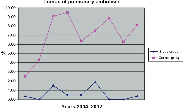
There was a significant difference in the number of cases
between the two groups during the study period (P = 0.0003,
Figure 1). The mean age of the patients in the study group
was 91.6 ± 1.67 years compared with 84.0 ± 2.59 years in
the control group (P , 0.0001). There was no significant
difference in gender ratio between the two groups (P = 0.584).
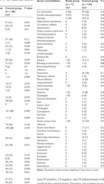
There were also no significant differences in the diagnostic procedures, clinical features of PE subtypes, and medical therapy administered (Table 1). The study group was more likely to experience minor bleeding at the extremities
(P = 0.016, Table 2). There was no significant difference in
cardiovascular risk factors (Table 3). Multiple significant differences were found in chronic comorbidities between
the two groups (P , 0.001, Table 4). Pulmonary embolism
(P = 0.365) and recurrences of deep vein thrombosis (P = 0.64) were detected in the two groups but without any statistically
significant differences (Table 4).The mean d-dimer value was
9.24 ± 12.8 mg/L (range 0.4–34.9 mg/L; mode 0.4 mg/L) in
the study group and 5.57 ± 7.73 mg/L (range 0.2–34.9 mg/L;
mode 0.5 mg/L) in the control group (Figure 2); this difference was not statistically significant (P = 0.67). In the study group, there were four positive, four negative, and
three indeterminate d-dimer results. In comparison, there
Trends of pulmonary embolism
Years 2004–2012
Study group Control group 10.00
9.00
8.00
7.00
6.00
5.00
4.00
3.00
2.00
1.00
0.00
%
Figure 1 Trends in rates of pulmonary embolism in the very elderly and elderly.
Dovepress Relative risk of pulmonary embolism in the very elderly
Clinical Interventions in Aging downloaded from https://www.dovepress.com/ by 118.70.13.36 on 20-Aug-2020
Table 1 Comparison of clinical presentation, diagnosis, and treatment for pulmonary embolism in the study and control groups
Study group (n = 11) (%)*
Control group (n = 148) (%)*
P value
Electrocardiogram
Sinus rhythm 10 (91) 91 (61) 0.051
Sinus tachycardia 0 20 (14) 0.192
Tachyarrhythmia absoluta in atrial fibrillation
1 (9) 8 (5) 0.61
Atrial fibrillation 0 27 (18) 0.12 Atrial flutter 0 2 (1) 0.698
T-negativity 3 (27) 24 (16) 0.346
S1Q3 1 (9) 11 (7) 0.841
Pacemaker 1 (9) 3 (2) 0.149
Clinical symptoms
Dyspnea 5 (45) 83 (56) 0.494
Chest pain 1 (9) 31 (21) 0.344
Cough 0 9 (6) 0.4
Hemoptysis 0 0 1.0
Tachypnea 0 0 1.0
Cervical venous obstruction
0 1 (1) 0.785
Hypotension 0 10 (7) 0.373
Shock 0 10 (7) 0.373
Cyanosis 0 4 (3) 0.581
Blood gas analysis
Hyperoxia 1 (9) 13 (9) 0.972
Hypoxia 4 (36) 78 (53) 0.296
Hypercapnia 0 0 1.0
Imaging modality Chest computed
tomography
9 (21) 131 (89) 0.509
Ventilation perfusion scintigraphy
2 (18) 11 (7) 0.209
Echocardiography 3 (27) 74 (50) 0.146 Venous duplex
ultrasound, legs
8 (73) 100 (68) 0.724
Clinical diagnosis Deep vein
thrombosis, right
4 (36) 40 (27) 0.504
Deep vein thrombosis, left
3 (27) 26 (18) 0.421
Location of pulmonary embolism
Central right 0 23 (16) 0.157
Central left 0 8 (5) 0.429
Fulminant 2 (18) 28 (19) 0.952
Peripheral right 5 (45) 62 (42) 0.817 Peripheral left 2 (18) 46 (31) 0.369
Segmental 2 (18) 18 (12) 0.561
Hemodilution treatment
Full heparinization 3 (27) 61 (41) 0.363 Low molecular heparin 8 (73) 99 (67) 0.691
Warfarin 7 (64) 70 (47) 0.296
Argatroban 0 2 (1) 0.698
Alteplase 0 4 (3) 0.581
Note: *Rounded to whole numbers.
Table 2 Acute comorbidities in the study and control groups
Acute comorbidities Study group (n = 11) (%)*
Control group (n = 148) (%)*
P value
Cor pulmonale 4 (36) 39 (26) 0.471
Cardiac decompensation 3 (27) 15 (10) 0.084
Syncope 2 (18) 18 (12) 0.561
Hypertensive derailment 0 7 (5) 0.461
Circulatory collapse 0 4 (3) 0.581
Hypotension 0 8 (5) 0.429
Acute coronary syndrome 0 1 (1) 0.785
Thrombocytopenia 0 3 (2) 0.634
Heparin-induced thrombocytopenia
0 3 (2) 0.634
Atrial thrombus 0 4 (3) 0.581
Sepsis 0 9 (6) 0.4
Takotsubo cardiomyopathy
0 1 (1) 0.785
Bradycardia 0 2 (1) 0.699
Anemia 1 (9) 16 (11) 0.859
Bleeding in extremities 1 (9) 1 (1) 0.016 Bronchopulmonary
infection
1 (9) 6 (4) 0.432
Pneumonia 0 26 (18) 0.129
Pulmonary edema 0 4 (3) 0.581
Pleural effusion 0 12 (8) 0.326
Lung atelectasis 0 6 (4) 0.496
Gastrointestinal hemorrhage
1 (9) 8 (5) 0.610
Gastritis 1 (9) 12 (8) 0.909
Esophagitis 1 (9) 6 (4) 0.432
Glucose derailment 0 1 (1) 0.785
Ileus 0 2 (1) 0.698
Gastric ulcer 0 4 (3) 0.581
Cholangitis 0 3 (2) 0.634
Constipation 0 6 (4) 0.496
Diarrhea 0 4 (3) 0.581
Ascites 0 1 (1) 0.785
Acute urinary tract infection
1 (9) 15 (10) 0.912
Acute renal failure 0 17 (11) 0.234
Overflow incontinence 0 4 (3) 0.581
Anuria 0 4 (3) 0.581
Electrolyte disturbance 0 10 (7) 0.373
Hematuria 0 2 (1) 0.698
Mental confusion 0 1 (1) 0.785
Vaginal thrush 0 1 (1) 0.785
Contusion 0 5 (3) 0.536
Exsiccosis 0 8 (5) 0.429
Erysipelas 0 2 (1) 0.698
Reanimation 0 1 (1) 0.785
Delirium 0 3 (2) 0.634
Somnolence 0 1 (1) 0.785
Notes: *Rounded to whole numbers; Significant P value shown in bold.
were 87 positive, 23 negative, and 38 indeterminate d-dimer
results in the control group. Therefore, the d-dimer level does
not have a significant predictive value in the very elderly
(P = 0.059). All seven troponin tests drawn in the study group
were negative. Of the 96 troponin tests drawn in the controls,
Dovepress Yayan
Clinical Interventions in Aging downloaded from https://www.dovepress.com/ by 118.70.13.36 on 20-Aug-2020
Table 3 Cardiovascular risk factors in the study and control groups
Cardiovascular risk factors
Study group (n = 11) (%)*
Control group (n = 148) (%)*
P value
Hypertension 6 (55) 76 (51) 0.838
Diabetes 2 (18) 23 (16) 0.816
Hyperlipidemia 1 (9) 20 (14) 0.676
Obesity 0 10 (7) 0.373
nicotine abuse 0 10 (7) 0.373
Note: *Rounded to whole numbers.
there were 21 positive and 75 negative results. Similar to
d-dimer, troponin has limited predictive value for PE in the
very elderly (P = 0.166).
None of the study patients were treated in the intensive care unit, whereas 30 (20.3%) control patients were; regard-less, there was no significant difference between the numbers
of critical care and ward treatments (P = 0.097). The duration
of hospitalization was not significantly different (P = 0.387)
between the study patients (15.6 ± 11.9 days) and control
patients (12.0 ± 10.3 days). There was no significant
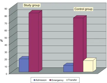
dif-ference in the number of hospital admissions between the
two groups (P = 0.578, Figure 3). Although there were
no deaths during the study, there were 16 (10.8%, 95% CI 5.81–15.8) deaths in the control group, including nine (59.25%) with fatal PE; however, a statistically significant difference in fatal outcomes between the groups could not
be detected (P = 0.735). Thus, the survival rate was 100%
in the study group and 89.2% (95% CI 83.9–94.5) in the control group.
Discussion
This study did not find an increased relative risk for PE in the very elderly compared with the elderly. PEs are
com-mon acom-mong the elderly,3 but are the least often accurately
diagnosed cardiovascular disease.3 The annual incidence
rates for PE and deep vein thrombosis have been reported to increase steadily with advancing age.4 However, there
was a significantly lower proportion of very elderly patients enrolled in this study, and these patients did not demonstrate an increased incidence of PE compared with the elderly population. Moreover, no increased relative risk for PE was detected in the very elderly. Women have also been reported to have lower rates of PE compared with men,4 but both
groups in this study contained more affected women, with no statistically significant difference found between the groups. These findings could be due to the small size of the study group and because of the study being conducted in
a single department. Further, numbers of patients become smaller with advancing biological age. Recurrent PEs were observed, but with no statistically significant difference in rate of recurrence between the two groups during this investigation.
Risk factors for PE include congestive heart failure, cancer, orthopedic conditions such as hip fracture, and other
debilitating diseases.5 We observed an increased prevalence
of congestive heart failure among the very elderly, but the difference relative to the elderly population was not statisti-cally significant. Further, using the example of renal tumors, cancer among those of very advanced age was not observed in this study. Bone disorders were found to be significantly more prevalent among the very elderly than in the elderly. There were other multiple chronic diseases affecting a significantly higher proportion of the very elderly in this study.
The most commonly reported comorbidities in elderly patients with PE are cardiomegaly (22%–64%), pleural effusion (15.8%–57%), right heart overload (50%), syncope (8%–62%), shock (5%–31%), cough (12%–43%), hemop-tysis (3%–14%), deep vein thrombosis (15%–50%), cancer (4%–32%), surgery (5%–44%), heart failure (5%–33%), previous deep vein thrombosis or PE (18%–41%), stroke (3%–13.5%), acute myocardial infarction (3%–11%), and
chronic obstructive pulmonary disease (2%–27%).6–17 The
most acute disorders documented in our study were cor pul-monale, cardiac decompensation, syncope, minor bleeding in the extremities, bronchopulmonary infection, gastrointestinal hemorrhage, gastritis, and glucose derangement in the very elderly, but not at significantly higher rates compared with the general elderly population. There was no correlation between these acute illnesses and PE in the very elderly, with these diseases appearing to be separate entities. A number of studies have shown that elderly patients over 80 years have a
higher incidence of bleeding complications.18 In our study,
the very elderly had more minor bleeding complications, but we did not examine this relationship further.
The clinical presentation of PE can be subtle and
atypical,5 as this was also noted in our study. Traditional
abnormalities in vital signs in the patient with PE can be missing, and syncope without chest pain or dyspnea is a frequent clinical symptom in the elderly.5 In our study,
although not statistically significant, the very elderly were more likely to experience syncope. Therefore, the differential diagnosis of syncope in the elderly should always include PE. Previous studies have reported that the main symptoms in the elderly with PE are dyspnea (59%–91.5%), tachypnea (46%–74%), tachycardia
Dovepress Relative risk of pulmonary embolism in the very elderly
Clinical Interventions in Aging downloaded from https://www.dovepress.com/ by 118.70.13.36 on 20-Aug-2020
Table 4 Chronic comorbidities in the study and control groups
Organ Chronic disease Study group
(n = 11) (%)*
Control group (n = 148) (%)*
P value
Heart and circulatory diseases Prior pulmonary embolism 1 (9) 7 (5) 0.365
Prior deep vein thrombosis 0 8 (5) 0.649
Congestive heart failure 1 (9) 7 (5) 0.523
Coronary heart disease 4 (36) 31 (21) 0.234
Hypertensive heart disease 1 (9) 5 (3) 0.337
Valvular heart disease 3 (27) 40 (27) 0.986
Atrioventricular block 1 (9) 1 (1) 0.016
Dilated cardiomyopathy 0 3 (2) 0.634
Aneurysm 0 5 (3) 0.536
State post heart attack 0 21 (14) 0.429
Peripheral arterial occlusive disease 0 8 (5) 0.429
State after syncope 0 2 (1) 0.698
Total 11 138 ,0.001
Lung diseases Chronic obstructive pulmonary disease 3 (27) 22 (15) 0.275
Bronchial asthma 0 1 (2) 0.785
Obstructive sleep apnea syndrome 1 (9) 9 (6) 0.692
Lung tumors 0 15 (10) 0.267
Pulmonary emphysema 0 5 (3) 0.536
Pulmonary fibrosis 0 8 (5) 0.429
Pulmonary tuberculosis 0 4 (3) 0.580
Pneumothorax 0 1 (1) 0.785
Total 4 65 0.002
Thyroid diseases Hyperthyroidism 0 11 (7) 0.349
Hypothyroidism 0 3 (2) 0.634
Struma nodosa 0 5 (3) 0.536
Strumectomy 0 3 (2) 0.634
Total 0 22 0.021
Liver diseases Chronic hepatitis B 0 5 (3) 0.536
Liver cirrhosis 0 1 (1) 0.785
Liver metastasis 0 4 (3) 0.581
Total 0 10 0.0495
Gallbladder diseases Gallbladder stones 0 5 (3) 0.536
Cholecystectomy 0 10 (7) 0.373
Gallbladder tumors 0 3 (2) 0.634
Total 0 18 0.0495
Gastrointestinal diseases Esophageal cancer 0 1 (1) 0.785
Hernia 1 (9) 6 (4) 0.432
Appendectomy 0 5 (3) 0.536
Colitis 0 4 (3) 0.581
Diverticulosis 0 11 (7) 0.349
Polypectomy 0 8 (5) 0.429
Pancreatic tumors 0 6 (4) 0.496
Colon tumor 0 25 (17) 0.138
Stoma 0 2 (1) 0.698
Fecal incontinence 0 3 (2) 0.634
Total 1 71 ,0.001
Lymphatic diseases non-hodgkin’s lymphoma 0 5 (3) 0.536
Various organs Cystic diseases 0 13 (9) 0.305
Total 0 18 0.121
Urogenital disorders Renal insufficiency 4 (36) 18 (12) 0.025
nephrectomy 1 (9) 1 (1) 0.016
nephrolithiasis 1 (9) 2 (1) 0.069
Kidney tumors 1 (9) 4 (3) 0.242
Urinary incontinence 0 4 (3) 0.581
Bladder tumors 0 3 (2) 0.634
Benign prostatic hyperplasia 4 (36) 20 (14) 0.041
(Continued)
Dovepress Yayan
Clinical Interventions in Aging downloaded from https://www.dovepress.com/ by 118.70.13.36 on 20-Aug-2020
Table 4 (Continued)
Organ Chronic disease Study group
(%)*
Control group (%)*
P value
Prostate carcinoma 0 8 (5) 0.429
Epididymitis 0 1 (1) 0.785
Total 11 61 0.015
neurological disorders State post intracranial hemorrhage 1 (9) 3 (2) 0.149
State post transient cerebral ischemia 0 7 (5) 0.461
State post cerebral ischemia 0 13 (9) 0.305
Parkinson disease 0 4 (3) 0.581
Hydrocephalus 0 2 (1) 0.698
Polyneuropathy 1 (9) 2 (1) 0.069
Restless legs syndrome 0 1 (1) 0.785
Meningioma 0 1 (1) 0.785
Multiple sclerosis 0 1 (1) 0.785
Total 3 41 0.0006
Psychiatric disorders Dementia 0 20 (14) 0.192
Alzheimer’s disease 0 2 (1) 0.698
Depression 0 5 (3) 0.536
Various 0 9 (6) 0.400
Total 0 36 0.021
Ear nose and throat diseases Ménière’s disease 1 (9) 3 (2) 0.149
Gynecological diseases Hysterectomy 0 9 (6) 0.400
Uterine polyps 0 2 (1) 0.698
Adnexal tumor 0 3 (2) 0.634
Ovarian cancer 0 1 (1) 0.785
Breast cancer 0 6 (4) 0.496
Mastectomy 1 (9) 0 0.0002
Total 2 24 0.015
Orthopedic conditions Hip endoprosthesis 0 8 (5) 0.429
Bone fracture 4 (36) 18 (12) 0.025
Knee replacement 0 6 (4) 0.496
Vertebral degeneration 3 (27) 15 (10) 0.084
Amputation 0 2 (1) 0.698
Rheumatism 0 8 (5) 0.429
Osteoporosis 0 9 (6) 0.400
Spinal stenosis 0 5 (3) 0.536
Herniated disc 0 3 (2) 0.634
State post fall 0 10 (7) 0.373
Plasmacytoma 0 1 (1) 0.785
Total 7 85 0.0003
Ophthalmologic diseases Various 4 (36) 14 (9) 0.007
Dermatological diseases Various 0 9 (6) 0.400
Allergy 0 4 (3) 0.581
Drug overdose 0 1 (1) 0.785
Autopsy 0 1 (1) 0.785
Total 4 29 0.037
Total diagnosis = 1289 82 1207 ,0.001
Notes: *Rounded to whole numbers; Significant P values are shown in bold.
(29%–76%), and chest pain (26%–59%).6–17 In our study,
dyspnea (45%–56%), chest pain (9%–21%), and cough (6%) were more frequent symptoms in the very elderly and elderly, although not significantly so.
The most common electrocardiographic changes reported in previous studies included sinus tachycardia (18%–62.5%), right bundle branch block (4.5%–40.5%), ST-T abnormalities
(9%–37%), and S1Q3 (8%).6–17 In this study, the most
common electrocardiographic abnormalities were tachyar-rhythmia absoluta in atrial fibrillation, T-negativity, and S1Q3, but these did not occur at significantly high rates com-pared with the elderly population. Further studies are needed to examine electrocardiographic changes in the elderly and very elderly with PE.
Dovepress Relative risk of pulmonary embolism in the very elderly
Clinical Interventions in Aging downloaded from https://www.dovepress.com/ by 118.70.13.36 on 20-Aug-2020
Value of D-dimer in both groups
Number of cases 10
0 5 10 15 20 25 30 35 40
19 28 37 46 55 64 73 82 91 100 109 118 127 130 145 1
Study group Control group
mg/L
Figure 2 Comparison of D-dimer values in control and study groups.
Allocation of patients to the hospital in both groups
Admission Emergency Transfer 90
80
70
60
50 %
40
30
20
10
0
Study group
Control group
Figure 3 Comparison of hospital admissions between the control and study groups.
Hypoxia has often been described in blood-gas analysis in previous studies of patients with PE.6,8,19 Similarly, hypoxia
was frequently observed in both groups in our study, but we found no significant difference between the elderly and very elderly in this regard. Respiratory and metabolic acidosis has also been reported to be more frequent in elderly patients with
PE than in their younger counterparts .6,8,19 However, respiratory
and metabolic acidosis was not detected in either group in our study. Nevertheless, we noted hypoxia in more than one third of our very elderly study patients. These respiratory changes and their implications for the diagnosis and treatment of PE require further evaluation in future prospective studies.
Dovepress Yayan
Clinical Interventions in Aging downloaded from https://www.dovepress.com/ by 118.70.13.36 on 20-Aug-2020
The specificity of d-dimer values in patients suspected
of having PE decreases with advancing age.20,21 Two studies
have examined the specificity of d-dimer for diagnosis of PE
in the elderly and reported similar specificities (5%) for PE diagnosis in the elderly.20,21 This very low specificity has led
to the proposal of an augmented cutoff value for d-dimer.6
An elevated cutoff would reduce the number of false posi-tives but also increase the proportion of false negaposi-tives.6
We found a higher mean d-dimer value in the study group;
however, there was no statistically significant difference in
d-dimer values between the two groups. Hence, d-dimer
has very limited predictive value for diagnosing PE in the very elderly.
The prognostic value of troponin has been demonstrated
in high-risk patients with PE.22 Troponin can improve the risk
stratification of patients with PE and help to identify patients
who may require aggressive treatment.22,23 Although troponin
was not routinely tested, the findings of our study suggest that troponin also has limited predictive value for PE in the very elderly. Further, we did not observe a direct relationship between PE and release of troponin.
Aging did not affect the diagnostic quality of single
detec-tor or multidetecdetec-tor pulmonary angio-CT for PE.24,25 Use of
lung scintigraphy for diagnosing PE is limited by pre-existing lung disease or an abnormal chest radiograph.26,27 In our study,
chest CT was used almost equally in both groups. Although not statistically significant, the elderly were more likely to undergo ventilation perfusion scintigraphy.
The sensitivity of Doppler ultrasound for detecting deep
vein thrombosis increases with age, but not the specificity.27
Because more Doppler ultrasound examinations were per-formed in the very elderly in our study, more deep venous thromboses were found in this age group. Pulmonary angiography for the diagnosis of PE has the same diagnostic
value in the elderly as in young age groups.15 However, this
test was not available for our study, so its utility and diag-nostic value cannot be commented upon.
Population-based studies have identified a greater risk of death from PE in the elderly.28 The mortality rates in this study
were surprising, in that there were more deaths observed in controls than in patients from the study group. Our findings do not provide a reasonable explanation for this discrepancy, and further research is required.
Study limitations
This study examined all patients with PE treated in a depart-ment of internal medicine, but did not investigate patients with
PE diagnosed in other departments. d-dimer, conventional
troponin T, and highly sensitive troponin T levels were not routinely tested in all patients with PE, including those with malignancy. The differences in our descriptive results may be due to the age difference between the study population groups and individual biological variations with regard to limited life expectancy. Other limitations of this study were its small size (particularly that of the study group), its retrospective nature, and the fact that it was a single-center analysis.
Conclusion
An increased incidence of PE was not observed among the very elderly compared with the elderly. The clinical presentation of PE in the very elderly was subtle and there were no clear clinical symptoms in this group. Blood gas
analyses were nonspecific, and d-dimer and troponin had
minimal predictive value in this population. Further, there were no typical electrocardiographic changes, and an ideal radiological modality for diagnosing PE in the very elderly could not be identified.
Disclosure
The author reports no conflicts of interest in this work.
References
1. Hirsh J, Hoak J. Management of deep vein thrombosis and pulmonary embolism. A statement for healthcare professionals. Council on Throm-bosis (in consultation with the Council on Cardiovascular Radiology), American Heart Association. Circulation. 1996;93:2212–2245. 2. Righini M, Le Gal G, Perrier A, Bounameaux H. The challenge of
diagnosing pulmonary embolism in elderly patients: influence of age on commonly used diagnostic tests and strategies. J Am Geriatr Soc. 2005;53:1039–1045.
3. Weberová D, Weber P, Kubesová H, et al. Occurrence of pulmonary embolism among 260 in-patients of acute geriatric department aged 65+ years in 2005–2010. Adv Gerontol. 2012;25:506–512.
4. Kniffin WD Jr, Baron JA, Barrett J, Birkmeyer JD, Anderson FA Jr. The epidemiology of diagnosed pulmonary embolism and deep venous thrombosis in the elderly. Arch Intern Med. 1994;154:861–866. 5. Rogers RL. Venous thromboembolic disease in the elderly patient:
atypical, subtle, and enigmatic. Clin Geriatr Med. 2007;23:413–423. 6. Masotti L, Ray P, Righini M, et al. Pulmonary embolism in the elderly:
a review on clinical, instrumental and laboratory presentation. Vasc
Health Risk Manag. 2008;4:629–636.
7. Busby W, Bayer A, Pathy J. Pulmonary embolism in the elderly. Age
Ageing. 1988;17:205–209.
8. Ceccarelli E, Masotti L, Barabesi L, Forconi S, Cappelli R. Pulmonary embolism in very old patients. Aging Clin Exp Res. 2003;15: 117–122.
9. Gisselbrecht M, Diehl JL, Meyer G, Collignon MA, Sors H. Clinical presentation and results of thrombolytic therapy in older patients with massive pulmonary embolism: a comparison with non-elderly patients.
J Am Geriatr Soc. 1996;44:189–193.
10. Kokturk N, Oguzulgen IK, Demir N, Demirel K, Ekim N. Differences in clinical presentation of pulmonary embolism in older vs younger patients. Circ J. 2005;69:981–986.
11. Le Gal G, Righini M, Roy PM, et al. Differential value of risk factors and clinical signs for diagnosing pulmonary embolism according to age. J Thromb Haemost. 2005;3:2457–2464.
Dovepress Relative risk of pulmonary embolism in the very elderly
Clinical Interventions in Aging downloaded from https://www.dovepress.com/ by 118.70.13.36 on 20-Aug-2020
Clinical Interventions in Aging
Publish your work in this journal
Submit your manuscript here: http://www.dovepress.com/clinical-interventions-in-aging-journal
Clinical Interventions in Aging is an international, peer-reviewed journal focusing on evidence-based reports on the value or lack thereof of treat-ments intended to prevent or delay the onset of maladaptive correlates of aging in human beings. This journal is indexed on PubMed Central, MedLine, the American Chemical Society’s ‘Chemical Abstracts
Service’ (CAS), Scopus and the Elsevier Bibliographic databases. The manuscript management system is completely online and includes a very quick and fair peer-review system, which is all easy to use. Visit http://www.dovepress.com/testimonials.php to read real quotes from published authors.
12. Masotti L, Ceccarelli E, Cappelli R, Guerrini M, Forconi S. Pulmonary embolism in the elderly: clinical, instrumental and laboratory aspects.
Gerontology. 2000;46:205–211.
13. Punukollu H, Khan IA, Punukollu G, Gowda RM, Mendoza C, Sacchi TJ. Acute pulmonary embolism in elderly: clinical characteristics and outcome. Int J Cardiol. 2005;99:213–216.
14. Ramos A, Murillas J, Mascías C, Carretero B, Portero JL. Influence of age on clinical presentation of acute pulmonary embolism. Arch
Gerontol Geriatr. 2000;30:189–198.
15. Stein PD, Gottschalk A, Saltzman HA, Terrin ML. Diagnosis of acute pulmonary embolism in the elderly. J Am Coll Cardiol. 1991;18: 1452–1457.
16. Timmons S, Kingston M, Hussain M, Kelly H, Liston R. Pulmonary embolism: differences in presentation between older and younger patients. Age Ageing. 2003;32:601–605.
17. Chung T, Emmett L, Khoury V, et al. Atrial and ventricular echocardio-graphic correlates of the extent of pulmonary embolism in the elderly.
J Am Soc Echocardiogr. 2006;19:347–1353.
18. Poli D, Antonucci E, Testa S, Cosmi B, Palareti G, Ageno W; The FCSA (Italian Federation of Anticoagulation Clinics). The predictive ability of bleeding risk stratification models in very old patients on VKA treatment for venous thromboembolism. Results of the Prospective Collaborative EPICA study. J Thromb Haemost. April 11, 2013. [Epub ahead of print.]
19. Jones JS, Van Deelen N, White L, Dougherty J. Alveolar-arterial oxygen gradients in elderly patients with suspected pulmonary embolism. Ann
Emerg Med. 1993;22:1177–1181.
20. Harper PL, Theakston E, Ahmed J, Ockelford P. D-dimer concentration increases with age reducing the clinical value of the D-dimer assay in the elderly. Intern Med J. 2007;37:607–613.
21. Righini M, de Moerloose P, Reber G, Perrier A, Bounameaux H. Should the D-dimer cut-off value be increased in elderly patients suspected of pulmonary embolism? Thromb Haemost. 2001;85:744.
22. Giannitsis E, Müller-Bardorff M, Kurowski V, et al. Independent prog-nostic value of cardiac troponin T in patients with confirmed pulmonary embolism. Circulation. 2000;102:211–217.
23. Enea I, Ceparano G, Mazzarella G, Di Sarno R, Cangiano G, Busino CA. Biohumoral markers and right ventricular dysfunction in acute pulmonary embolism: the answer to thrombolytic therapy. Ital
Heart J Suppl. 2004;5:29–35. Italian.
24. Righini M, Le Gal G, Perrier A, Bounameaux H. Effect of age on the assessment of clinical probability of pulmonary embolism by prediction rules. J Thromb Haemost. 2004;2:1206–1208.
25. Stein PD, Woodard PK, Weg JG, et al; PIOPED II investigators. Diagnostic pathways in acute pulmonary embolism: recommendations of the PIOPED II investigators. Am J Med. 2006;119:1048–1055. 26. Stein PD, Hull RD, Kayali F, Ghali WA, Alshab AK, Olson RE. Venous
thromboembolism according to age: the impact of an aging population.
Arch Intern Med. 2004;164:2260–2265.
27. Righini M, Goehring C, Bounameaux H, Perrier A. Effects of age on the performance of common diagnostic tests for pulmonary embolism.
Am J Med. 2000;109:357–361.
28. Sakuma M, Nakamura M, Takahashi T, et al. Pulmonary embolism is an important cause of death in young adults. Circ J. 2007;71: 1765–1770.
Dovepress
Dovepress
Yayan
Clinical Interventions in Aging downloaded from https://www.dovepress.com/ by 118.70.13.36 on 20-Aug-2020