biology
ISSN 2079-7737 www.mdpi.com/journal/biology Review
Methods for Differentiating Prion Types in
Food-Producing Animals
Kevin C. Gough 1,*, Helen C. Rees 2, Sarah E. Ives 1 and Ben C. Maddison 2
1 School of Veterinary Medicine and Science, The University of Nottingham, Sutton Bonington Campus, College Road, Sutton Bonington, Leicestershire LE12 5RD, UK;
E-Mail: [email protected]
2 ADAS UK, School of Veterinary Medicine and Science, The University of Nottingham, Sutton Bonington Campus, College Road, Sutton Bonington, Leicestershire LE12 5RD, UK; E-Mails: [email protected] (H.C.R.); [email protected] (B.C.M.)
* Author to whom correspondence should be addressed; E-Mail: [email protected]; Tel.: +44-115-951-6272; Fax: +44-115-951-6440.
Academic Editor: Christian D. Doerig
Received: 31 August 2015 / Accepted: 4 November 2015 / Published: 13 November 2015
Keywords: prion; transmissible spongiform encephalopathy; scrapie; BSE; CWD; strain typing
1. Introduction
1.1. Transmissible Spongiform Encephalopathies, and the Concept of Prions as an Infectious Agent The transmissible spongiform encephalopathies (TSEs), commonly referred to as prion diseases, are a group of infectious, fatal neurodegenerative disorders that affect a number of mammalian species, including man. The prion hypothesis proposes that the infectious agent is largely comprised of the PrPSc conformer of a benign host encoded membrane glycoprotein (PrPC) in the absence of any coding nucleic acid [1]. The conformational rearrangements (protein misfolding) that accompany conversion of PrPC to PrPSc result in increased insolubility in detergents [2] and protease resistance [3]. PrPC has a wide tissue distribution and is present as di-, mono- and un-glycosylated forms. It is thought that PrPSc is responsible for the conversion of additional molecules of PrPC into PrPSc and that this is the central molecular event that occurs during TSE disease. This conversion event is largely restricted to those cells of the lymphoreticular tissues (LRS) and central nervous system (CNS), and the end product has a propensity to aggregate and form amyloid. It is unclear as to the effect of the loss of function of PrPC that occurs after conversion to PrPSc as the physiological function of PrPC remains a controversial matter. However, it is thought that either this PrPSc end product, or a conformer of this protein formed along the folding pathway is toxic to cells of the CNS and is responsible for the characteristic brain pathology including the spongiosis, astrocytosis and neuronal loss that are commonly associated with these diseases.
1.2. Examples of Prion Diseases
and BSE, illustrate the requirement for continued research and surveillance into TSEs of food-producing animals. This includes the development of methods for their detection and differentiation.
Table 1. Prion types affecting farmed ruminants.
Prion types * Host Species
Classical BSE (bovine spongiform encephalopathy) Cattle, Goats #
Atypical BSE: H-BSE, L-BSE/bovine amyloidotic spongiform
encephalopathy (BASE) Cattle
Classical Scrapie^ Sheep, Goats
CH1641 Scrapie+ Sheep
Atypical/Nor98 Scrapie Sheep, Goats
CWD (chronic wasting disease) White-tailed deer, Elk, Mule Deer, Moose
* Types as defined through analysis of tissue taken from food production animals. # BSE in goats is very rare,
to date only 2 cases of BSE have been recorded in this species. ^ Further characterisation of classical scrapie
isolates in inbred strains of mice have demonstrated that the classical scrapie category can comprise over 15
distinct scrapie strains. + CH1641 scrapie is a rare form of scrapie found in sheep that shares similar
biochemical features to that of BSE.
1.3. TSE Diagnosis
TSEs often present as a spectrum of different clinical signs in the infected animal. In scrapie-affected sheep and goats, infection may show as pruritus, weight loss, wool pulling, the swaying of hips and hind limbs, and animals can develop a body tremor and have an increased sensitivity to noise and movement. As disease progresses animals can develop an inability to stand, and exhibit other behavioural changes. In BSE affected cattle clinical observations may be subtle or obvious and can include changes in temperament such as nervousness or aggression, animals can also be seen to develop an abnormal posture, demonstrate a lack of coordination and have difficulty in rising. There is decreased milk production, and animals often show weight loss. With CWD, clinical signs may include emaciation, an excessive salivation, ataxia, the drooping of head and ears, weakness and behavioural changes.
Figure 1. (A) Example western blot using a core antibody to detect prion protein (PrPSc) after proteinase K digestion showing the typical di- mono- and unglycosylated protein banding pattern. Ovine scrapie, ovine bovine spongiform encephalopathy (BSE) and bovine classical BSE (c-BSE) are shown, M, molecular mass markers 20 and 30kDa. Evident are the lower molecular weight of the unglycosylated protein band in ovine or bovine BSE compared to scrapie. Also, the di-glycan band is more dominant in BSE compared to the scrapie sample. A schematic diagram (B) to illustrate examples of the different electrophoretic glycoform profiles of the PrPSc from different prion types following Proteinase K digestion and western blotting using an antibody to the core region of the prion protein. Ovine SSBP1 is readily differentiated from both CH1641 scrapie and BSE, by glycoform ratio and molecular weight of the unglycosylated PrPSc species. Atypical forms of scrapie are less protease resistant than classical scrapie and typically show lower molecular weight species. The unglycosylated PrPSc band in classical bovine BSE is intermediate in size between that of L-type and H-type atypical forms of bovine BSE. M.W. Molecular mass markers in kDa. It should be noted that for ovine CH1641and bovine H-BSE, evidence for two different conformer populations are present within these profiles. These are revealed upon independently probing blots with the core antibody SAF84 and a second antibody that binds more towards the N terminus (for example L42), and evidenced by differences within the glycoprofiles and banding patterns between these blot [8,9], also see sections 4.1 and 4.2. This is a unique trait of these two prion types.
1.4. The Concept of Prion Strains and Species Barrier
[image:4.595.93.507.77.226.2]typing is often used to refer to the description of the biochemical properties of PrPSc from a particular isolate, however the true identification of a TSE strain can only be confirmed by determining a stable disease phenotype in specific lines of inbred rodent strains.
The ability of a TSE to infect a distinct host species is governed by a phenomenon commonly referred to as the species barrier [13]. When prion-containing material is inoculated experimentally from one species into another, for example an isolate of sheep scrapie being inoculated into an inbred mouse line, the incubation time for the development of TSE will often be long and there will be an incomplete attack rate (only a proportion of the mice inoculated will develop disease). This inefficient transmission is the species barrier. However, if brain material from these infected animals from this first passage is then further inoculated into more animals of the same genetic background, the attack rate will increase and the incubation time to disease will reduce. On subsequent passage this incubation time will stabilise to produce consistent and reproducible incubation times, hit rates and other phenotypic properties. The apparent “species barrier” between the inoculum from the donor species and the recipient species will have been overcome. Species barriers can also be overcome where homologous PrPC is expressed in the host and the infecting inoculum, as is the case when using transgenic rodents for bioassay [14]. The nature of the TSE agent also contributes to the species barrier. An example is BSE, this TSE of cattle is experimentally transmissible to a wide variety of species with diverse PrPC primary structures [15] and in natural transmissions it has caused prion disease in a number of species including humans, domestic cats (FSE) and exotic ungulates [16], presumably as a result of ingestion of BSE-contaminated food. This is in contrast to scrapie, where a similar variety of species would have been exposed to the scrapie agent, yet this disease has only been seen in natural infections of sheep and goats.
1.5. The Emergence of “New” Prion Strains
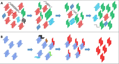
Whilst it is easy to regard prion strains as a fixed entity, several lines of experimental evidence suggest that this is an oversimplification. ‘New’ strains with different properties to that in the inoculum have been seen to emerge after transmission to different species, even when the inoculum had been cloned by limiting dilution [17]. The emerging view is that a prion strain may exist much like a quasispecies for viral and bacterial pathogens. The cloud hypothesis [13] proposes that a wide range of PrPSc conformers are possible within a given strain, but that within each prion ‘strain’ only a subset of those conformers are able to interact with the host PrPC that is presented within any given infection. Thus there will be a range of PrPSc infectiousconformers that are able to become the dominant species upon transmission within a new host. For an efficient infection to proceed the PrPC from the host has to be capable of forming a conformation that is compatible with the infecting PrPSc strain. Thus the selection of the new dominant species is dependent on the appropriate conditions being present for its selection. The selection of a minor conformer within a newly infected host and its preferential replication over the previous dominant ‘strain’ would go some way to explaining how prion strains can apparently mutate (Figure 2).
conformations (Figure 2). It is likely that the cloud hypothesis and deformed template theory are both relevant, the main differences between the two theories are that in the deformed template theory the environment has an active part in the creation of novel PrPSc conformations and that the original or “parent” prions act as a template for the creation of such de novo prion strains [18].
Studies have suggested that novel synthetic prions can be created de novo; a transmissible prion strain being generated after inoculation of rodents with recombinant-PrP amyloid fibrils. This could give further support to the deformed template theory with PrPSc possibly being formed from the original (non-PrPSc) fibrils [19–21]. Using an in vitro prion replication method (protein misfolding cyclic amplification; PMCA) to amplify prions from a scrapie-infected brain sample, another study showed that when RNA was removed from and then returned to the replication environment, a novel prion strain developed which was not present in the original brain sample, further supporting the deformed template theory [22].
Figure 2. (A) The premise of the “cloud” hypothesis is that a prion type is comprised of a heterogeneous mix of PrPSc variants made up of a dominant conformer (the example given is depicted in red) and minor populations of PrPSc variants (other colours). A change in the replication environment or an environmental pressure can then select for a minor variant within the “cloud” to become the dominant type over time (the example of a new dominant PrPSc conformer is depicted in green). (B) The deformed template theory proposes that new PrPSc structural variants are created directly from the original dominant conformer (blue) due to a new environment for replication. Variants are generated from a number of trial and error seeding events and one that is particularly suited to the new replication environment becomes the new dominant conformer (red) replacing the original variant (blue) (figure adapted from Makarava and Baskakov [18]).
A
[image:6.595.67.555.292.555.2]1.6. The Significance of New Strain Emergence
Prion strains can appear to undergo Darwinian selection given the right selective pressures. Ghaemmaghami and co-workers [23] have demonstrated the in vitro and in vivo selection of novel prions in both cell culture and in mice, using biologically cloned murine adapted prions. In these experiments prions could be seen to adapt to the selection pressures of either brain (infection of mice) or cell culture replication where the action of the anti-prion compound quinacrine could select for resistant prions. In addition, Li and co-workers [24] demonstrated that the rodent scrapie strain 22L changed its dominant conformer and concomitant disease phenotype due to selective pressure exerted by the presence of swainsonine which disrupts N-linked glycosylation. These observations support the notion of a prion quasispecies and prion strain selection; they are also extremely important to our understanding of these diseases and the potential zoonotic risk that these diseases may pose in the future.
A recent study used transgenic mouse bioassay to examine a mixture of ovine scrapie isolates before and after incubation on soil for 13 months to determine whether the dominant types were consistent during this incubation period within an environmental matrix [25]. The data showed that prolonged ageing of scrapie prions within the soil resulted in changes in the dominant scrapie pathologies and concomitant PrPSc biological/biochemical properties. This data indicates that environmental reservoirs of ruminant prions may be dynamic and the dominant “strain” may change over time potentially even leading to the emergence of novel prions. Understanding how TSE strains are maintained within populations and how they might have the ability to cause new infections within new hosts or after prolonged incubation in environmental reservoirs given the correct selective pressures is of importance if we are to minimise the potential for these agents to affect human health.
1.7. Prion Diseases that Affect Food-Producing Animals 1.7.1. BSE
1.7.2. Scrapie
Classical scrapie in sheep and goats is the prototype TSE and is thought to have existed for hundreds of years. An atypical scrapie type in sheep was found in Norway in 1998 and designated Nor98, now commonly known as “atypical scrapie" or atypical/Nor98. This differs from classical scrapie in several ways, including the neuroanatomical distribution of histopathological lesions and the pattern of PrPSc deposits in the brain [33]. In 2002, an obligatory active surveillance programme for scrapie in the sheep population submitted for abattoir slaughter was implemented and has resulted in an increased diagnosis of classical scrapie across the EU. Additionally, scrapie cases in sheep with the newly discovered atypical/Nor98 characteristics have been widely reported [7,34,35].
In 2007, an atypical/Nor98 scrapie case was found in a Swiss goat [36] the findings of the study suggested that cases in goats may have been missed in the past and may continue to be under-represented due to the routine sampling and analysis of caudal brainstem and cerebellum. The latter is a major site of PrPSc deposition in atypical/Nor98 scrapie of sheep but not of goats. Recently a study has proposed the co-existence of classical and atypical/Nor98 scrapie types due to the finding of an unusual case in Italy in which a sheep showed immunohistochemical and PrPSc features different from other atypical/Nor98 cases [37].
1.7.3. CWD
CWD was first identified in captive deer in Colorado in 1967 and has now been described in both captive and wild cervid populations across the US, in three Canadian provinces, and in the Republic of Korea. Two strains of CWD have been reported in mule deer, Rocky Mountain Elk, white-tailed deer, and moose [38,39].
2. Typing of TSEs in Food-Producing Animals
Overall, there are a considerable number of TSE types that infect ruminants (Table 1). Whilst scrapie, BSE and CWD appear to be mostly constrained to ovines, bovines and caprines respectively, the occurrence of natural infections of BSE in multiple mammalian species [32,40–42] indicates the potential for these infectious agents to cross species barriers. In addition, there are multiple types of each of these TSEs. The ability to define and diagnose individual types of TSE agent is of importance to understand the epidemiology, transmission and pathology of these diseases in farmed animals. Prion typing also underpins the testing of food products to protect human health by ensuring zoonotic BSE prions are not present and by monitoring for the emergence of any novel TSE types. This review will report the most up to date advances in the prion typing of natural TSE infections of ruminants.
2.1. Rodent Bioassay: Wild-Type Mouse Lines
within a particular TSE strain, the genetic background of the mouse model used leads to different incubation periods. Thus, consistency in incubation time becomes a good marker for a particular TSE strain within a particular genetic background of murine host [43]. In terms of the observed neuropathology, different TSE strains can cause marked differences in the severity and distribution of the pathological changes within the brain tissue, due to the different neuroanatomical targeting of each strain. As a semi-quantitative measure of strain discrimination, the severity of the tissue vacuolation as observed by hematoxylin and eosin staining is generally scored from sections of nine grey matter [44] and three white matter brain areas [43]. This data is usually plotted to form a lesion profile, reporting the severity of lesions being scored against each brain area. The plot that is generated shows characteristics of that particular TSE strain within that mouse line.
A more detailed analysis of the neuropathology and the cell types that are involved can be carried out by immunostaining sections of brain for PrPSc within areas of vacuolation. TSE strains demonstrate reproducible differences from each other in the extent of this staining [45]. Accumulation of PrPSc at different foci within the tissue may be in the form of diffuse deposits within the areas of vacuolation or may appear as amyloid plaques [10]. The basis of the differences in the observed neuropathology is thought to be due to the differences in the ability of each prion strain conformer to replicate within the different cell populations, and this in part may be coincident with subtle differences in the structure of PrPC within these sites.
Without doubt, mouse bioassays have provided a validated method for prion strain discrimination, including the analysis of a range of isolates from natural ruminant hosts. The biggest drawback of using wild-type mice for the identification of distinct prion strains from natural hosts are the long incubation periods and incomplete attack rates that are often associated with the species barrier between the inoculums and the murine host on primary passage.
2.2. Rodent Bioassay: Transgenic Mouse Lines
Mice transgenic for bovine PrPC have been used to demonstrate the specific features of L-BSE, H-BSE and classical BSE (C-BSE). Masujin and co-workers [49] describe the inoculation of bovinised mice with either L- or C-BSE. Whilst wild type mice were refractory to infection with L-BSE, the transgenic mice developed clinical disease to both L- and C-BSE, with the retention of the same biochemical features that distinguish these two prion types in cattle brain. Similarly, H-BSE has been used to successfully infect bovinised mice and retained its characteristic molecular phenotype [50]. Together, data demonstrate that the three BSE types can be differentiated in bovinised mice [48,49,51], providing a method to type BSE material from suspect atypical bovine cases.
Transmission of CWD prions to wild type mice lines is inefficient. However, mice expressing cervid PrPC [52] have high attack rates and low incubation times with cervid derived CWD material allowing the analysis of this disease in a rodent model.
2.3. Bioassay in Other Model Species
Alternate rodent models have been developed for the monitoring for prion infectivity and prion type. In recent years the bank vole has been demonstrated to have a high susceptibility to different prion types from a variety of sources [53,54]. The bank vole model is often more sensitive than murine models and can have very short incubation times. This model has been demonstrated to be useful in cases where a scrapie isolate was not able to infect conventional or transgenic murine lines [54]. The brain lesion scoring profiles could again differentiate between two different scrapie isolates and these profiles were maintained upon sub-passage of the inoculums. Recently the bank vole has been used to isolate a type of CWD exhibiting an extremely low incubation time on secondary passage, with incubation periods down to 25-28 days [53]. In a study by Raymond and co-workers [55], multiple CWD isolates were inoculated into both transgenic mice and also different species of hamster. The study demonstrated that after serial passage in Syrian golden hamsters disease incubation periods of CWD isolates could stabilise to either CWD with a short or long incubation period, which was indicative of two different CWD strains. More recently, data using a ferret bioassay also demonstrated the hallmarks of two different CWD types in material originating from mule deer [56]. Apparent differences included incubation time and PrPSc deposition within the CNS and periphery.
3. Histological and Immunohistochemical Analysis of Tissue from Natural Hosts
3.1. Histology
cattle BSE but this is not restricted to BSE infections [62]. The lesion profile is consistent for natural infections of cattle with classical BSE indicating a single prion type is involved in these infections [63,64]. However, there are higher levels of vacuolation and PrPSc accumulation in caudal brainstem in clinically affected compared to asymptomatic BSE-infected cattle [65].
In goats, BSE and CH1641 (an experimental type of scrapie with a PrPSc molecular phenotype very similar to BSE PrPSc; [66]) produce similar vacuolation patterns to the classical scrapie SSBP1 [57,67].
For CWD infections of deer or elk, again lesions are routinely found in various brain regions although within elk the lesions are generally less severe. In deer, pathology is usually found in the medulla oblongata, olfactory bulb, cortex and hypothalamus (reviewed in [63]).
Overall, whilst vacuolation patterns are a diagnostic feature of a prion strain/type in rodent bioassays, they have very limited application in the definition of prion types in natural infections due to high variability [58,68,69].
3.2. Immunohistochemistry
With BSE in cattle the accumulation of PrPSc is largely consistent between individuals when analysing distinct neuroanatomical sites and distinct patterns are observed for different BSE types [63,64]. L-BSE (or BASE) produces characteristic amyloid plaques as well as a distinct distribution of PrPSc compared to C-BSE: L-BSE PrPSc is present at comparatively lower levels in brainstem and higher levels within more rostral sites of the brain such as the olfactory bulb and olfactory cortex as well as the hippocampus [29]. At present it is not possible to distinguish C-BSE and H-BSE based on PrPSc deposition [65].
Considerable research effort has gone into using IHC to distinguish experimental BSE infections in sheep from other scrapie types. There is no one anatomical location/PrPSc type that can diagnose BSE from classical scrapie [70]; however, the defining of PrPSc morphological types, cell-type associations as well as levels of PrPSc accumulation across multiple neuroanatomical sites allows the discrimination of ovine BSE, CH1641, atypical/Nor98 scrapie and classical scrapie. This technique has been termed PrPSc profiling and has been pioneered by Jeffrey and co-workers [66,68,71]. Seven distinct morphological types of PrPSc accumulation have been described, each associated with distinct cell types: four associated with cell surface or extracellular PrPSc and three with intracellular PrPSc (reviewed in [68]). Ovine BSE infections could be distinguished from CH1641 and classical scrapie infections with BSE producing relatively high levels of intraneuronal, intramicroglial and neuropil-associated PrPSc [68,72]. The distinctive BSE profile was not affected by the sheep breed, PRNP genotype or route of inoculation of the agent; however, the latter two parameters affected the overall levels of PrPSc accumulation [72]. Certain classical scrapie isolates/experimental types can also be differentiated via PrPSc profiling due to distinct cell tropisms and PrP processing [71]. However, scrapie type and host PRNP genotype affect the PrPSc profile and the extent of PrPSc accumulation [68,71,73].
neurones and glia in the brain [75]. In addition, intraneuronal levels of PrPSc were consistently higher for ovine BSE than scrapie across multiple brain regions when detected with a range of antibodies [74]. Interestingly, the exact N-terminal cleavage site for PrPSc for ovine BSE appeared to be determined by the cell type with three distinct sites being identified, all of which result in smaller PrP fragments compared to classical scrapie PrPSc [75]. The scrapie type CH1641 displays a ‘peptide map' for intraglial and intraneuronal PrPSc that is distinct from both classical scrapie and ovine BSE as it is truncated even further from the N-terminus than BSE [66]. However, an independent study indicated that experimental CH1641 and CH1641-like field isolates may well differ [76].
With atypical/Nor98 scrapie the pattern of PrPSc accumulation was distinct from classical scrapie with increased accumulation in the cerebellar cortex and no accumulation in the medulla oblongata [59]. Such cerebellar PrPSc staining was of a diffuse granular type that has not been seen with classical scrapie [59]. In addition, for atypical/Nor98 scrapie there was no accumulation of PrPSc in the LRS [33].
With goat scrapie, very little PrPSc staining was seen with SSBP1 or CH1641 infections, whereas goats infected with BSE show clear PrPSc accumulation particularly in the thalamic, hypothalamic nuclei and in the basal ganglia [57]. Almost identical peptide mapping was observed for intracellular PrPSc when comparing goat and sheep infections, with intraneuronal PrPSc for BSE being truncated further from the N-terminus compared to classical scrapie [32]. Interestingly, this peptide mapping technique revealed a natural case of goat BSE [32].
3.3. Diagnostic Applications of Histology and Immunohistochemistry
For classical BSE, classical scrapie and CWD the presence of spongiform lesions and PrPSc accumulation in the medulla oblongata at the level of the obex make this region important for the diagnoses of prion disease. The caveat to this is that this region is less involved in pathology in atypical/Nor98 diseases and therefore other brain regions require examination, such as the cerebellum in sheep [35].
4. Protease Fragmentation of PrPSc and Glycoform Ratios
One of the main properties that can differentiate PrPSc from PrPC is the high proteolytic resistance of PrPSc. Of course, this trait is not absolute and it is likely that for all prion diseases PrPSc will exist as both protease-resistant (PrPSc-res) and protease-sensitive (PrPSc-sen) conformers. At one extreme are 263K scrapie infection in hamsters and human Gerstmann–Sträussler–Scheinker syndrome P102L infection of transgenic 101LL mice, where both infection models accumulate high infectivity titres in the absence of PrPSc-res [78]. However, in all other cases, prion diseases produce levels of PrPSc-res that are detectable by conventional immunoassays. As such, immunoassay detection of PrPSc-res has become the mainstay of prion diagnostics including defining prion types in ruminant diseases. When detected in western blots, the pattern of the PrPSc-res fragments can be used to differentiate prion types as it is thought that their distinct PrPSc conformers will present distinct cleavage sites to the protease (Figure 1).
4.1. Differentiating Prion Isolates in Small Ruminants
By far the most common protease used in prion typing is PK, which digests PrPC fully but only digests the N-terminal region of PrPSc-res resulting in un-, mono- and di-glycosylated protease resistant core. Hill and co-workers reported [79] that BSE in cattle, when passaged in sheep, displayed a lower molecular weight unglycosylated PrPSc band compared to classical scrapie. One aspect that needs to be taken into account when applying this method is that the metal occupancy of the PrPSc [80] and the pH of the sample [81] can influence the apparent molecular weight of the banding pattern. It has also been noted that CH1641 scrapie produces a banding pattern indistinguishable from ovine and caprine BSE [79,82,83]. This method was further developed by Stack and co-workers [84] who used the antibody P4 to detect cleavage products. This antibody binds in the region that is largely removed by PK cleavage of ovine BSE PrPSc (and CH1641 PrPSc) but retained during scrapie PrPSc digestion. When compared to an antibody binding further C-terminally in the protease resistant core, ratios of P4: “core antibody” are much higher for classical scrapie that for BSE or CH1641.
Screening of small ruminant prion diseases has mainly been designed to distinguish the zoonotic agent BSE from the non-zoonotic agents of scrapie. Most studies use a combination of molecular weight of PK-resistant PrPSc, the ratio of P4:‘core antibody’ binding and glycoform profiling to search for BSE infections. Samples displaying one or more traits of BSE are usually subsequently analysed by mouse bioassay for confirmatory prion typing [84,89,91]. Given the experimental designs, it is still largely unknown what molecular variations exist for classical scrapie in the natural hosts [82,83,91].
In addition to PK cleavage, an alternative protease thermolysin can also be applied to distinguish BSE from classical scrapie in small ruminants. Digestion resulted in distinct cleavage patterns of PrPSc for ovine BSE compared to classical scrapie that was not influenced by host genotype or scrapie type [92].
Recently, there have been further developments in the differentiation of BSE from natural scrapie with particular emphasis on differentiating BSE from CH1641 which is not possible using any of the criteria described above [84]. Pirisinu and co-workers [93] demonstrated that ovine BSE was more stable to guanidinium hydrochloride (GndHCl) than classical scrapie or CH1641 scrapie and that treatment with 3.5 mol/L GndHCl before digestion in combination with P4/’core antibody’ binding ratios allowed the discrimination of both CH1641 and classical scrapie from ovine BSE. Alternatively, the probing of a single western blot with differently labelled SAF84 and L42 antibodies revealed a so-called dual glycotype aspect at the size of the monoglycosylated band which distinguished CH1641 from BSE in sheep; as CH1641 had a relatively high SAF84 to L42 binding ratio compared to BSE [8]. In addition, a microwell immunofluorometric assay has been developed to differentiate between classical scrapie, atypical scrapie, CH1641 scrapie, experimentally infected ovine BSE and naturally infected bovine BSE [94]. The assay is based on the capture of PrPSc-res with three distinct antibodies that bind to different regions on PrP. Due to distinct PK-cleavage sites for PrPSc from different TSE types, all three antibodies capture PrPSc-res from classical scrapie, two bind PrPSc-res from atypical scrapie and a different combination of two antibodies bind to PrPSc-res from BSE and CH1641. BSE and CH1641 can then be differentiated due to the distinct binding ratios of these two antibodies.
Endogenous proteolysis also results in PrPSc fragments that may also be indicative of prion type in ruminant, such as the so-called C2 fragment in scrapie cases but not in experimental BSE or CH1641 cases [92] and the diagnostic 14kDa fragment (so called CTF14) that is detected with the antibody SAF84 in experimental CH1641 and naturally occurring CH1641-like isolates [95].
Atypical/Nor98 scrapie cases were first noted by Bernestad and co-workers [33], this can occur in both sheep and goats [36]. Atypical/Nor98 scrapie produces PrPSc-res with a lower protease resistance than classical scrapie and PK digestion patterns for atypical/Nor98 scrapie often (but not always, [34]) also contain diagnostic fragments between 6.5 and 12 kDa [96].
4.2. Differentiating Cattle BSE Types
differences in PK cleavage sites, L-BSE can be distinguished from C-BSE and H-BSE by a distinct glycoform pattern [27,28] although as mentioned previously, glycoform profiling of prion types can be difficult to perform reproducibly [83,85,86,88]. Also, the H-BSE appears to contain two distinct glycoform profiles depending on the antibody used to detect PrP [27] that can be exploited to diagnose this type when using the so called dual glycotype aspect for detection [8]. H-BSE also produces a unique fragment of 6-10kDa [98]. Whilst the molecular weight and antibody recognition of H-BSE PrPSc-res is sufficient to diagnose such infections, the difference in molecular weight between L-BSE and C-BSE PrPSc-res is just 0.3kDa for the unglycosylated band and is not suitable for routine prion typing [27]. However, Jacobs and co-workers [27] demonstrated that both H-BSE and L- BSE display enhanced protease digestion at pH8 relative to pH6.5 compared to C-BSE which may provide a more robust biochemical test for distinguishing L-BSE and C-BSE types.
5. Differential Stability of Prion Types
The underlying theory that prion characteristics are defined by the conformational make-up of the PrPSc molecules led to the development of tests to characterise the stability of these conformers. These methods were developed into two complementary assays that quantified the disruption of prion conformations with GndHCl [12,99]. The conformation-dependent immunoassay (CDI) analysed the exposure of epitopes within PrPSc molecules upon denaturation and the conformational stability assay (CSA) measured the loss of PK-resistance of the PrPSc molecules upon denaturation.
The CDI was developed and applied to rodent passaged strains and could distinguish multiple prion strains in hamsters [12]. Despite this obvious promise, the application of the assay for typing of ruminant prions has not been pursued.
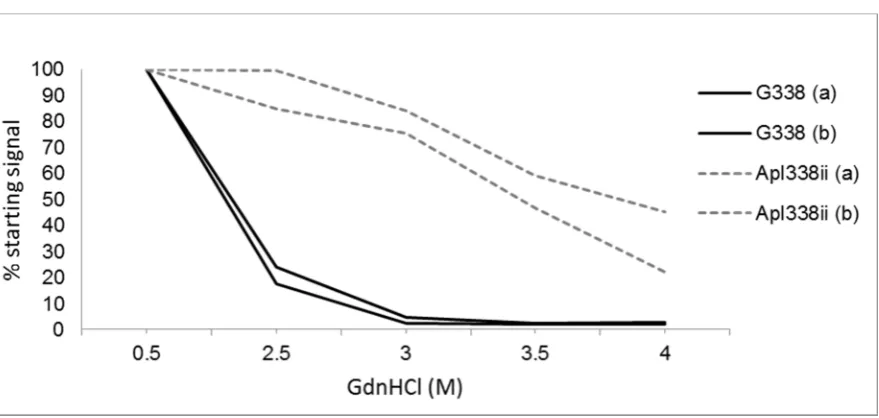
The CSA measures the stability of PrPSc-res (Figure 3) and a particular prion type can be defined as the [GndHCl]1/2 value: the concentration of GndHCl that results in the denaturation of half of the PrPSc. The determination of such values could differentiate some but not all hamster passaged prion strains [100]. With respect to ruminant prions, CSA analysis has shown that cattle BSE was less stable than a single scrapie isolate but conversely mouse-passaged BSE prion was more stable than passaged scrapie [12]. A further study demonstrated that BSE passaged in sheep was more stable than classical and CH1641 ovine scrapie [93]. It therefore seems that stability is dictated by both the prion type and host species.
Pirisinu and co-workers [99] also adapted the CSA assay to analyse the detergent solubility of PrPSc upon treatment with a range of GndHCl concentrations, a process termed the conformational solubility and stability assay (CSSA). With this CSSA assay, different ovine classical scrapie isolates with a range of PRNP genotypes could be differentiated from a similarly diverse set of atypical/Nor98 scrapie isolates.
Figure 3. Conformational stability assay of two scrapie strains (as determined in Tg338 mice). Two isolates of each strain G338 and Apl338ii were compared by western blotting after treatment with increasing concentrations of the denaturant Guanidine hydrochloride (GdnHCl). Denatured samples were treated with proteinase K and then western blotted using a core antibody. Total PrPSc signal was calculated by densitometry and plotted as a % of the PrPSc-res signal after treatment with 0.5M GdnHCl. These two scrapie strains have very different sensitivities to GdnHCl, G338 being less stable than apl338.
6. Cell Culture Methods
A number of cell lines permissive to prion multiplication have been developed including established cell lines, neuronal stem cells, and primary neuronal cells. Whilst initial cell lines were only permissive to rodent-adapted prion strains, the rabbit epithelial RK13 cell line expressing an ovine PrPC gene was permissive to ovine scrapie and was found to accumulate high levels of PrPSc [101]. Since this study, prion replication has been demonstrated in a variety of cell systems. An isolate from CWD has been successfully transmitted to a mule deer cell line [102], and cell lines expressing ovine PrP (Rov9 and MovS6) have been infected with experimental and natural ovine scrapie agents [103]. Cell models of prion infection are comprehensively reviewed by Vilette [104].
7. In Vitro Replication of Prion Types
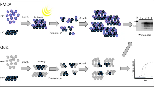
A non-radioactive cell free conversion assay that used recombinant mouse PrP as the substrate to examine the replication of murine passaged scrapie strains and BSE demonstrated that the conversion efficiency differed between some strains [106]. More recently, the protein misfolding cyclic amplification (PMCA) methodology has demonstrated distinct replication traits for prion types across multiple studies. PMCA is an in vitro prion replication assay that relies on iterative rounds of sonication and incubation [107] (Figure 4). The sonication step is thought to break prion aggregates into smaller units that can efficiently seed the conversion of PrPC into PrPSc during the incubation period. The technique allows the amplification of minute amounts of PrPSc when spiked into normal brain homogenate substrate where the prion is amplified over multiple (24–48) cycles of sonication/incubation (a round) and the substrate is then replenished by diluting the amplification products into fresh substrate. Any number of rounds can then be carried out. When analysing a range of rodent passaged scrapie strains or synthetic prions, Gonzales-Montalban and co-workers [108] demonstrated that the addition of beads to the PMCA reaction (PMCAb) had distinct effects on replication efficiency that was strain dependent. Strains with higher conformational stability displayed more enhancement of replication efficiency with the addition of beads compared to strains with lower conformational stability. Further analyses demonstrated that the prion strain influenced both the growth rate of PrPSc particles and also the total yield of PrPSc produced in the assay [109]. Other studies using rodent passaged prion strains have shown that changes in the substrate/cofactor availability during the PMCA/PMCAb reactions can also influence the amplification efficiency in a strain-specific manner. Such changes have included the use of RNase treated substrate or supplementation of substrate with polyA, polyG or DNA [110], as well as the glycosylation state of the PrPC substrate [111].
these two substrates. Overall, PMCA has shown application in distinguishing ovine prion types by analysing amplification efficacy in PrPC substrate with distinct PRNP genotypes.
Figure 4. Protein Misfolding Cyclic Amplification (PMCA) is an in vitro reaction thought to reproduce the molecular events of the PrPC to PrPSc conversion that occur in vivo with transmissible spongiform encephalopathies (TSE) diseases. Here a PrPC substrate (a healthy brain homogenate) is seeded with a PrPSc source, which causes conversion of PrPC to further aggregates of PrPSc. These aggregates are fragmented into a number of smaller seeds by sonication and the process of prion growth is reinitiated. Over time enough PrPSc can be produced to be detected by western blot. Quaking induced conversion (Quic) is an alternative in vitro assay that is also based on the seeding ability of PrPSc. A PrPSc seed is used to convert a recombinant PrPC substrate into an “amyloid” form. These large aggregates are fragmented into smaller seeds by vigorous shaking followed by further re-seeding and growth. Quic reactions are generally monitored by Thioflavin T fluorescence over time but the products can also be measured by western blot.
Together, the data supports the hypothesis that a range of substrate/co-factor conditions in tandem with a range of amplification conditions could provide a panel of defined PMCA conditions capable of defining prion types in the natural host; effectively producing an “amplification profile” for each ruminant prion type.
Recently, the use of recombinant bank vole PrP as a universal substrate for a similar in vitro replication assay known as quaking induced conversion or “Quic” has been described [116] (Figure 4). Quic differs from PMCA in that fibrils of recombinant prion are generated during cycles of incubation followed by shaking which is thought to break growing fibrils and form the new nucleation sites for fibril formation. The products of the reaction are formed from the seeding effect of prion containing
PrPC
PrPSc
Sonica+on Growth
Fragmenta+on
Growth
WesternBlot
PMCA
Quic
Shaking Growth
Fragmenta+on
Growth rPrPC
PrPSc
Time
T>
Fl
uo
re
sc
ence
material; however unlike PMCA the products of this reaction are non-infectious. The authors demonstrate some specific differences in the PK digestion profiles on western blots of Quic reaction products that were seeded by different ruminant prion types, demonstrating that in vitro amplification and prion typing of unknown samples may be possible using this rapid and sensitive technique.
8. Conclusions and Future Perspectives
Whilst the cases of BSE in ruminants is now very low and the associated concern for the contamination of the human food chain with the zoonotic BSE agent has eased, there are still concerns surrounding the exposure of humans to prions from food-producing animals. The more recent description of atypical/Nor98 scrapie in goats/sheep and atypical bovine BSE as well as the discovery of two distinct types of CWD all raise the possibility that further types of prions are circulating in ruminants that are not detected and/or defined by current assay methods. An additional concern is that novel types may emerge in these animals. One diagnostic challenge in prion biology is to develop and apply prion typing tests to fully elucidate the range of existing prion types in ruminants and to monitor for the emergence of novel types. This is a significant challenge as it is unknown what molecular and pathological differences any novel type will have compared to those already described. Therefore, assays that have a wide range of distinct measurements that describe a PrPSc type or in vivo pathology will be best suited for diagnosing new prion types.
could detect prion types at very low concentration would therefore be advantageous. PMCA may provide such a method. It has been reported that PMCA usually maintains the PrPSc molecular profile of the seed and is exquisitely sensitive in detecting PrPSc. A combination of PMCA amplification followed by analysis of the PrPSc product by an assay such as CSA may allow a robust prion typing assay to detect novel types even when PrPSc is present at low levels in the tissue sample. Alternatively, PMCA methods have now also been directly applied to prion typing in ruminants demonstrating that different prion types display different replication efficiencies under defined conditions. The data supports the hypothesis that a range of substrate/co-factor conditions in tandem with a range of amplification conditions could provide a panel of defined PMCA conditions capable of defining prion types from the natural host; effectively producing an ‘amplification profile’ for each type.
For all in vitro prion typing assays the definition of a novel prion type would need to be assessed as being representative of an inheritable in vivo strain phenotype rather than of a quasispecies that was selected/identified during the in vitro analyses. Therefore, any novel type would require analysis within rodent bioassay to define a novel and consistent pathology that is stable upon sub-passage and can therefore be described as a novel prion strain. The recent advent of transgenic murine models and a range of rodent host species have extended the use of rodent bioassay to a relatively wide range of natural ruminant isolates making this a much more realistic possibility.
Author Contributions
Kevin C. Gough and Ben C. Maddison designed and wrote the manuscript. Helen C. Rees and Sarah E. Ives wrote the manuscript. All authors read and approved the final manuscript.
Conflicts of Interest
The authors declare no conflict of interest. Abbreviations
Prion protein: PrP; disease associated prion protein: PrPSc; cellular prion protein: PrPC; variant Creutzfeldt Jakob disease: vCJD; bovine spongiform encephalopathy:BSE; chronic wasting disease: CWD; transmissible spongiform encephalopathy:TSE; central nervous system:CNS; lymphoreticular system: LRS; proteinase K: PK; immunohistochemistry: IHC; bovine amyloidotic spongiform encephalopathy: BASE; guanidinium hydrochloride: GndHCl; conformation-dependent immunoassay: CDI;conformational stability assay: CSA;the concentration of GndHCl that results in the denaturation of half of the PrPSc: [GndHCl]1/2 value;conformational solubility and stability assay: CSSA;Protein Misfolding Cyclic Amplification: PMCA; PMCA including beads: PMCAb.
References
1. Prusiner, S.B. Prions. Proc. Natl. Acad. Sci. USA 1998, 95, 13363–13383.
3. Bolton, D.C.; McKinley, M.P.; Prusiner, S.B. Identification of a protein that purifies with the scrapie prion. Science 1982, 218, 1309–1311.
4. Sala, C.; Morignat, E.; Oussaid, N.; Gay, C.; Abrial, D.; Ducrot, C.; Calavas, D. Individual factors associated with l- and h-type bovine spongiform encephalopathy in france. BMC Vet. Res. 2009, 8, e74.
5. Biacabe, A.; Morignat, E.; Vulin, J.; Calavas, D.; Baron, T. Atypical bovine spongiform encephalopathies, France, 2001–2007. Emerg. Infect. Dis. 2008, 14, 298–300.
6. Simmons, H.A.; Simmons, M.M.; Spencer, Y.I.; Chaplin, M.J.; Povey, G.; Davis, A.; Ortiz-Pelaez, A.; Hunter, N.; Matthews, D.; Wrathall, A.E. Atypical scrapie in sheep from a uk research flock which is free from classical scrapie. BMC Vet. Res. 2009, 5, doi:10.1186/1746-6148-5-8.
7. Fediaevsky, A.; Maurella, C.; Noremark, M.; Ingravalle, F.; Thordeirsdottir, S.; Orge, L.; Poizat, R.; Hautaniemi, M.; Liam, B.; Calavas, D.; et al. The prevalence of atypical scrapie in sheep from positive flocks is not higher than in the general sheep population in 11 European countries. BMC Vet. Res. 2010, 6, doi:10.1186/1746-6148-6-9.
8. Jacobs, J.G.; Sauer, M.; van Keulen, L.J.M.; Tang, Y.; Bossers, A.; Langeveld, J.P.M. Differentiation of ruminant transmissible spongiform encephalopathy isolate types, including bovine spongiform encephalopathy and ch1641 scrapie. J. Gen. Virol. 2011, 92, 222–232.
9. Langeveld, J.P.; Erkens, J.H.; Rammel, I.; Jacobs, J.G.; Davidse, A.; van Zijderveld, F.G.; Bossers, A.; Schildorfer H. Four independent molecular prion protein parameters for discriminating new cases of C, L, and h bovine spongiform encephalopathy in cattle. J. Clin. Microbiol. 2011, 49, 3026–3028.
10. Bruce, M.E. Tse strain variation. Br. Med. Bull. 2003, 66, 99–108.
11. Stack, M.; Jeffrey, M.; Gubbins, S.; Grimmer, S.; Gonzalez, L.; Martin, S.; Chaplin, M.; Webb, P.; Simmons, M.; Spencer, Y.; et al. Monitoring for bovine spongiform encephalopathy in sheep in great britain, 1998–2004. J. Gen. Virol. 2006, 87, 2099–2107.
12. Safar, J.; Wille, H.; Itrri, V.; Groth, D.; Serban, H.; Torchia, M.; Cohen, F.E.; Prusiner, S.B. Eight prion strains have prpsc molecules with different conformations. Nat. Med. 1998, 4, 1157–1165.
13. Collinge, J.; Clarke, A.R. A general model of prion strains and their pathogenicity. Science 2007, 318, 930–936.
14. Scott, M.R.; Peretz, D.; Nguyen, H.O.B.; DeArmond, S.J.; Prusiner, S.B. Transmission barriers for bovine ovine, and human prions in transgenic mice. J. Virol. 2005, 79, 5259–5271.
15. Foster, J.D.; Hope, J.; Fraser, H. Transmission of bovine spongiform encephalopathy to sheep and goats. Vet. Rec. 1993, 133, 339–341.
16. Kirkwood, J.K.; Cunningham, A.A. Epidemiologic observations on spongiform encephalopathies in captive wild animals in the british-isles. Vet. Rec. 1994, 135, 296–303.
17. Kimberlin, R.H.; Walker, C.A.; Fraser, H. The genomic identity of different strains of mouse scrapie is expressed in hamsters and preserved on reisolation in mice. J. Gen. Virol. 1989, 70, 2017–2025.
19. Legname, G.; Baskakov, I.V.; Nguyen, H.O.B.; Riesner, D.; Cohen, F.E.; DeArmond, S.J.; Prusiner, S.B. Synthetic mammalian prions. Science 2004, 305, 673–676.
20. Colby, D.W.; Wain, R.; Baskakov, I.V.; Legname, G.; Palmer, C.G.; Nguyen, H.-O.B.; Lemus, A.; Cohen, F.E.; DeArmond, S.J.; Prusiner, S.B. Protease-sensitive synthetic prions. PLoS Pathog. 2010, 6, e1000736.
21. Makarava, N.; Kovacs, G.G.; Bocharova, O.; Savtchenko, R.; Alexeeva, I.; Budka, H.; Rohwer, R.G.; Baskakov, I.V. Recombinant prion protein induces a new transmissible prion disease in wild-type animals. Acta Neuropathol. 2010, 119, 177–187.
22. Gonzalez-Montalban, N.; Lee, Y.J.; Makarava, N.; Savtchenko, R.; Baskakov, I.V. Changes in prion replication environment cause prion strain mutation. FASEB J. 2013, 27, 3702–3710.
23. Ghaemmaghami, S.; Ahn, M.; Lessard, P.; Giles, K.; Legname, G.; DeArmond, S.J.; Prusiner, S.B. Continuous quinacrine treatment results in the formation of drug-resistant prions. PLoS Pathog. 2009, 5, e1000673.
24. Li, J.L.; Browning, S.; Mahal, S.P.; Oelschlegel, A.M.; Weissmann, C. Darwinian evolution of prions in cell culture. Science 2010, 327, 869–872.
25. Maddison, B.C.; Spiropoulos, J.; Vickery, C.M.; Lockey, R.; Owen, J.P.; Bishop, K.; Baker, C.A.; Gough, K.C. Incubation of ovine scrapie with environmental matris results in biological and biochemical changes of prpsc over time. Vet. Res. 2015, 46, doi: 10.1186/s13567-015-0179-y.
26. Wells, G.A.H.; Scott, A.C.; Johnson, C.T.; Gunning, R.F.; Hancock, R.D.; Jeffrey, M.; Dawson, M.; Bradley, R. A novel progressive spongiform encephalopathy in cattle. Vet. Rec. 1987, 121, 419–420.
27. Jacobs, J.G.; Langeveld, J.P.M.; Biacabe, A.G.; Acutis, P.L.; Polak, M.P.; Gavier-Widen, D.; Buschmann, A.; Caramelli, M.; Casalone, C.; Mazza, M.; et al. Molecular discrimination of atypical bovine spongiform encephalopathy strains from a geographical region spanning a wide area in europe. J. Clin. Microbiol. 2007, 45, 1821–1829.
28. Buschmann, A.; Gretzschel, A.; Biacabe, A.G.; Schiebel, K.; Corona, C.; Hoffmann, C.; Eiden, M.; Baron, T.; Casalone, C.; Groschup, M.H. Atypical BSE in Germany—Proof of transmissibility and biochemical characterization. Vet. Microbiol. 2006, 117, 103–116.
29. Casalone, C.; Zanusso, G.; Acutis, P.; Ferrari, S.; Capucci, L.; Tagliavini, F.; Monaco, S.; Caramelli, M. Identification of a second bovine amyloidotic spongiform encephalopathy: Molecular similarities with sporadic creutzfeldt-jakob disease. Proc. Natl. Acad. Sci. USA 2004, 101, 3065–3070.
30. Biacabe, A.G.; Laplanche, J.L.; Ryder, S.; Baron, T. Distinct molecular phenotypes in bovine prion diseases. EMBO Rep. 2004, 5, 110–114.
31. Eloit, M.; Adjou, K.; Coulpier, M.; Fontaine, J.J.; Hamel, R.; Lilin, T.; Messiaen, S.; Andreoletti, O.; Baron, T.; Bencsik, A.; et al. BSE agent signatures in a goat. Vet. Rec. 2005, 156, 523–524.
33. Benestad, S.L.; Sarradin, P.; Thu, B.; Schonheit, J.; Tranulis, M.A.; Bratberg, B. Cases of scrapie with unusual features in norway and designation of a new type, nor98. Vet. Rec. 2003, 153, 202–208.
34. Everest, S.J.; Thorne, L.; Barnicle, D.A.; Edwards, J.C.; Elliott, H.; Jackman, R.; Hope, J. Atypical prion protein in sheep brain collected during the british scrapie-surveillance programme. J. Gen. Virol. 2006, 87, 471–477.
35. Benestad, S.L.; Arsac, J.N.; Goldmann, W.; Noremark, M. Atypical/nor98 scrapie: Properties of the agent, genetics, and epidemiology. Vet. Res. 2008, 39, 1–14.
36. Seuberlich, T.; Botteron, C.; Benestad, S.L.; Brunisholz, H.; Wyss, R.; Kihm, U.; Schwermer, H.; Friess, M.; Nicolier, A.; Heim, D.; et al. Atypical scrapie in a swiss goat and implications for transmissible spongiform encephalopathy surveillance. J. Vet. Diagn. Investig. 2007, 19, 2–8. 37. Mazza, M.; Iulini, B.; Vaccari, G.; Acutis, P.L.; Martucci, F.; Esposito, E.; Peletto, S.; Barocci, S.;
Chiappini, B.; Corona, C.; et al. Co-existence of classical scrapie and nor98 in a sheep from an italian outbreak. Res. Vet. Sci. 2010, 88, 478–485.
38. Baeten, L.A.; Powers, B.E.; Jewell, J.E.; Spraker, T.R.; Miller, M.W. A natural case of chronic wasting disease in a free-ranging moose (Alces alces shirasi). J. Wildl. Dis. 2007, 43, 309–314. 39. Spraker, T.R.; Miller, M.W.; Williams, E.S.; Getzy, D.M.; Adrian, W.J.; Schoonveld, G.G.;
Spowart, R.A.; Orourke, K.I.; Miller, J.M.; Merz, P.A. Spongiform encephalopathy in free-ranging mule deer (Odocoileus hemionus), white-tailed deer (Odocoileus virginianus) and rocky mountain elk (Cervus elaphus nelsoni) in northcentral colorado. J. Wildl. Dis. 1997, 33, 1–6. 40. Kirkwood, J.K.; Cunningham, A.A.; Wells, G.A.; Wilesmith, J.W.; Barnett, J.E. Spongiform
encephalopathy in a herd of greater kudu (Tragelaphus strepsiceros): Epidemiological observations. Vet. Rec. 1993, 133, 360–364.
41. Bruce, M.E.; Will, R.G.; Ironside, J.W.; McConnell, I.; Drummond, D.; Suttie, A.; McCardle, L.; Chree, A.; Hope, J.; Birkett, C.; et al. Transmissions to mice indicate that “new variant” CJD is caused by the BSE agent. Nature 1997, 389, 498–501.
42. Bons, N.; Mestre-Frances, N.; Belli, P.; Cathala, F.; Gajdusek, D.C.; Brown, P. Natural and experimental oral infection of nonhuman primates by bovine spongiform encephalopathy agents. Proc. Natl. Acad. Sci. USA 1999, 96, 4046–4051.
43. Bruce, M.E.; McConnell, I.; Fraser, H.; Dickinson, A.G. The disease characteristics of different strains of scrapie in sinc congenic mouse lines—Implications for the nature of the agent and host control of pathogenesis. J. Gen. Virol. 1991, 72, 595–603.
44. Fraser, H.; Dickinson, A.G. Distribution of experimentally induced scrapie lesions in the brain. Nature 1967, 216, 1310–1311.
45. Bruce, M.E.; McBride, P.A.; Farquhar, C.F. Precise targeting of the pathology of the sialoglycoprotein, PrP, and vacuolar degeneration in mouse scrapie. Neurosci. Lett. 1989, 102, 1–6.
47. Le Dur, A.; Beringue, V.; Andreoletti, O.; Reine, F.; Lai, T.L.; Baron, T.; Bratberg, B.; Vilotte, J.L.; Sarradin, P.; Benestad, S.L.; et al. A newly identified type of scrapie agent can naturally infect sheep with resistant PrP genotypes. Proc. Natl. Acad. Sci. USA 2005, 102, 16031–16036.
48. Thackray, A.M.; Hopkins, L.; Lockey, R.; Spiropoulos, J.; Bujdoso, R. Propagation of ovine prions from “poor” transmitter scrapie isolates in ovine PrP transgenic mice. Exp. Mol. Pathol. 2012, 92, 167–174.
49. Masujin, K.; Shu, Y.J.; Yamakawa, Y.; Hagiwara, K.; Sata, T.; Matsuura, Y.; Iwamaru, Y.; Imamura, M.; Okada, H.; Mohri, S.; et al. Biological and biochemical characterization of l-type-like bovine spongiform encephalopathy (BSE) detected in japanese black beef cattle. Prion 2008, 2, 123–128.
50. Okada, H.; Masujin, K.; Iwamaru, Y.; Imamura, M.; Matsuura, Y.; Mohri, S.; Czub, S.; Yokoyama, T. Experimental transmission of h-type bovine spongiform encephalopathy to bovinized transgenic mice. Vet. Pathol. 2011, 48, 942–947.
51. Wilson, R.; Hart, P.; Piccardo, P.; Hunter, N.; Casalone, C.; Baron, T.; Barron, R.M. Bovine PrP expression levels in transgenic mice influence transmission characteristics of atypical bovine spongiform encephalopathy. J. Gen. Virol. 2012, 93, 1132–1140.
52. Browning, S.R.; Mason, G.L.; Seward, T.; Green, M.; Eliason, G.A.J.; Mathiason, C.; Miller, M.W.; Williams, E.S.; Hoover, E.; Telling, G.C. Transmission of prions from mule deer and elk with chronic wasting disease to transgenic mice expressing cervid PrP. J. Virol. 2004, 78, 13345–13350.
53. Di Bari, M.A.; Nonno, R.; Castilla, J.; D’Agostino, C.; Pirisinu, L.; Riccardi, G.; Conte, M.; Richt, J.; Kunkle, R.; Langeveld, J.; et al. Chronic wasting disease in bank voles: Characterisation of the shortest incubation time model for prion diseases. PLoS Pathog. 2013, 9, e1003219.
54. Di Bari, M.A.; Chianini, F.; Vaccari, G.; Esposito, E.; Conte, M.; Eaton, S.L.; Hamilton, S.; Finlayson, J.; Steele, P.J.; Dagleish, M.P.; et al. The bank vole (Myodes glareolus) as a sensitive bioassay for sheep scrapie. J. Gen. Virol. 2008, 89, 2975–2985.
55. Raymond, G.J.; Raymond, L.D.; Meade-White, K.D.; Hughson, A.G.; Favara, C.; Gardner, D.; Williams, E.S.; Miller, M.W.; Race, R.E.; Caughey, B. Transmission and adaptation of chronic wasting disease to hamsters and transgenic mice: Evidence for strains. J. Virol. 2007, 81, 4305–4314.
56. Perrott, M.R.; Sigurdson, C.J.; Mason, G.L.; Hoover, E.A. Evidence for distinct chronic wasting disease (CWD) strains in experimental CWD in ferrets. J. Gen. Virol. 2012, 93, 212–221.
57. Foster, J.; Goldmann, W.; Parnham, D.; Chong, A.; Hunter, N. Partial dissociation of prpsc deposition and vacuolation in the brains of scrapie and BSE experimentally affected goats. J. Gen. Virol. 2001, 82, 267–273.
59. Gavier-Widen, D.; Noremark, M.; Benestad, S.; Simmons, M.; Renstrom, L.; Bratberg, B.; Elvander, M.; af Segerstad, C.H. Recognition of the nor98 variant of scrapie in the swedish sheep population. J. Vet. Diagn. Investig. 2004, 16, 562–567.
60. Ligios, C.; Dexter, G.; Spiropoulos, J.; Maestrale, C.; Carta, A.; Simmons, M.M. Distribution of vascular amyloid in scrapie-affected sheep with different genotypes. J. Comp. Pathol. 2004, 131, 271–276.
61. Wells, G.A.H.; Wilesmith, J.W. The neuropathology and epidemiology of bovine spongiform encephalopathy. Brain Pathol. 1995, 5, 91–103.
62. Gavier-Widen, D.; Wells, G.A.H.; Simmons, M.M.; Wilesmith, J.W.W.; Ryan, J. Histological observations on the brains of symptomless 7-year-old cattle. J. Comp. Pathol. 2001, 124, 52–59. 63. Gavier-Widen, D.; Stack, M.J.; Baron, T.; Balachandran, A.; Simmons, M. Diagnosis of
transmissible spongiform encephalopathies in animals: A review. J. Vet. Diagn. Investig. 2005, 17, 509–527.
64. Stack, M.J.; Moore, S.J.; Davis, A.; Webb, P.R.; Bradshaw, J.M.; Lee, Y.H.; Chaplin, M.; Focosi-Snyman, R.; Thurston, L.; Spencer, Y.I.; et al. Bovine spongiform encephalopathy: Investigation of phenotypic variation among passive surveillance cases. J. Comp. Pathol. 2011, 144, 277–288.
65. Siso, S.; Doherr, M.G.; Botteron, C.; Fatzer, R.; Zurbriggen, A.; Vandevelde, M.; Seuberlich, T. Neuropathological and molecular comparison between clinical and asymptomatic bovine spongiform encephalopathy cases. Acta Neuropathol. 2007, 114, 501–508.
66. Jeffrey, M.; Gonzalez, L.; Chong, A.; Foster, J.; Goldmann, W.; Hunter, N.; Martin, S. Ovine infection with the agents of scrapie (CH1641 isolate) and bovine spongiform encephalopathy: Immunochemical similarities can be resolved by immunohistochemistry. J. Comp. Pathol. 2006, 134, 17–29.
67. Foster, J.; McKelvey, W.; Fraser, H.; Chong, A.; Ross, A.; Parnham, D.; Goldmann, W.; Hunter, N. Experimentally induced bovine spongiform encephalopathy did not transmit via goat embryos. J. Gen. Virol. 1999, 80, 517–524.
68. Jeffrey, M.; Gonzalez, L. Classical sheep transmissible spongiform encephalopathies: Pathogenesis, pathological phenotypes and clinical disease. Neuropathol. Appl. Neurobiol. 2007, 33, 373–394.
69. Gonzalez, L.; Siso, S.; Monleon, E.; Casalone, C.; van Keulen, L.J.M.; Balkema-Buschmann, A.; Ortiz-Pelaez, A.; Iulini, B.; Langeveld, J.P.M.; Hoffmann, C.; et al. Variability in disease phenotypes within a single PRNP genotype suggests the existence of multiple natural sheep scrapie strains within europe. J. Gen. Virol. 2010, 91, 2630–2641.
70. Lezmi, S.; Seuberlich, T.; Oevermann, A.; Baron, T.; Bencsik, A. Comparison of brain PrPd distribution in ovine BSE and scrapie. Vet. Pathol. 2011, 48, 1101–1108.
71. Gonzalez, L.; Martin, S.; Jeffrey, M. Distinct profiles of PrPd immunoreactivity in the brain of scrapie- and BSE-infected sheep: Implications for differential cell targeting and PrP processing. J. Gen. Virol. 2003, 84, 1339–1350.
73. Spiropoulos, J.; Casalone, C.; Caramelli, M.; Simmons, M.M. Immunohistochemistry for PrPSc in natural scrapie reveals patterns which are associated with the PrP genotype. Neuropathol. Appl. Neurobiol. 2007, 33, 398–409.
74. Jeffrey, M.; Martin, S.; Gonzalez, L.; Ryder, S.J.; Bellworthy, S.J.; Jackman, R. Differential diagnosis of infections with the bovine spongiform encephalopathy (BSE) and scrapie agents in sheep. J. Comp. Pathol. 2001, 125, 271–284.
75. Jeffrey, M.; Martin, S.; Gonzalez, L. Cell-associated variants of disease-specific prion protein immunolabelling are found in different sources of sheep transmissible spongiform encephalopathy. J. Gen. Virol. 2003, 84, 1033–1045.
76. Lezmi, S.; Martin, S.; Simon, S.; Comoy, E.; Bencsik, A.; Deslys, J.P.; Grassi, J.; Jeffrey, M.; Baron, T. Comparative molecular analysis of the abnormal prion protein in field scrapie cases and experimental bovine spongiform encephalopathy in sheep by use of western blotting and immunohistochemical methods. J. Virol. 2004, 78, 3654–3662.
77. Siso, S.; Jeffrey, M.; Martin, S.; Chianini, F.; Dagleish, M.P.; Gonzalez, L. Characterization of strains of ovine transmissible spongiform encephalopathy with a short PrPd profiling method. J. Comp. Pathol. 2010, 142, 300–310.
78. Barron, R.M.; Campbell, S.L.; King, D.; Bellon, A.; Chapman, K.E.; Williamson, R.A.; Manson, J.C. High titers of transmissible spongiform encephalopathy infectivity associated with extremely low levels of prpsc in vivo. J. Biol. Chem. 2007, 282, 35878–35886.
79. Hill, A.F.; Sidle, K.C.L.; Joiner, S.; Keyes, P.; Martin, T.C.; Dawson, M.; Collinge, J. Molecular screening of sheep for bovine spongiform encephalopathy. Neurosci. Lett. 1998, 255, 159–162.
80. Wadsworth, J.D.F.; Hill, A.F.; Joiner, S.; Jackson, G.S.; Clarke, A.R.; Collinge, J. Strain-specific prion-protein conformation determined by metal ions. Nat. Cell Biol. 1999, 1, 55–59.
81. Notari, S.; Capellari, S.; Giese, A.; Westner, I.; Baruzzi, A.; Ghetti, B.; Gambetti, P.; Kretzschmar, H.A.; Parchi, P. Effects of different experimental conditions on the prpsc core generated by protease digestion—Implications for strain typing and molecular classification of CJD. J. Biol. Chem. 2004, 279, 16797–16804.
82. Hope, J.; Wood, S.C.E.R.; Birkett, C.R.; Chong, A.; Bruce, M.E.; Cairns, D.; Goldmann, W.; Hunter, N.; Bostock, C.J. Molecular analysis of ovine prion protein identifies similarities between BSE and an experimental isolate of natural scrapie, ch1641. J. Gen. Virol. 1999, 80, 1– 4.
83. Sweeney, T.; Kuczius, T.; McElroy, M.; Parada, M.G.; Groschup, M.H. Molecular analysis of irish sheep scrapie cases. J. Gen. Virol. 2000, 81, 1621–1627.
84. Stack, M.J.; Chaplin, M.J.; Clark, J. Differentiation of prion protein glycoforms from naturally occurring sheep scrapie, sheep-passaged scrapie strains (CH1641 and SSBP1), bovine spongiform encephalopathy (BSE) cases and romney and cheviot breed sheep experimentally inoculated with BSE using two monoclonal antibodies. Acta Neuropathol. 2002, 104, 279–286. 85. Somerville, R.A. Host and transmissible spongiform encephalopathy agent strain control
86. Somerville, R.A.; Hamilton, S.; Fernie, K. Transmissible spongiform encephalopathy strain, PrP genotype and brain region all affect the degree of glycosylation of prpsc. J. Gen. Virol. 2005, 86, 241–246.
87. Madec, J.Y.; Groschup, M.H.; Calavas, D.; Junghans, F.; Baron, T. Protease-resistant prion protein in brain and lymphoid organs of sheep within a naturally scrapie-infected flock. Microbiol. Pathog. 2000, 28, 353–362.
88. Kuczius, T.; Haist, I.; Groschup, M.H. Molecular analysis of bovine spongiform encephalopathy and scrapie strain variation. J. Infect. Dis. 1998, 178, 693–699.
89. Nonno, R.; Esposito, E.; Vaccari, G.; Conte, M.; Marcon, S.; di Bari, M.; Ligios, C.; di Guardo, G.; Agrimi, U. Molecular analysis of cases of italian sheep scrapie and comparison with cases of bovine spongiform encephalopathy (BSE) and experimental BSE in sheep. J. Clin. Microbiol. 2003, 41, 4127–4133.
90. Race, R.E.; Raines, A.; Baron, T.G.M.; Miller, M.W.; Jenny, A.; Williams, E.S. Comparison of abnormal prion protein glycoform patterns from transmissible spongiform encephalopathy agent-infected deer, elk, sheep, and cattle. J. Virol. 2002, 76, 12365–12368.
91. Gretzschel, A.; Buschmann, A.; Eiden, M.; Ziegler, U.; Luhken, G.; Erhardt, G.; Groschup, M.H. Strain typing of german transmissible spongiform encephalopathies field cases in small ruminants by biochemical methods. J. Vet. Med. Ser. B 2005, 52, 55–63.
92. Owen, J.P.; Rees, H.C.; Maddison, B.C.; Terry, L.A.; Thorne, L.; Jackman, R.; Whitelam, G.C.; Gough, K.C. Molecular profiling of ovine prion diseases by using thermolysin-resistant PrPSc and endogenous C2 PrP fragments. J. Virol. 2007, 81, 10532–10539.
93. Pirisinu, L.; Migliore, S.; di Bari, M.A.; Esposito, E.; Baron, T.; D’Agostino, C.; de Grossi, L.; Vaccari, G.; Agrimi, U.; Nonno, R. Molecular discrimination of sheep bovine spongiform encephalopathy from scrapie. Emerg. Infect. Dis. 2011, 17, 695–698.
94. Tang, Y.; Gielbert, A.; Jacobs, J.G.; Baron, T.; Andreoletti, O.; Yokoyama, T.; Langeveld, J.P.M.; Sauer, M.J. All major prion types recognised by a multiplex immunofluorometric assay for disease screening and confirmation in sheep. J. Immunol. Meth. 2012, 380, 30–39.
95. Vulin, J.; Biacabe, A.G.; Cazeau, G.; Calavas, D.; Baron, T. Molecular typing of protease-resistant prion protein in transmissible spongiform encephalopathies of small ruminants, france, 2002–2009. Emerg. Infect. Dis. 2011, 17, 55–63.
96. Gretzschel, A.; Buschmann, A.; Langeveld, J.; Groschup, M.H. Immunological characterization of abnormal prion protein from atypical scrapie cases in sheep using a panel of monoclonal antibodies. J. Gen. Virol. 2006, 87, 3715–3722.
97. Polak, M.P.; Zmudzinski, J.F.; Jacobs, J.G.; Langeveld JP. Atypical status of bovine spongiform encephalopathy in Poland: A molecular typing study. Arch. Virol. 2008, 153, 69–79.
98. Terry, L.A.; Jenkins, R.; Thorne, L.; Everest, S.J.; Chaplin, M.J.; Davis, L.A.; Stack, M.J. First case of h-type bovine spongiform encephalopathy identified in great britain. Vet. Rec. 2007, 160, 873–875.
100. Peretz, D.; Scott, M.R.; Groth, D.; Williamson, R.A.; Burton, D.R.; Cohen, F.E.; Prusiner, S.B. Strain-specified relative conformational stability of the scrapie prion protein. Protein Sci. 2001, 10, 854–863.
101. Vilette, D.; Andreoletti, O.; Archer, F.; Madelaine, M.F.; Vilotte, J.L.; Lehmann, S.; Laude, H. Ex vivo propagation of infectious sheep scrapie agent in heterologous epithelial cells expressing ovine prion protein. Proc. Natl. Acad. Sci. USA 2001, 98, 4055–4059.
102. Raymond, G.J.; Olsen, E.A.; Lee, K.S.; Raymond, L.D.; Bryant, P.K.; Baron, G.S.; Caughey, W.S.; Kocisko, D.A.; McHolland, L.E.; Favara, C.; et al. Inhibition of protease-resistant prion protein formation in a transformed deer cell line infected with chronic wasting disease. J. Virol. 2006, 80, 596–604.
103. Neale, M.H.; Mountjoy, S.J.; Edwards, J.C.; Vilette, D.; Laude, H.; Windl, O.; Saunders, G.C. Infection of cell lines with experimental and natural ovine scrapie agents. J. Virol. 2010, 84, 2444–2452.
104. Vilette, D. Cell models of prion infection. Vet. Res. 2008, 39, 1–14.
105. Mahal, S.P.; Baker, C.A.; Demczyk, C.A.; Smith, E.W.; Julius, C.; Weissmann, C. Prion strain discrimination in cell culture: The cell panel assay. Proc. Natl. Acad. Sci. USA 2007, 104, 20908–20913.
106. Eiden, M.; Palm, G.J.; Hinrichs, W.; Matthey, U.; Zahn, R.; Groschup, M.H. Synergistic and strain-specific effects of bovine spongiform encephalopathy and scrapie prions in the cell-free conversion of recombinant prion protein. J. Gen. Virol. 2006, 87, 3753–3761.
107. Castilla, J.; Saa, P.; Soto, C. Detection of prions in blood. Nat. Med. 2005, 11, 982–985.
108. Gonzalez-Montalban, N.; Makarava, N.; Savtchenko, R.; Baskakov, I.V. Relationship between conformational stability and amplification efficiency of prions. Biochemistry 2011, 50, 7933–7940. 109. Gonzalez-Montalban, N.; Baskakov, I.V. Assessment of strain-specific prpsc elongation rates
revealed a transformation of prpsc properties during protein misfolding cyclic amplification. PLoS ONE 2012, doi: 10.1371/journal.pone.0041210.
110. Saa, P.; Sferrazza, G.F.; Ottenberg, G.; Oelschlegel, A.M.; Dorsey, K.; Lasmezas, C.I. Strain-specific role of RNAs in prion replication. J. Virol. 2012, 86, 10494–10504.
111. Makarava, N.; Savtchenko, R.; Baskakov, I.V. Selective amplification of classical and atypical prions using modified protein misfolding cyclic amplification. J. Biol. Chem. 2013, 288, 33–41. 112. Taema, M.M.; Maddison, B.C.; Thorne, L.; Bishop, K.; Owen, J.; Hunter, N.; Baker, C.A.;
Terry, L.A.; Gough, K.C. Differentiating ovine BSE from CH1641 scrapie by serial protein misfolding cyclic amplification. Mol. Biotechnol. 2012, 51, 233–239.
113. Gough, K.C.; Bishop, K.; Maddison, B.C. Highly sensitive detection of small ruminant bovine spongiform encephalopathy within transmissible spongiform encephalopathy mixes by serial protein misfolding cyclic. J. Clin. Microbiol. 2014, 52, 3863–3868.
115. Thorne, L.; Holder, T.; Ramsay, A.; Edwards, J.; Taema, M.M.; Windl, O.; Maddison, B.C.; Gough, K.C.; Terry, L.A. In vitro amplification of ovine prions from scrapie-infected sheep from great britain reveals distinct patterns of propagation. BMC Vet. Res. 2012, doi:10.1186/1746-6148-8-223.
116. Orru, C.D.; Groveman, B.R.; Raymond, L.D.; Hughson, A.G.; Nonno, R.; Zou, W.Q.; Ghetti, B.; Gambetti, P.; Caughey, B. Bank vole prion protein as an apparently universal substrate for RT-QUIC-based detection and discrimination of prion strains. PLoS Pathog. 2015, 11, e1004983.