Ankylosing Spondylitis*
JOHN BAUM, M.D.
Professor of Medicine, Pediatrics, and Preventive Medicine and
Community Health, University of Rochester School of Medicine,
Rochester, New York
Within the last few years, the study of
anky-losing spondylitis has produced some of the more remarkable new developments in the field of the rheumatic diseases. In modern days the disease has been described in Germany by Striimpell and in France by Marie. As a result, in Germany it is known as Striimpell's disease and in France as
Marie's disease. Physicians in the United States and England, to be fair, call it Marie-Striimpell disease.
It should be emphasized at this point that ankylosing spondylitis is not a variant of rheumatoid arthritis as it had been considered for a number of years. The term rheumatoid spondylitis is still occasionally used but should be completely abandoned.
The initial diagnosis of ankylosing spondylitis is often a difficult one to make. The best criteria we have are quite simple. They are 1) limitation of motion of the lumbar spine; 2) pain in the low back; and 3) limitation of chest expansion. As you can see, no laboratory tests except the x-ray film are
used and in the early stages, x-ray film examination
may not be helpful.
Examination of the lumbar spine in some studies shows that limitation of extension may actually be a better way of separating normal from abnormal
spines ( 1), although limitation of flex ion (flattening of the lumbar curve) is usually what we look for in the back examination. Moll and Wright (2) have shown that lateral spinal flexion is the best way
to distinguish between spondylitis and lumbar disc disease. Spondylitis will also cause limitation in this direction.
The erythrocyte sedimentation rate can be normal in patients with ankylosing spondylitis.
Even patients with rather marked degrees of pain * Presented by Dr. Baum at the 45th Annual McGuire Lecture Series, November 9, 1973, at the Medical College of Virginia, Richmond.
MCV QUARTERLY 10(2): 71-75, 1974
can have normal sedimentation rates while patients who may just show changes in the sacroiliac, hips, or back without pain can have a significantly elevated sedimentation rate. Of course, rheumatoid factor is not found in these patients and can be of diagnostic help only by its absence.
The patient with ankylosing spondylitis is usually a young white male who gives a fairly
typical story. Back pain usually wakes him up in the small hours of the morning. It is rare that he will complain of being unable to go to sleep because of pain. He will usually awake because of discom-fort in the lower back and get out of bed. He may get relief (and go back to sleep) lying on the floor or sitting up in a hard back chair or sitting on the floor with his back against the wall. The pain fre-quently, as with sciatica, goes down the back of the legs. Alternation of the radiating pain from one side
to the other is a typical feature which helps to distinguish the symptoms from those produced by a
lumbar disc protrusion.
Examination of the x-ray films may initially
show erosions in the sacroiliac joints and finally fusion, and of course, the "bamboo" spine of late disease is well known. The development of the changes in the spine can be shown diagrammatically
so one can differentiate the syndesmophyte of
anky-losing spondylitis from the osteophyte of degenera-tive joint disease (Fig. 1). This is taken from a
recent article of Riley, Ansell, and Bywaters (3). On the left in the figure is seen the progression of osteophytosis and on the right, the progression of
syndesmophytosis. The osteophyte is associated with
disc narrowing. The bony plate is marginal and most importantly is horizontal as seen in stages three and
four. It is built up at the base with subperiosteal bone. The base appears to lie against protruded disc substance. In ankylosing spondylitis, there is no
OSTEOPHYTOSIS SYNDESMOPHYTOSIS
Reprinted by permission from Ann Rheum Dis (3 ). Fi 1.
disc narrowing or protrusion. A vertical plate is built up in the outer layer of the annulus fibrosis. These syndesmophytes will ultimately bridge in stage IV of the disease.
Ankylosing spondylitis is a disease of young males, seen in Figure 2 where the peak in a large series is found to occur in the late teens and early 20's (4, pg. 10). There is, however, a group of ankylosing spondylitics who start at an earlier age. Barbara Ansell and Eric Bywaters in Taplow, Eng·· land followed up 139 patients with juvenile rheuma -toid arthritis (JRA), 55 males and 84 females, for at least 15 years into adult life. What was striking about the 55 males was that 9% of them, as they
went on to adulthood, developed typical ankylosing spondylitis.
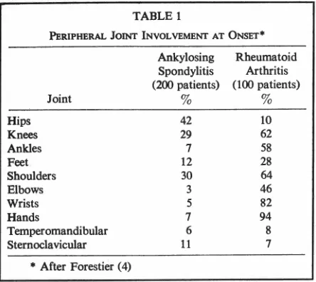
Joint involvement in ankylosing spondylitis is, of course, different from that seen in patients with rheumatoid arthritis. Forestier ( 4) showed (Table 1) the high frequency of hip involvement in this disease when compared to patients with rheumatoid arthritis. In the rheumatoid, there is much more peripheral joint involvement.
There are a number of complications which are more specific and more frequent in ankylosing spondylitis than in many of the other forms of arthritis. Most of the complications that have been noted are listed in Table 2. Iritis is said to occur in up to 20% of patients. Aortic insufficiency is usually seen quite late in the disease. Heart block is rarely seen but also probably represents the same type of inflammatory action in the myocardium as is seen in the aorta. Amyloidosis, again, is not seen frequently but is a recognized cause of death in ankylosing spondylitis-certainly more so than with rheumatoid arthritis. Atlantoaxial subluxation is a constant threat to these people with spine fusion and can sometimes be fatal after only moderate degrees of trauma. Cauda equina involvement is rarely seen but, again, is a well-recognized problem. Pulmonary fibrosis of the upper lobe is of interest because this fibrosis, which may be typical of spondylitics, has been mistaken for tumor, tuberculosis, and the like.
NUMBER OF CASES 40
35
30
!5
20
15
10
5
AGE
0 16 21 26 .31 36 41 46 51 56
5 10 15 20 25 30 35 40 45 50 55 60
From Forestier, J. et al., ( 4). Courtesy of Charles C Thomas, Publisher, Springfield, Illinois.
TABLE 1
PERIPHERAL JOINT INVOLVEMENT AT ONSET*
Ankylosing Rheumatoid
Spondylitis Arthritis (200 patients) (100 patients)
Joint % %
Hips 42 10
Knees 29 62
Ankles 7 58
Feet 12 28
Shoulders 30 64
Elbows 3 46
Wrists 5 82
Hands 7 94
Temperomandibular 6 8
Sternoclavicular 11 7
* After Forestier ( 4)
It probably accounts for some of the earlier state-ments made about the higher frequency of tubercu-losis in these patients. It is remarkable how good pulmonary function is in these men with limited chest expansion. A recent study indicates that im-pairment of lung function is minimal in most of the patients with this disease ( 5).
The variants of ankylosing spondylitis are shown in Table 3. All of these conditions can show
sacro-iliitis and indeed in some cases, go on to more exten-sive spinal involvement quite similar to those seen in ankylosing spondylitis. I will mention some of these again later because of an interesting relationship.
For many years, it has been felt that there were strong genetic features connected with ankylosing spondylitis. Several years ago, when I was still in Dallas, Morris Ziff and I, seeing our patients at the Veterans Administration Hospital, were struck by the relative rarity of ankylosing spondylitis among our black patients. (One of the earliest clinicians to note this rarity was Elam Toone in 1949.) He found, in a group of patients that he was studying in the McGuire Veterans Administration Hospital in Richmond, 26 white and only three black patients with ankylosing spondylitis ( 6). Table 4 shows what we found in Dallas at the Veterans Hospital when we looked at the frequency of anky-losing spondylitis in relation to a number of other disease admissions rates and the male population of Dallas ( 7). As you can see, although the admission rate to the Veterans Hospital clearly reflected the racial distribution of the population, there was a marked discrepancy in the relative ratio of patients
TABLE 2
COMPLICATION~ OF ANKYLOSING SPONDYLITIS
Iritis
Aortic Insufficiency (Aortitis) Heart Block (Myocarditis) Amyloidosis
Atlantoaxial Subluxation Cauda Equina Involvement Upper Lobe Fibrosis
with ankylosing spondylitis. We were not satisfied with this view, thinking perhaps we might have an unusual situation. When data were pooled from a number of VA hospitals, we found a fourfold dif-ference in the frequency of ankylosing spondylitis between whites and blacks. A search of the African literature showed that there are virtually no patients with ankylosing spondylitis in Africa among the black native population. A review of genetic studies in the United States indicated that the black popula-tion of the United States has a 20-25% admixture of white genes. This admixture of white genes would very nicely account for the fact that ankylosing spondylitis was only found in about 20-25% of the expected frequency, if it had the same distribu-tion in the blacks as it did in whites.
This information was subsequently utilized by Schlosstein, Terasaki, Blues tone, and Pearson ( 8) at the Wadsworth VA Hospital in Los Angeles. The transplantation antigen known as W-27 was found in 8% of a caucasian population but was found in 88% of a group of patients with ankylosing spondylitis. It was also interesting to note that W-27 is not found in Black Africans and is present in an approximate frequency of 4% in Black Americans. To further support the relationship of this antigen to ankylosing spondylitis, W-27 was found in eight of ten Black Americans with ankylosing spondylitis.
Another and perhaps more striking correlary of
TABLE 3
VARIANTS OF ANKYLOSING SPONDYLITIS
TABLE4
RACIAL DISTRIBUTION OF ADMISSIONS AND OF ANKYLOSING SPONDYLITIS, RHEUMATOID ARTHRITIS, AND REITER'S SYNDROME AT DALLAS VETERANS ADMINISTRATION HOSPITAL
Number White
Males-Dallas, Texas (1960 census) 263,354
Male Admissions (10 months, 1966) 5, 182
Ankylosing Spondylitis (1959-1966) 41
Rheumatoid Arthritis (1959-1966) 90
Reiter's Syndrome (1959-1966) 7
this data was recently presented by Rodney Blue-stone of this group at the XIIIth International Con-gress of Rheumatology in Kyoto, Japan (9). In Table 5 are seen their most recent data. As you can see, W-27 is found in 14 of 156 patients with psoriasis alone. This is just about the frequency found in the normal population; however, in those patients with psoriasis who develop spondylitis it has been found in four of six individuals. In patients with colitis who developed spondylitis, this high frequency was again found. Perhaps even more remarkable are 15 out of 16 juvenile rheumatoid arthritics with W-27 who had spondylitis. Chronic Reiter's syndrome had the W-27 antigen in every case. Perhaps more astonishing were the studies they subsequently did looking at patients with acute Reiter's syndrome. Even those who do not have spondylitis showed W-27 in a remarkably high frequency.
We now seem to have a new tool for the diag-nosis of some forms of arthritis. For example, in male children who develop what appears to be juvenile rheumatoid arthritis, looking for W-27 might give us a strong clue as to the future course of their disease. A similar use could be in suspected Reiter's syndrome. This has certainly opened the door to a most fascinating series of studies and conjectures
TABLE 5
Psoriasis with Spondylitis Psoriasis Alone
Colitis with Spondylitis JRA with Spondylitis
Chronic Reiter's with Spondylitis
Acute and Chronic Reiter's
Frequency of W-27
4/6 14/156
4/6 15/16
8/8
22/23
% % White-Black
Number Black White Black Ratio
61, 911 81 19 4.2
1,227 81 19 4.2
3 93 7 13.7
16 85 15 5.6
4 I. 8
about the relationship of the W-27 antigen and
spondylitis.
In the treatment of ankylosing spondylitis, radiation therapy was at an early date, found to be effective for relief of pain; it's use was given up because of a reported high frequency of blood dyscrasias following its application. There are still some authors who feel that its limited use in patients who are having pain in spite of other therapy is of distinct benefit and of low risk ( 10). Indomethacin and phenylbutazone are the most frequently used drugs in the treatment of this disease. An observa-tion was made recently that a patient with severe ankylosing spondylitis who did not respond to the above mentioned drugs responded quite well to penicillamine therapy ( 11). Steroids are of little use in this group of patients. Teaching the patients
exercises in an attempt to increase diaphragmatic
breathing is of benefit and exercises may sometimes help to maintain the spine in better position.
It is worth noting that what we have learned about this disease in recent years has provided us with better methods of establishing the diagnosis and prognosis of ankylosing spondylitis.
REFERENCES
1. STURROCK RD, WoJTULEWSKI JA, HART FD:
Spondy-lometry in a normal population and in ankylosing spondylitis. Rheumatol Rehab 12: 135, 1973.
2. MOLL JMH, WRIGHT V: The pattern of chest and spinal mobility in ankylosing spondylitis. An objective clinical study of 106 patients. Rheumatol Rehab 12: 115, 1973.
3. RILEY MJ, ANSELL BM, BYWATERS EGL: Radiological
4. FORESTIER J, JACQUELINE F, ROTES-QUEROL J: Anky-/osing spondylitis. Charles C Thomas, publisher, 1956.
5. GACAD G, HAMOSH P: The lung in anklosing spondylitis. Am Rev Res Dis 107:286, 1973.
6. TooNE EC JR: Rheumatoid spondylitis: Observations on
the incidence and response to therapy among veterans
of the recent war. Ann Intern Med 30:733, 1949.
7. BAUM J, ZIFF M: The rarity of ankylosing spondylitis
in the black race. Arth Rheum 14: 12, 1971.
8. SCHLOSSTEIN L, TERASAKI Pl, BLUESTONE R, PEARSON
CM: High association of an HL-A antigen, W-27, with
ankylosing spondylitis. N Engl I Med 288:704, 1973.
9. 8LUESTONE RH, ScHLOSSTEIN LH, TERASAKI P, PEARSON
CM: High association of an HL-A antigen, W-27, with
ankylosing spondylitis. XIII International Congress of
Rheumatology, Kyoto, Japan, September 30-0ctober 6,
1973. Excerpta Medica International Congress Series No. 299.
10. SINCLAIR RJG: Treatment of rheumatic disorders with
special reference to ankylosing spondylitis. Proc R Soc Med 64:1031, 1971.
11. GOLDING DN: o-Penicillamine in rheumatoid arthritis.