145
AGE-RELATED RESPONSE OF IL-4/LUC/CNS-1 TRANSGENIC MICE TO PHTHALIC
ANHYDRIDE EXPOSURE
Ji Eun Sung, Ji Eun Kim, Jun Go, Eun Kyoung Koh, Sung Hwa Song, Hyun Ah Lee and Dae Youn Hwang*
Department of Biomaterials Science, College of Natural Resources & Life Science/Life and Industry Convergence Research Institute, Pusan National University, Miryang 627-706, Korea
*Corresponding author: [email protected]
Received: April 17, 2015; Revised: April 28, 2015; Accepted: April 30, 2015; Published online: December 14, 2015
Abstract:Age-related changes are associated with susceptibility to infection, malignancy, autoimmunity, response to vac-cination and wound healing. To investigate the relationship of several pathological phenotypes of allergic inflammation to age, alterations in the IL-4 derived luciferase signal and general phenotype biomarkers were measured in young (2-month-old) and old (12-month-(2-month-old) IL-4/Luc/CNS-1 transgenic (Tg) mice with phthalic anhydride (PA)-induced allergic inflam-mation for 2 weeks. There was no difference in the ear phenotypes and thickness between young and old mice, although these levels were higher in the PA-treated group than the acetone-olive oil (AOO)-treated group. The luciferase signal was detected in the mesenteric lymph node (ML), thymus and pancreas of both young and old PA-treated mice, but showed a greater increase in old Tg mice (except in the thymus). A greater increase in the epidermal thickness and dermal thickness was measured in old PA-treated mice than young PA-treated mice, while total mast cell number remained constant in both groups. Furthermore, the concentration of IgE was greater in young PA-treated mice than in old PA-treated mice, as was the expression of VEGF and IL-6. Taken together, the results of this study showed that an animal’s age is an important factor that must be considered when PA-induced allergic inflammation in IL-4/Luc/CNS-1 Tg mice are investigated to screen for allergens and therapeutic compounds.
Key words: aging; skin inflammation; IL-4/Luc/CNS-1 transgenic mice; phthalic anhydride; IgE
INTRODUCTION
Several important changes in the innate and adaptive response associated with increasing age are defined as immunosenescence (Busse and Mathur, 2010; Mil-grom and Huang, 2014). The clinical consequences of immunosenescence include enhanced susceptibility to various infections, increased rate of malignancies and autoimmune disease, decreased response to vaccina-tion and delayed wound healing (Busse and Mathur, 2010; Franceschi et al., 1995). Immunosenescence also influences the development of three allergic dis-orders including asthma, allergic rhinitis and atopic dermatitis, as well as common complications such as comorbidities, polypharmacy and adverse effects of drugs (Milgrom and Huang, 2014).
individuals, although this cell number in bronchoal-veolar lavage fluid increased (Milgrom and Huang, 2014). However, the number of mast cells in tissue was shown to decrease in an elderly group when compared with a young group (Gunin et al., 2011). Moreover, age influenced the anti- and proinflammatory cyto-kine expression pattern and final outcome, including reaction intensity and scale of fibrosis, in a hypersen-sitivity pulmonitis (HP) model of 3- and 18-month-old mice (Lemieszek et al., 2013). Moreover, greater elevation of pulmonary inflammation and IFN-γ and IL-5 expression occurred in older mice, while airway hyperresponsiveness (AHR) and IL-4 and IL-13 ex-pression was higher in young mice (Busse et al., 2007). However, no other studies have investigated whether the allergic responses to PA sensitization are affected by aging in IL-4/Luc/CNS-1 Tg mice.
Therefore, we attempted to evaluate alterations in allergic phenotypes, including the IL-4-derived lucif-erase signal, in response to skin inflammation induced by repeated dermal exposure to PA using young and old IL-4/Luc/CNS-1 Tg mice. The results of the pres-ent study suggest that several pathological phenotypes of allergic inflammation and IL-4 responses to PA ap-plication can be affected by aging in IL-4/Luc/CNS-1 Tg mice.
MATERIALS AND METHODS Experimental animals
The animal protocols used in this study were reviewed and approved for ethical and scientific care procedures by the Pusan National University-Institutional Animal Care and Use Committee (PNU-IACUC; Approval Number PNU-2013-0410). The young (2-month-old) and old (12-month-old) IL-4/Luc/CNS-1 Tg mice used in this study were kindly provided by the Nation-al Institute of Food and Drug Safety EvNation-aluation of the Korea Food and Drug Administration (FDA) (Osong, Korea). All mice were provided with ad libitum access to a standard irradiated chow diet (Samtako, Korea) and water throughout the 2-week feeding study. This diet was composed of moisture (12.5%), crude
pro-tein (25.43%), crude fat (6.06%), crude fiber (3.9%), crude ash (5.31%), calcium (1.14%) and phosphorus (0.99%), as well as corn (546 g/kg), vegetable protein (316 g/kg), fish meal (34 g/kg), beet pulp (30 g/kg), animal fat and oil (36 g/kg), lysine (2 g/kg), choline bi-tartrate (2 g/kg), 7.17% methionine solution (2 g/kg), limestone (10 g/kg), calcium/phosphate supplement (13 g/kg), salt (5 g/kg), mineral mix (2 g/kg) and vita-min mix (2 g/kg). During the experiment, mice were maintained in a specific pathogen-free state under a strict light cycle (lights on at 08:00 and off at 20:00) at 23±2°C and 50±10% relative humidity. The mice were housed in the Pusan National University-Laboratory Animal Resources Center accredited by the Korea FDA in accordance with the Laboratory Animal Act (Accredited Unit Number-000231) and AAALAC International according to the National Institutes of Health guidelines (Accredited Unit Number; 001525). Young and old IL-4/Luc/CNS-1 Tg mice (n=5-6 per group) were randomly divided into two groups. In the first group (AOO, n=4-5), 100 μl of AOO (4:1 acetone:olive oil, v/v) was repeatedly spread on the dorsum of the ears three times a week for 2 weeks. In the second group (PA, n=4-5), 100 μl of 15% PA solution in AOO was repeatedly spread on the dor-sum of the ears three times a week for 2 weeks. After final treatment, the animals in each group were eu-thanized using a chamber filled with CO2 gas. Ear tissue samples and whole blood were then collected for further analysis.
Production and identification of IL-4/Luc/CNS-1 Tg mice
ge-nomic DNA template mixture. Amplification was then conducted in a T100 thermal cycler (BioRad Labo-ratories Inc., Hercules, CA, USA) by subjecting the samples to 35 cycles of 1 min at 94°C, 1 min at 62°C and 1 min at 72°C. The amplified PCR products were subsequently separated by 1% agarose gel electropho-resis, after which the band patterns were detected us-ing a UV-transilluminator (ATTO, Tokyo, Japan).
Observation of ear morphology and measurement of ear thickness
Changes in ear color, ear vein and other morphologi-cal characteristics were analyzed by evaluation of pho-tographs. Additionally, ear thickness was measured to determine the degree of skin inflammation induced by AOO and PA topical application using a thickness gauge (Digimatic Indicator, Matusutoyo Co., Tokyo, Japan).
Measurement of body and organ weight
The body weights of all animals in the subset groups were measured using an electronic balance (Met-tler Toledo, Greifensee, Switzerland) throughout the experimental period. In addition, the weight of the lungs, kidneys, spleen, heart, ML, thymus and pan-creas was determined using the same method.
Bioluminescence imaging analysis
In vivo imaging analysis was conducted using an IVIS imaging system (Xenogen, Oakland, CA, USA) as pre-viously described (Kwak et al., 2013). Briefly, IL-4/ Luc/CNS-1 Tg mice were anesthetized with Zoletil 50 (Virbac, Carros, France) and then injected intraperito-neally with 150 mg/kg D-luciferin (Sigma-Aldrich, St. Louis, MO, USA). Ten min after D-luciferin injection, whole body and organ images of mice were taken for 3 min using an IVIS imaging system, after which the photons emitted from specific regions were quantified using the Living Image software (Xenogen). The in vivo luciferase activity was then expressed in photons per second.
Enzyme-linked immunosorbent assay (ELISA) for detection of serum IgE concentration
The serum IgE concentration was measured using an ELISA kit (Shibayagi Inc., Gunma, Japan) according to the manufacturer’s instructions. Briefly, wells coated with antibody were washed three times with wash-ing solution (50 mM Tris, 0.14 M NaCl, 0.05% Tween 20, pH 8.0), after which 50 µl of serum samples and standards were added to the wells and the plate was incubated for 2 h. Following washing with the above solution, 50 µl of biotin-conjugated avidin (1000-fold dilution) was added and the samples were then in-cubated for 2 h. Horseradish peroxidase-conjugated detection antibodies (2000-fold dilution) were then transferred to each well. The plates were subsequently incubated at room temperature for 1 h, after which they were washed three times with washing solution. Next, an enzyme reaction was initiated by adding substrate solution and incubating the plate at room temperature for 20 min. Finally, the reaction was ter-minated by adding stop solution (1 M H2SO4 solution) and the absorbance was measured at 450 nm.
Histological analysis
specific area was determined using the Leica Applica-tion Suite (Leica Microsystems).
Western blotting
Ear tissues collected from a subset of the groups were homogenized using a PRO-PREPTM Solution Kit (iN-tRON Biotechnology Inc., Sungnam, Korea), then centrifuged at 13000xg for 10 min. The prepared pro-teins were subsequently subjected to 10% SDS-PAGE, after which they were transferred to a nitrocellulose membrane (GE Healthcare, Little Chalfont, UK) for 2 h at 40 V in transfer buffer (25 mM Trizma base, 192 mM glycine and 20% methanol). Appropriate dilutions of primary antibodies including anti-IL-6 antibody (Santa Cruz Biotechnology, Santa Cruz, TX, USA), anti-VEGF antibody (Pepro Tech., Rckyhill, NJ, USA) and anti-β-actin (Sigma-Aldrich Co.) were added to the membranes and allowed to hybridize overnight at 4°C. After the antibodies were removed, the membrane was washed three times in a solution composed of 10 mM Trizma base (150 mM NaCl and 0.05% Tween-20) for 10 min. The membrane was then incubated with horseradish peroxidase-conjugated secondary antibody for 1 h at room temperature, fol-lowed by washing as described above and developed using an enhanced chemiluminescence reagent plus kit (Amersham Biosciences). The results were quan-tified using the Image Analyzer System (Fluorchem FC2, Alpha Innotech, CA, USA) and expressed as the fold-increase over control values.
Statistical analysis
One-way ANOVA was used to determine whether or not significant differences existed between the PA-treated and AOO-PA-treated groups (SPSS for Windows, Release 10.10, Standard Version, Chicago, IL, USA). Additionally, differences in response between the young and old groups were evaluated by a post hoc test (SPSS for Windows, Release 10.10, Standard Ver-sion) of the variance and significance levels. All values were expressed as the means±SD. A p value of <0.05 was considered significant.
RESULTS
Difference in ear morphology and thickness between young and old IL-4/Luc/CNS-1 Tg mice
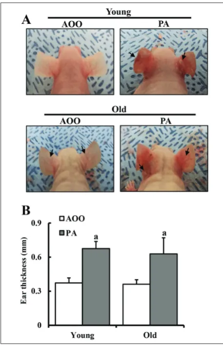
To investigate the effects of aging on ear phenotypes after PA application, we first evaluated changes in the ear morphology and thickness in young and old IL-4/Luc/CNS-1 Tg mice after PA treatment for 2 weeks. During morphological analysis, the ear color of IL-4/Luc/CNS-1 Tg mice changed significantly from a fleshy tint to dark brown in the PA-treated group compared to the AOO-treated group, while the outline of the ear vein became thickened. However, there were no significant differences in ear morphol-ogy between young and old IL-4/Luc/CNS-1 Tg mice (Fig. 1A). Conversely, the ear thickness was elevated in the young and old PA-treated group compared to the AOO-treated group. However, this increase (81%) in the young PA-treated group was not significantly different from that (74%) of the old PA-treated group (Fig. 1B). Thus, these results demonstrate that there is no correlation between ear morphology response to PA treatment and age in IL-4/Luc/CNS-1 Tg mice.
Difference in IL-4-derived luciferase signal between young and old IL-4/Luc/CNS-1 Tg mice
Difference in organ weight between young and old IL-4/Luc/CNS-1 Tg mice
To measure the difference in PA application between young and old IL-4/Luc/CNS-1 Tg mice, body and organ weight were measured in the subset groups after PA treatment for 2 weeks. The whole body weight in both groups was maintained at a constant level throughout the experimental periods (data not shown). However, only three of the seven organs in-vestigated showed significant alteration after PA
treat-Fig. 1. Morphological analysis of the ears of IL-4/Luc/CNS-1 Tg mice. (A) Ear vein (arrow) and other morphological character-istics were analyzed in photographs of mice. (B) Ear thickness of mice in four groups was measured using a thickness gauge and phenotypes were observed as described in the Materials and Methods after the application of PA solution. Data shown are the means±SD (n=5). a, p<0.05 compared to the AOO-treated group.
Fig. 2. Detection of luciferase signal in seven organs of IL-4/ Luc/CNS-1 Tg mice.Mice were treated with AOO and PA for 2 weeks and imaged at 24 h after final treatment using Living Image software. The color overlay on the image represents the photons per second emitted from the organs in accordance with the pseudocolor scale shown next to the image. In this image, red indicates the highest number of photons per second, while blue indicates the lowest. L − lung; K − kidney; S − spleen; H − heart; ML − mesenteric lymph node; T − thymus; P − pancreas. a, p<0.05 compared to the AOO-treated group.
Difference in histological structure of young and old Tg mice
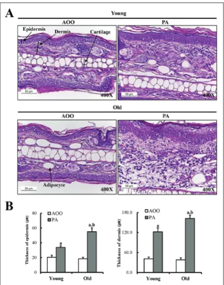
Differences in the histological structure of ear tissue between young and old PA-treated Tg mice were evalu-ated by histological analysis of ear tissue of IL-4/Luc/ CNS-1 Tg mice after PA treatment for 2 weeks. The epidermis and dermis of the ear tissue in the mice was thicker in the PA-treated group than in the AOO-treated group. However, in the PA-AOO-treated group, the increase in epidermal and dermal thickness was greater; increased thickness (120-125%) was observed in old Tg mice as compared to young Tg mice (Fig. 3). Taken together, these results showed that age may be closely correlated with increasing epidermal and dermal thick-ness in IL-4/Luc/CNS-1 Tg mice after PA treatment.
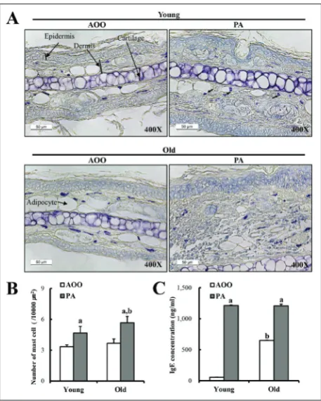
Mast cells play important roles in asthma, eczema, itch, allergic rhinitis, allergic conjunctivitis and skin inflammation (Prussin et al., 2003). Therefore, ear skin sections were stained with toluidine blue and mast cells were measured to investigate the effects of aging on the infiltration of mast cells after PA treat-ment. The total number of mast cells in the dermis region increased significantly in the PA-treated group compared to the AOO-treated group. However, the fold change in the total number of mast cells was sig-nificantly greater, with 25.1% more observed in old Tg mice than in young Tg mice after PA application
Table 1. Comparison of organ weights between young and old group
Categories Weight of young mice (g) Weight of old mice (g)
AOO PA AOO PA
Lung 0.21±0.028 0.18±0.007 0.26±0.031 0.24±0.035 Kidney 0.23±0.057 0.29±0.099 0.37±0.116 0.39±0.244 Spleen 0.11±0.028 0.20±0.023* 0.20±0.057 0.21±0.014 Heart 0.16±0.007 0.17±0.007 0.20±0.322 0.21±0.055 Mesenteric
lymph
node 0.19±0.028 0.31±0.021* 0.32±0.102 0.27±0.020 Thymus 0.07±0.000 0.03±0.007* 0.06±0.007 0.03±0.014* Pancreas 0.20±0.028 0.17±0.050 0.29±0.023 0.25±0.080
*, p<0.05 compared to the AOO-treated group.
Fig. 3. Histological analysis of ear tissue. PA solution was re-peatedly applied to the dorsum of the ear of IL-4/Luc/CNS-1 Tg mice. After 2 weeks, ear tissues were collected from AOO- and PA-treated IL-4/Luc/CNS-1 Tg mice. Histological changes in the slide sections of ear tissue were identified by staining with H&E followed by observation at 400x magnification. Data shown are the means±SD (n=5). a, p<0.05 compared to the AOO-treated group. b, p<0.05 compared to the young Tg mice.
(Figs. 4A and B). These results suggest that aging can affect the levels of PA-induced mast cells infiltration in the dermis of IL-4/Luc/CNS-1 Tg mice.
Difference in IgE concentration between young and old Tg mice
PA treatment, the change in this level was greater in the young PA-treated group (1100%) than in the old PA-treated group (85%) (Fig. 4C). These results sug-gest that IgE concentration may be more sensitive to PA topical application in young IL-4/Luc/CNS-1 Tg mice.
Cytokine expression in ear tissue
To understand the mechanism by which ear thickness and cytokine expression increased, we investigated the ear tissue of the subset groups. VEGF is known to be a multifunctional proinflammatory cytokine responsible for erythema and edema in atopic
der-matitis (Kasperska-Zaiac and Koczy-Baron, 2011). Moreover, IL-6 is widely known as an important pro-inflammatory mediator in allergic inflammation and atopic dermatitis (Zhao et al., 2005). The expression of VEGF and IL-6 was significantly enhanced in the PA-treated group compared to the AOO-PA-treated group. However, the increase in VEGF and IL-6 expression was greater in the young PA-treated group (1650% and 1500%, respectively) than the old-PA treated group (362% and 125%, respectively) (Fig. 5). Overall, the above results indicate that the increase in IL-6 and VEGF expression in the skin inflammation induced by PA treatment may be suppressed by aging in IL-4/ Luc/CNS-1 Tg mice.
Fig. 4. Analysis of mast cells and IgE concentration. (A and B) Infiltration of mast cells in the slide sections of ear tissue was identified by staining with toluidine blue followed by observation at 400x magnification. Arrows indicate the infiltrated mast cells in the dermis of the ear tissue. (C) The serum used to measure the IgE concentration was prepared from blood samples collected from the abdominal veins of mice. The serum IgE concentration was quantified by an enzyme-linked immunosorbent assay. Data shown are the means±SD (n=5). a, p<0.05 compared to the AOO-treated group. b, p<0.05 compared to the young Tg mice.
Fig. 5. Analysis of cytokine expression in ear tissues. The levels of IL-6 and VEGF in the ear tissue of IL-4/Luc/CNS-1 Tg mice from subset groups were detected by Western blot analysis using specific antibodies. The intensity of each band was determined using an imaging densitometer and the relative level of each pro-tein was calculated based on the intensity of actin propro-tein as an endogenous control. Data shown are the means±SD (n=5). a, p
DISCUSSION
The changes in the immune system associated with age include a significant reduction in responsiveness, dysregulation of immune effector cells and remod-eling of the cytokine network (Albright et al., 2004; Linton and Dorshkind, 2004). However, there is still a poor correlation between immunosenescence and the clinical outcome of many diseases that preferen-tially affect elderly individuals (Shurin et al., 2007). Therefore, in the present study, we investigated the age-related alterations in skin inflammation of IL-4/ Luc/CNS-1 Tg mice during PA application. The re-sults demonstrated that aging can affect several path-ological phenotypes, as well as the luciferase signal derived from the IL-4 promoter in response to PA topical application.
Th2 cytokines including 4, 5, 10 and IL-13 mediate immune responses against a large num-ber of pathogens and regulate allergic inflammation through the stimulation of IgE class switching and eo-sinophil activation (Romagnani, 2001). Among these cytokines, IL-4 stimulation of B cells to produce IgE is primarily responsible for allergic inflammatory and atopic dermatitis responses (Khodoum et al., 2004). However, it is not clear if the IL-4 response against respiratory sensitizers is affected by age. Indeed, sev-eral studies have shown opposite results regarding age-related changes in IL-4 response to some aller-gens. The IL-4 concentration in the supernatants of peripheral mononuclear cells after 48 h of stimula-tion with phytohemagglutinin did not differ signifi-cantly between two groups of young (20-64 years) and old humans (70-93 years) (Di Lorenzo et al., 2003). However, semiquantitative RT-PCR analysis of bron-choalveolar lavage (BAL) cells revealed that cells from young Brown Norway rats (8- to 10-wk-old) sensitized with ovalbumin preferentially expressed the mRNA of Th2-type cytokines, including IL-4 and IL-5 (Ide et al., 1999). Juvenile mice infected with the parasitic nematode Nippostrongylus brasiliensis showed signifi-cantly reduced IL-4 expression in the early stages of infection (day 5 and 9) relative to adult mice (6-8-wk-old) (Hendrik et al., 2011). In this study, old Tg mice showed a greater increase in IL-4-derived luciferase
than young Tg mice in the PA-treated group (Fig. 2). The results of the present study are in agreement with those of several previous studies of juvenile mice, although the levels of change vary. However, our re-sults differ from those of previous studies that showed IL-4 expression was higher in young Brown Norway rats sensitized with OVA. This difference was likely due to differences in the properties of the sensitizing chemicals and the specificity of the target organs of the inflammatory response.
It is well known that the hyperproduction of IgE is characteristic of type 1 hypersensitivity and an in-dicator of the magnitude of allergic immune response (Gao et al., 2004; Dearman et al., 2003). However, there are conflicting results regarding the effects of aging on serum total IgE concentration after allergen sensitization. In 20- to 93-year-old humans, serum total IgE concentration did not differ significantly between the young and old (Di Lorenzo et al., 2003). However, a significantly lower concentration of total IgE was measured in juvenile mice infected with N. brasiliensis than in adult mice (Hendrik et al., 2011). In the present study, a high fold change of IgE con-centration was observed in young Tg mice after PA topical application. These findings differ from those of previous reports where IgE was preferentially sup-pressed in a young group after allergen infection. Therefore, the results presented herein provide ad-ditional evidence that aging affects IgE production in PA-treated IL-4/Luc/CNS-1 Tg mice.
months) mice. However, they showed similar mast cell degranulation in the ear tissue after IgE-dependent passive sensitization (Nguyen et al., 2005). In this study, alterations in the number of mast cells in the ears of IL-4/Luc/CNS-1 Tg mice were investigated in young and old Tg mice after PA treatment. As shown in Fig. 4, the total number of mast cells that infiltrated the dermis was significantly higher in the PA-treated group than in the AOO-treated group, although the fold change was greater in old Tg mice. These results are not directly comparable to those of previous stud-ies because of differences in the type of sensitizing chemical and the specificity of analysis factor of the inflammatory response. However, our results provide novel scientific evidence that the infiltration of mast cells into ear tissue after PA treatment may be affected by aging in IL-4/Luc/CNS-1 Tg mice.
Overall, we investigated the relationship between aging and allergic phenotypes including IL-4 response against PA application using IL-4/Luc/CNS-1 Tg mice. Differences in the IL-4-derived luciferase sig-nal as well as general phenotypes of skin inflamma-tion induced by PA applicainflamma-tion was detected in both young and old Tg mice as shown Table 2. The results presented herein suggest that animal age should be considered when using IL-4/Luc/CNS-1 Tg mice for the investigation of PA-induced skin inflammatory responses.
Acknowledgments: We thank Jin Hyang Hwang and the animal technicians for directing the Animal Facility and Care at the Laboratory Animal Resources Center. This work was supported for two years by a Pusan National University Research Grant.
Authors’ contribution: JES, JEK, JG, EKK, SUS and DYH participated in the design of the study, sample prepara-tion, animal experiments and data analyses. SKP and HAL helped with data analysis and manuscript preparation. All authors read and approved the final manuscript.
Conflict of interest disclosure: The authors have no com-peting interests to declare.
REFERENCES
Albright, J.W., Bream, J.H., Bere, E.W., Young, H.A., Winkler-Pick-ett, R. and J.R.Ortaldo (2004). Aging of innate immunity: functional comparisons of NK/LAK cells obtained from bulk cultures of young and aged mouse spleen cells in high concentrations of interleukin-2. Exp. Gerontol.39, 73-82.
Bae, C.J., Lee, J.W., Bae, H.S., Shim, S.B., Jee, S.W., Lee, S.H., Shim, S.B., Jee, S.W., Lee, S.H., Lee, C.K., Hong, J.T. and D.Y. Hwang (2011). Detection of allergenic compounds using an IL-4/Luciferase/CNS-1 transgenic mice model. Toxicol. Sci.120, 349-359.
Busse, P.J. and S.K.Mathur (2010). Age-related changes in immune function: effect on airway inflammation. J. Allergy Clin. Immunol.126, 690-699.
Busse, P.J., Zhang, T.F., Srivastava, K., Schofield, B. and X.M.Li
(2007). Effect of ageing on pulmonary inflammation, air-Table 2. Quantitative comparison of allergic phenotypes between the young and old groups.
Categories Response magnitude against PA(change rate to vehicle group) young and old groupDifference between
Young Old
Ear thickness
Luciferase signal +0.302 +0.267 11.6% lower in old group
Mesenteric lymph node +1.29×10⁴ +2.59×10⁴ 101% higher in old group
Thymus +6.32×10³ +4.94×10³ 21.8% lower in old group
Pancreas +1.50×10⁴ +2.58×10⁴ 72% higher in old group
Thymus weight (g) - 0.04 - 0.03 25% lower in old group
IgE concentration (ng/mL) +1,157.30 +558.96 51.8% lower in old group
Epidermal thickness (μm) +6.39 +36.64 473.4% higher in old group
Dermal thickness (μm) +80.82 +123.79 53.8% higher in old group
Mast cells number (ea) +1.33 +2 50.4% higher in old group
way hyperresponsiveness and T and B cell responses in antigen-sensitized and -challenged mice. Clin. Exp. Allergy.
37, 1392-1403.
Dearman, R.J., Skinner, A., Humphreys, N.E. and I. Kimber (2003). Methods for the identification of chemical respiratory aller-gens in rodents: comparisons of cytokine profiling with induced changes in serum IgE. J. Appl. Toxicol.23, 199-207.
Di Lorenzo, G., Pacor, M.L., Esposito Pellitteri, M., Listì, F., Colombo, A., Candore, G., Mansueto, P., Lo Bianco, C., Ditta, V., Battista Rini, G. and C.Caruso (2003). A study of age-related IgE pathophysiological changes. Mech. Ageing Dev.124, 445-448.
Franceschi, C., Capri, M., Monti, D., Giunta, S., Olivieri, F., Sevini, F., Panourgia, M.P., Invidia, L., Celani, L., Scurti, M., Cev-enini, E., Castellani, G.C. and S. Salvioli (2007). Inflammag-ing and anti-inflammagInflammag-ing: a systemic perspective on agInflammag-ing and longevity emerged from studies in humans. Mech. Age-ing Dev.128, 92-105.
Franceschi, C., Monti, D., Sansoni, P. and A. Cossarizza (1995). The immunology of exceptional individuals: the lesson of centenarians. Immunol. Today.16, 12-16.
S.J. Galli (2000). Mast cells and basophils. Curr. Opin. Hematol. 7, 32-39.
Gao, X.K., Nakamura, N., Fuseda, K., Tanaka, H., Inagaki, N. and
H. Nagai (2004). Establishment of allergic dermatitis in NC-Nga mice as a model for severe atopic dermatitis. Biol. Pharm. Bull.27, 1376-1381.
Gunin, A.G., Kornilova, N.K., Vasilieva, O.V. and V.V. Petrov
(2011). Age-related changes in proliferation, the numbers of mast cells, eosinophils, and cd45-positive cells in human dermis. J. Gerontol. A Biol. Sci. Med. Sci.66, 385-392.
Hannoun, C., Megas, F. and J. Piercy (2004). Immunogenicity and protective efficacy of influenza vaccination. Virus Res.103, 133-138.
Hasegawa, A., Miki, T., Hosokawa, H., Hossain, M.B., Shimizu, C., Hashimoto, K., Kimura, M.Y., Yamashita, M. and T. Nakayama (2006). Impaired GATA3-dependent chro-matin remodeling and Th2 cell differentiation leading to attenuated allergic airway inflammation in aging mice. J. Immunol.176, 2546-2554.
Ide, K., Hayakawa, H., Yagi, T., Sato, A., Koide, Y., Yoshida, A., Uchijima, M., Suda, T., Chida, K. and H. Nakamura (1999). Decreased expression of Th2 type cytokine mRNA contrib-utes to the lack of allergic bronchial inflammation in aged rats. J. Immunol.163, 396-402.
Kasperska-Zajac, A. and E. Koczy-Baron (2011). Etiopathogenesis of atopic dermatitis. Pol. Merkur. Lekarski.31, 309-312.
Kawakami, T., Ando, T., Kimura, M., Wilson, B.S. and Y. Kawakami (2009). Mast cells in atopic dermatitis. Curr. Opin. Hematol.21, 666-678.
Khodoun, M.V., Orekhova, T., Potter, C., Morris, S. and F.D. Fin-kelman (2004). Basophils initiate IL-4 production during a memory T-dependent response. J. Exp. Med.200, 857-870.
Kim, J.E., Lee, Y.K., Nam, S.H., Choi, S.I., Goo, J.S., Jang, M.J., Lee, H.S., Son, H.J., Lee, C.Y. and D.Y.Hwang (2010). The symp-toms of atopic dermatitis in NC/Nga mice were signifi-cantly relieved by the water extract of Liriope platyphylla.
Lab. Anim. Res.26, 377-384.
Kwak, M.H., Kim, J.E., Hwang, I.S., Lee, Y.J., An, B.S., Hong, J.T., Lee, S.H. and D.Y. Hwang (2013). Quantitative evaluation of therapeutic effect of Liriope platyphylla on phthalic anhydride-induced atopic dermatitis in IL-4/Luc/CNS-1 Tg mice. J. Ethnopharmacol.148, 880-889.
Lemieszek, M.K., Chilosi, M., Golec, M., Skórska, C., Dinnyes, A., Mashayekhi, K., Vierlinger, K., Huaux, F., Wielscher, M., Hofner, M., Yakoub, Y., Pastena, C., Daniele, I., Cholewa, G., Sitkowska, J., Lisowska, W., Zwoliński, J., Milanowski, J., Mackiewicz, B., Góra-Florek, A., Ziesche, R. and J. Dutkie-wicz (2013). Age influence on hypersensitivity pneumonitis induced in mice by exposure to Pantoea agglomerans. Inhal. Toxicol.25, 640-650.
Linton, P.J. and K.Dorshkind (2004). Age-related changes in lymphocyte development and function. Nat. Immunol.5, 133-139.
Milgrom, H. and H. Huang (2014). Allergic disorders at a vener-able age: a mini-review. Gerontology.60, 99-107.
Nel, H.J., Hams, E., Saunders, S.P., Mangan, N.E., Smith, P., Atz-berger, A., Flavell, R.A., Akira, S., McKenzie, A.N. and P.G. Fallon (2011). Impaired basophil induction leads to an age-dependent innate defect in type 2 immunity during helminth infection in mice. J. Immunol.186, 4631-4639.
Nguyen, M., Pace, A.J. and B.H.Koller (2005). Age-induced reprogramming of mast cell degranulation. J. Immunol.
175, 5701-5707.
Prussin, C. and D.D. Metcalfe (2003). IgE, mast cells, basophils, and eosinophils. J. Allergy. Clin. Immunol.111, 486-494.
Romagnani, S. (2001). T-cell responses in allergy and asthma.
Curr. Opin. Allergy Clin.Immunol.1, 73-78.
Shurin, G.V., Yurkovetsky, Z.R., Chatta, G.S., Tourkova, I.L., Shurin, M.R. and A.E. Lokshin (2007). Dynamic alteration of soluble serum biomarkers in healthy aging. Cytokine.
39, 123-129.
Wu, D., Han, S.N., Bronson, R.T., Smith, D.E. and S.N. Meydani
(1998). Dietary supplementation with mushroom-derived protein-bound glucan does not enhance immune function in young and old mice. J. Nutr.128, 193-197.
Zhao, W., Oskeritzian, C.A., Pozez, A.L. and L.B. Schwartz (2005). Cytokine production by skin-derived mast cells: endoge-nous proteases are responsible for degradation of cytokines.