Ivan Gwilym Sabath
A dissertation submitted to the faculty at the University of North Carolina at Chapel Hill in partial fulfillment of the requirements for the degree of Doctor of Philosophy in the Department
of Biochemistry and Biophysics in the School of Medicine.
Chapel Hill 2014
Approved by: Zbigniew Dominski William Marzluff
Howard Fried Greg Matera
ii
iii
Ivan Gwilym Sabath: Characterization of Factors Required for 3’ End Processing
of Histone pre-mRNAs (Under the direction of Dr. Zbigniew Dominski)
Metazoan replication dependent histone mRNAs form their 3’ end by endonucleolytic cleavage of the pre-mRNA transcript without the subsequent addition of a poly(A) tail. These are the only cellular mRNAs that are not polyadenylated but instead end in a highly conserved stem-loop. Analogous to the poly(A) tail of canonical mRNAs, the terminal 3’ stem-loop of histone mRNAs is a crucial regulatory feature that provides an interface for mechanisms coupling the mRNA to cell-cycle regulation, stability, and translation. Histone pre-mRNA 3’ end processing requires the U7 snRNP, which interacts with a purine rich sequence called the histone
downstream element (HDE) located about 10 nts 3’ of the cleavage site. The U7 snRNP is composed of a small 60 nucleotide snRNA bound with a U7 specific heptameric ring resembling that of splicesomal snRNPs except that SmD1 and SmD2 are replaced by Lsm10 and Lsm11, respectively. Unlike other Lsm proteins, Lsm11 has an extended N-terminal domain, which interacts with FLASH. The interaction between the N-terminus of Lsm11 and FLASH is essential for processing. My research shows that the native U7 snRNP has a larger, more
iv
v Chapter
I. Introduction……….… 1
Role of Histones……….…. 1
Organization of Histone Genes and Nuclear Bodies……….….. 3
Canonical pre-mRNA 3’ end Processing……….… 6
Early Insights into Histone pre-mRNA 3’ end processing…………...………..…. 8
Role of RNA in Histone pre-mRNA 3’ end Processing…………..……...……… 10
Role of SLBP in Histone pre-mRNA 3’ end Processing ………..………. 14
Identification of Symplekin as a Heat Labile Factor and CPSF73 as the Endonuclease………...……….… 15
FLASH is Essential for Histone pre-mRNA 3’ end Processing ……….. 17
Summary………...……… 18
Thesis Goals……….. 19
II. In mammals, a subset of cleavage and polyadenylation factors including the endocuclease CPSF73 associate with the U7 snRNP in a FLASH dependent manner………. 31
vi
Materials and Methods………..……… 33
Results………..………. 36
Discussion………. 46
III. The U7 snRNP and FLASH associate with a modified subset of cleavage and polyadenylation factors lacking CPSF30 and Fip1 in Drosophila……… 65
Introduction……….……….….… 65
Materials and Methods…….……….……… 67
Results……….……….…. 72
Discussion………. 93
IV. C-terminal FLASH Interacts with NPAT and Contributes to Formation of Histone Locus Bodies……….……….. 127
Introduction………...…..…… 127
Materials and Methods……… 129
Results………. 132
Discussion………..…………. 143
V. Summary and Conclusions………. 164
Biochemical isolation and identification of the HCC ………....…… 165
Drosophila HCC contains a different set of polyadenylation factors ………...….. 167
Drosophila HCC differs from a larger super complex specific to cleavage and polyadenylation.……….... 167
Functional elements of FLASH required for 3’ end processing are conserved in Drosophila………...…… 168
FLASH is required for DCP degradation in Drosophila………....… 170
vii
MiniFLASH is a splice variant of FLASH that joins two regions essential for histone biogenesis………...… 172
viii
LIST OF FIGURES
Figure 1. Histone mRNA levels peak during S-phase……….………. 21
Figure 2. Organization of histone genes in Drosophila……… 23 Figure 3. 3’ end processing of canonical and histone pre-mRNAs
depend on different sequence elements……… 25
Figure 4. A large network of protein complexes positions the endonuclease in cleavage and polyadenylation………..….…… 27
Figure 5. Histone pre-mRNA 3’ end processing depends upon a unique set of factors……….……. 29
Figure 6. Biochemical assay to identify proteins interacting with a complex of FLASH and Lsm11………..…. 53
Figure 7. The Lsm11/FLASH complex binds a subset of factors involved in cleavage and polyadenylation………...… 55
Figure 8. The LDLY motif in FLASH is required for the interaction of the complex with polyadenylation factors……… 57
Figure 9. Sequences in Lsm11 required for binding the polyadenylation complex by the Lsm11/FLASH complex………. 59
Figure 10. Refined mapping of Lsm11 regions required for binding to FLASH………..………… 61
Figure 11. Polyadenylation factors bind to the endogenous U7 snRNP and histone pre-mRNA……….………… 63
x
Figure 13. The role of the Drosophila FLASH LDIY motif in processing…………...………. 104 Figure 14. Second conserved LDIY motif located between amino acids 45-48 has a minor role in processing efficiency in vitro……….………...… 107
Figure 15. A complex of the N-terminal FLASH and Lsm11 binds
Drosophila polyadenylation factors……….... 109
Figure 16. Degradation of the DCPin Drosophila requires FLASH……….… 111
Figure 17. Analyzing the involvement of Drosophila Ars2 in 3’ end
processing of histone pre-mRNAs………...………... 113
Figure 18. Composition of Drosophila U7 snRNP……….……… 115 Figure 19. Composition of processing complexes assembled on the dH3 pre-mRNA………...… 117
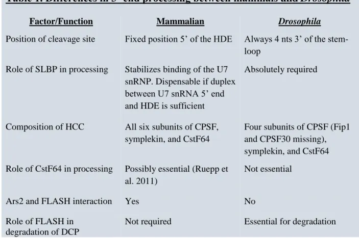
Figure 20. CstF64 is a component of the Drosophila U7 snRNP………..………. 120 Figure 21. Major differences in 3’ end processing of replication-dependent histone pre-mRNAs between mammals and Drosophila……… 123
Figure 22. Potential forms of the U7 snRNP in Drosophila………..…. 125
Figure 23. The C-terminal regions of FLASH and YARP interact with NPAT…….………… 152 Figure 24. The C-terminal domain shared by FLASH and YARP is sufficient for the interaction with NPAT……… 154
x
Figure 26. The C-terminal regions of FLASH targets SLBP to HLBs in HeLa cells…………. 158 Figure 27. YARP is a component of HLBs in HeLa cells……….. 160
xi
LIST OF ABBREVIATIONS CB cajal body
CPSF cleavage and polyadenylation specificity factor CStF cleavage stimulation factor
FLASH FLICE associated huge protein HCC histone pre-mRNA cleavage complex HDE histone downstream element
HLB histone locus body mRNA messenger RNA
PAS polyadenylation signal pre-mRNA precursor messenger RNA RBD RNA binding domain
RD replication dependent RNAi RNA interference
SL stem-loop
xii snRNA small nuclear RNA
snRNP small nuclear ribonucleoprotein particle
1
CHAPTER 1: INTRODUCTION
ROLE OF HISTONES
The eukaryotic genome is packaged in a tightly and highly ordered arrangement of nucleo-protein units called nucleosomes. These repeating units form the basis of chromatin. At the center of the nucleosome is an octameric core formed from two copies each of the four histone proteins H2A, H2B, H3 and H4. At the center of the core, H3 and H4 exist as dual heterodimers with two H2A/H2B heterodimers on either side of the H3/H4 tetramer. There are 147 base pairs of DNA wrapped 1.67 helical turns around each octamer with a stretch of linker DNA
approximately 10-80 base pairs between nucleosomes. While the ordered core of the histone proteins in the nucleosome make direct contact with the DNA, a significant portion of the histone proteins protrude out beyond the nucleosome as less ordered N-terminal tails available for a variety of regulatory post-translational modifications collectively termed the histone code (Strahl and Allis, 2000). The core nucleosome particle also interacts with the histone H1 linker protein. H1 binds the linker DNA to the octamer at the point at which 5’ and 3’ ends of the wrapped DNA make contact with the core nucleosome. H1 promotes compaction of nucleosomes to form a dense chromatin structure. This chromatin environment serves two critical functions: protecting the integrity of the genome and providing complex architecture for regulation of gene
expression.
3
core histone proteins H2A, H2B, H3, H4 and H1 that form the nucleosome. Expressed only when DNA is replicated, these five histones form the primary bulk of proteins constituting chromatin. Their expression is tightly coupled to cell-cycle progression and coordinated with DNA replication during S-phase when there is a need to package newly synthesized DNA. RD histone mRNA levels peak during S-phase with a 30 fold induction of available mRNA when the demand for histone proteins is greatest. In vertebrates, regulation of mRNA levels is the result of changes in rates and efficiency of transcription, mRNA 3’ end formation, and mRNA stability with 3’ end formation playing the most significant role (Fig. 1). In metazoans, RD histone mRNAs are the only messenger RNAs (mRNAs) that do not have poly(A) tails. Furthermore, their precursor messenger RNA (pre-mRNA) transcripts are monocistronic and lack introns.
Replication-independent histones, or histone replacement variants, are not cell-cycle regulated but instead expressed constitutively at basal levels throughout the cell-cycle. They are expressed from genes scattered through the genome and can substitute for RD histones under a number of circumstances including transcriptional activation, kinetochore assembly, and DNA repair (Sarma and Reinberg, 2005). A significant difference from the RD genes is that pre-mRNA transcripts from replication-independent genes often contain introns and the pre-mRNAs are polyadenylated.
4
histone genes: 49 for the core histones and 6 for the linker H1. Two smaller clusters, HIST2 and HIST3, lie on chromosome 1 and contain 6 and 3 histone genes respectively (Marzluff et al., 2002). In contrast, histone genes in Drosophila melanogaster are located at just one locus on chromosome 2L 39DE (Lifton et al., 1978). These genes are organized in 100 tandem 5.1kB repeating units each encoding one copy of all five RD histones (Fig. 2). The Drosophila H3 and H4 genes, and the H2a and H2b genes, are in pairs with divergent transcription from shared promoters while the histone H1 gene has its own promoter. Grouping large numbers of histone genes into distinct clusters is an evolutionary conserved feature unique to RD histone genes, unlike many other large copy number gene families that are found scattered throughout the genome. This clustered arrangement is ideal for localizing cis-acting transcription and pre-mRNA processing factors at concentrations sufficient for increased transcription rates and efficient biosynthesis of RD histone pre-mRNA during S-phase.
Eukaryotes have evolved a complex system of cellular compartmentalization in order to regulate and ensure the efficiency of distinct biochemical processes. While some of these compartments such as mitochondria, the nuclear envelope, lysosomes, peroxisomes, and the endoplasmic reticulum, are clearly defined by membranes, others, like the nucleolus, can be characterized by protein-protein and RNA-protein interactions that bring together factors in a macromolecular body lacking a membrane but still possessing a specialized function. Since these bodies lack a lipid membrane, constituent components can rapidly exchange between the
macromolecular structure and its surrounding nucleoplasm or cytoplasm environment.
5
role in the formation of the bodies (Liu et al., 2009) and which is used as a marker to identify CBs. These bodies play a key role in the biogenesis and organization of RNA processing machinery. Despite not being a center for splicing activity, the U1, U2, U4, U5 and U6 small nuclear RNAs (snRNAs) and their associated proteins that together form the splicesomal small nuclear ribonucleoprotein particles (snRNPs) can be found in CBs. Although the connection between CBs and splicing snRNPs is not completely understood, there are indications that upon return to the nucleus and association with CBs, the assembled snRNPs are subject to further modification. For example, the CB-specific RNA (scaRNA) U85 contains a C/D and H/ACA motif similar to that found in small nucleolar RNAs (snoRNAs) and in conjunction with the methyltransferase fibrillarin and pseudouridine synthase dyskerin/NAP57/CBF5 complex has been shown to guide 2′-O-methylation and pseudouridylation respectively of distinct nucleotide ribose moieties on the U5 snRNA (Matera and Ward, 1993; Almond and Carmo-fonseca, 1993). This could be considered analogous to the way ribosomal RNAs (rRNAs) are modified in the nucleolus by snoRNAs.
S-6
phase. Harmony Salzler showed the bi-directional promoter region located between H3 and H4 is sufficient to promote assembly of ectopic HLBs in Drosophila (Salzler et al., 2013).
The protein p220 nuclear protein ataxia-telengiectasia locus (NPAT) is also found constitutively in HLBs. It is required for the G1 to S-phase transition (Ma et al., 2000) and expression of histone genes during S-phase (Zhao et al., 2000). The C-terminal half of NPAT contains domains critical for G1 to S-phase transition. During the start of S-phase, NPAT is a substrate of cyclin E-cyclin dependent kinase 2 (CDK2/cyclin E) and is phosphorylated at five sites within its C-terminal half resulting in an increased level of histone gene expression (Ma et al., 2000). Regions in the N-terminal portion of NPAT are critical for its role as a histone gene co-activator. NPAT contains an approximately 30 amino acid LisH domain at its N-terminus, which is known to be essential for histone gene transcription. Mutations at conserved residues in this region block histone gene transcription but do not prevent localization to the HLB (Wei et al., 2003). NPAT has also been shown to bind histone nuclear factor P (HiNF-P), a transcription factor for H4 genes, and disrupting this interaction inhibits the transcription of the H4 gene at the G1 to S-phase transition (Miele et al., 2005).
CANONICAL PRE-MRNA 3’ END PROCESSING
The vast majority of mRNAs are polyadenylated at the 3’ end in a two-stage reaction. In the first stage, the cleavage and polyadenylation machinery recognizes a conserved AAUAAA
7
second reaction where the cleaved pre-mRNA 3’ end is appended with a poly(A) tail of approximately 200-250 adenosines added cotranscriptionally by poly(A) polymerase (PAP).
Cleavage coupled to polyadenylation requires at least five multi-subunit protein complexes to proceed including cleavage polyadenylation specificity factor (CPSF), cleavage stimulation factor (CstF), cleavage factor I (CF Im), cleavage factor II (CF IIm), in addition to
symplekin, PAP. CPSF consists of six protein subunits: CPSF30, CPSF73, CPSF100, CPSF160, Fip1, and WDR33. The CPSF complex recognizes the AAUAAA poly(A) signal through an interaction with CPSF160 (Mandel et al., 2008). The endonuclease that cleaves the transcript after a CA dinucleotide is CPSF73 (Mandel et al., 2006). CPSF30 binds a uridine rich sequence 5’ of the PAS (Barabino et al., 2000). Fip1 was identified as a member of CPSF that binds several cleavage and polyadenylation factors including CPSF30 and CstF77 (Helmling et al., 2001) and CPSF160 (Kaufmann et al., 2004). CPSF100, the catalytically inactive homologue of CPSF73, forms a heterodimer with the endonuclease (Dominski et al., 2005; Kolev et al., 2008). The protein WDR33 was also found to bind CPSF73 and co-purify with the CPSF complex (Shi et al., 2009).
CstF is a heterotrimeric RNA binding complex consisting of CstF77, CstF50, and CstF64 which recognizes and binds the downstream G/U rich element through an RNA binding domain (RBD) in CstF64 (Takagaki and Manley, 1997). CstF77 was shown to interact with both CstF64 and CstF50 in addition to self-associating through its C-terminus. CstF50 can also form a
8
inhibits the efficiency of CstF77 binding to CstF64, and the same region of CstF64 binds both CstF77 and symplekin.
In higher eukaryotes, CF Im is necessary for stimulating cleavage and polyadenylation
(Brown and Gilmartin, 2003). CF Im is a heterodimer consisting of eithera 59, 68, or 72 kDa
subunit (CF Im 68) and a 25 kDa subunit (CF Im 25) (Rüegsegger et al., 1998). CF Im 68 contains
an N-terminal RNA recognition motif (RRM) that mediates its interaction with CF Im 25 and
enhances the ability of the CF Im 25 RBD to bind a UGUA sequence element located 5’ of the
PAS. Evidence indicates CF Im plays a role in recognition of canonical and non-canonical
poly(A) sites (Venkataraman et al., 2005) and a role for CF Im in inhibiting usage of proximal
polyadenylation sites has been described as a mechanism for regulating alternative polyadenylation (Gruber et al., 2014).
The CF IIm heterodimer comprised of Clp1 and Pcf11 is also required for cleavage and
polyadenylation in higher eukaryotes. Clp1 is an RNA kinase that has been shown to be
essential for cleavage but dispensable for polyadenylation. Pcf11 binds pre-mRNA in proximity to the PAS (Kim et al., 2004) and associates with the C-terminal domain (CTD) of RNA
Polymerase II (Pol II) (Sadowski et al., 2003), linking transcription and 3’ end processing of canonical mRNAs. Mutations that affect the ability of Pcf11 to interact with the CTD result in transcription termination defects and are lethal yet do not impact normal 3’ end formation. The CF IIm complex co-purifies in relatively low amounts with the rest of the cleavage and
polyadenylation machinery and is not stably associated with a complex assembled on pre-mRNA, suggesting that it may play a regulatory role in 3’ end processing of pre-mRNAs.
9
endonucleolytic cleavage of the pre-mRNA (Fig 4). After the cleavage of the pre-mRNA, PAP generates the poly(A) tail that is essential for nuclear export, translational regulation, and the general stability of the mRNA. It is entirely possible that additional unknown protein factors that participate in 3’ end processing of canonical pre-mRNAs yet remain to be discovered.
EARLY INSIGHTS INTO HISTONE PRE-MRNA 3’ END PROCESSING
Similar to other eukaryotic mRNAs, histone mRNAs have a 7-methylguanosine cap and require two cis-acting sequence elements in the pre-mRNA for efficient formation of the mature
transcript 3’ end. But the similarities end there. Unlike other eukaryotic mRNAs, histone mRNAs lack introns and could not be purified by binding to poly(dT)-cellulose suggesting that this class of mRNA lacked a poly(A) tail. The RD histone pre-mRNA sequence elements
necessary for 3’ end processing are a stem-loop structure 5’ of the cleavage site and a purine-rich element 3’ of the cleavage site named the histone downstream element (HDE) (Fig. 3). These two elements differ drastically from those required for canonical pre-mRNA 3’ end formation.
10
does the 3’ end of H1 mRNA lack the stem-loop and is instead polyadenylated. Because of similarities to bacterial transcriptional termination signals, researchers initially thought the stem-loop structure might function as a transcription terminator. However, experiments using
synthetic chicken H2b histone pre-mRNA injected into Xenopus oocytes indicated the 3’ ends of mature histone mRNAs were formed by a processing reaction as opposed to transcription
termination (Krieg and Melton, 1984).
The mechanism of histone pre-mRNA processing was first investigated using a biochemical complementation assay. When injected into Xenopus oocytes, sea urchin histone genes are all expressed and properly processed with the exception of H3. Properly processed H3 histone mRNA was only formed if injected into the Xenopus oocyte in conjunction with extract from sea urchin embryos. Sucrose gradient fractionation of sea urchin embryo extract identified a fraction sedimenting at 12S that could restore normal processing to H3 pre-mRNA when co-injected into Xenopus oocytes, suggesting a key factor missing in the Xenopus oocyte was present in this fraction. The specific RNA molecule required to catalyze correct formation of H3 3’ ends was isolated by deproteinizing the poly(A)- RNAs of the 12S fraction and preparing a
second sucrose gradient from this pool. A small RNA of approximately 60 nucleotides in the 4S fraction was identified by S1 protection assays using 32P labeled RNAs to identify protected
regions of histone pre-mRNA (Strub and Birnstiel, 1986).
11
1987). Cleavage occurred about 4-5 nucleotides 3′ of the stem-loop and favored a CA dinucleotide substrate, similarly to the conditions in canonical cleavage/polyadenylation and cleavage occured in 20mM EDTA. Once cleavage occurs, the downstream cleavage product (DCP) 3’ of the cleavage site is degraded (Walther et al. 1998)
ROLE OF RNA IN HISTONE PRE-MRNA 3’ END PROCESSING
In eukaryotes, a number of small nuclear RNAs (snRNAs) are known to play key roles in RNA metabolism. U1, U2, U4, U5 and U6 snRNAs form RNA-protein complexes known as small nuclear ribonucleoprotein particles (snRNPs) that participate in the splicing reactions responsible for removing introns from pre-mRNA (Jurica and Moore, 2003; Nilsen, 2003). In a number of animal species, these snRNPs can be immunoprecipitated using sera from patients suffering from the auto-immune disease systemic lupus ertythermatosus. The anti-Sm sera specifically
recognize the protein component of the snRNP and not the RNA (Lerner and Steitz, 1979; Billings et al., 1982). Enrichment of snRNPs from sea urchin ovary extracts using anti-Sm sera resulted in the identification of a very low abundance small RNA that migrated at the same size as the active one identified in the 4S fraction in the sea urchin/Xenopus biochemical
complementation assay. When isolated, this RNA stimulated the formation of sea urchin mature H3 mRNA 3’ ends in Xenopus oocytes. cDNA copies of the RNA component were cloned for sequencing and the unique RNA named U7 snRNA (Strub et al., 1984). The authors noted that the 5’ end of U7 snRNA contained partial sequence complimentary to the conserved
12
The importance of sequence complementarity between sea urchin U7 snRNA 5’ end and the HDE of histone pre-mRNA was confirmed using complementation assays in Xenopus
oocytes. Plasmids containing the wild type sea urchin Psammechinus miliaris H3 genes or a variety of mutant H3 genes with alterations in the HDE sequence were injected into sea urchin Paracentrotus lividus eggs where they are transcribed and subjected to the same regulatory pathways as endogenous histone genes after fertilization. Mutations in the HDE of H3 resulted in a reduction or loss of correctly processed 3’ ends while compensatory changes in the sequence of the U7 snRNA restored processing to wild type levels (Schaufele et al., 1986). Kim Mowry and Joan Steitz identified and sequenced the U7 snRNA from humans. The region complementary to the histone pre-mRNA was identified in human U7 snRNA using HeLa cells to express mouse H2A with purine to pyrimidine block substitutions in the HDE, which abolished processing. These were then rescued by co-transfection of a synthetic U7 snRNA containing compensatory changes complementary to the mutated histone HDE (Bond et al., 1991).
There are two classes of snRNAs. Sm-class snRNAs have a 5’ trimethylguanosine cap, a 3′ stem-loop structure proceeded by a Sm site for binding a heptameric ring composed of Sm proteins B/B’, D1, D2, F, E, G, and D3. Sm-class snRNAs include the U1, U2, U4, U5, (U11, U12, U4atac). They are transcribed by RNA polymerase II, as suggested by the cap-structure. A
second class of snRNAs is transcribed by RNA polymerase III, and hence end in a short oligo(U) tail. Lsm-class snRNAs such as U6 have a 5’ monomethylphosphate cap, a 3′ stem-loop
13
strands. Between the two motifs is a linker of variable length. Contacts between the subunits of the heptameric ring are primarily mediated by the anti-parallel interactions of the fourth and fifth β strands in Sm2 although the α helix and other amino acid residues also contribute at the
interface of subunits (Khusial et al., 2005).
The U7 snRNA identified by Schaufle et al. contained a non-canonical Sm binding site with consensus of AAUUUGUCUAG which was similar to but distinct from those found in the Sm-class of splicesomal snRNAs (Grimm et al., 1993). The functionality of the Sm binding site for binding Sm proteins was confirmed in a series of experiments using plasmids expressing sea urchin U7 genes under a Xenopus U2 promoter that were injected into Xenopus oocytes. Anti-Sm sera precipitated the sea urchin U7 snRNA and associated proteins while key mutations or
deletions within the U7 Sm binding site disrupted the formation of the snRNP (Gilmartin et al., 1988).
The unique Sm binding site in U7 snRNA associates with an unusual Sm protein ring. In order to isolate and identify the protein components of the U7 snRNP, Pillai et al. successfully adapted a purification scheme in which Sm-class snRNPs are first enriched from HeLa nuclear extracts by immunoprecipitation using anti-trimethylguanosine cap antibodies followed by anion exchange column chromotography on a Resource Q column. After identifying a fraction
containing U1 and U7 snRNA, a biotinylated anti-sense oligonucleotide complementary to the 5’end of U7 snRNA was used to affinity purify the U7 snRNP. Although Sm proteins B/B’, D1, D2, F, G, and D3 were detected by Western blot analysis in the fraction containing U1 and U7 snRNA (no E antibody available), D1 and D2 were not present in the U7 anti-sense
14
new Sm-like protein termed Lsm10. Lsm10, but not SmD1 or SmD2, was shown to coprecipitate with U7 snRNA in mammalian (293T) cells in a manner dependent on the sequence of the Sm-site (Pillai et al., 2001a). A second peptide band migrating around 50 kDa from an U7 snRNP anti-sense oligonucleotide affinity purified fraction was later identified as Lsm11 and shown to (Parsons et al.)cross-link directly to U7 snRNA (Pillai et al., 2003). Sequence alignments
between known Sm/Lsm proteins and Lsm10/Lsm11 indicated key residues are conserved in the Sm1 and Sm2 domains.
The 170 amino acid N-terminal domain of Lsm11 is particular to this specific protein and not found in other Sm/Lsm family members. Processing assays in Xenopus oocytes indicated the unique N-terminus portion of Lsm11 preceding the two Sm domains was critical for processing. In vitro transcribed, capped, and polyadenylated mRNAs encoding a number of various HA-tagged N-terminal deletions of Lsm11 were injected into the oocytes followed by the subsequent injection of a labeled chimeric histone-U7 snRNA substrate (Stefanovic et al., 1995a; Stefanovic et al., 1995b). Immunoprecipitiation of cytoplasmic extracts prepared from the oocytes using an anti-HA antibody showed removing the first N-terminal 104 amino acids of Lsm11 resulted in a marked reduction of cleavage of the chimeric probe (Pillai et al., 2003). Additionally, when mouse nuclear extract was incubated with bacterially expressed recombinant N-terminally tagged 136 amino acids of Lsm11, processing efficiency of the extract diminished significantly
15
ROLE OF SLBP IN HISTONE PRE-MRNA 3’ END PROCESSING
The highly conserved stem-loop structure in histone pre-mRNA contains a six base pair stem and four nucleotide uridine loop. The 5’ and 3’ single stranded flanking regions are also conserved. A 32 kDa protein named the stem-loop binding protein (SLBP) binds the stem-loop and 3’ end processing in Drosophila critically depends on this interaction (Dominski et al., 2005b).
Sequestering SLBP using a stem-loop oligonucleotide competitor completely inhibits processing in vitro. In mammals, SLBP stabilizes the base pairing of the U7 snRNP with the HDE and is somewhat dispensable if the strength of the duplex formed between the HDE and 5’ end of U7 snRNA is sufficient in vitro (Dominski et al., 1999). SLBP levels increase with the onset of S-phase, when it is necessary to promote the upregulation of 3’ end processing (Whitfield et al., 2000). After cleavage, SLBP remains bound to the mature message and plays a key role in message stability (Zheng et al., 2003), nuclear export (Sullivan et al., 2009a), and translation (Gorgoni et al., 2005; Sanchez and Marzluff, 2002).
SLBP contains a conserved RBD domain (amino acids 125-200) (Wang et al., 1996) comprised of three helices, ɑA, ɑB, and ɑC (Tan et al., 2013). Only helices ɑA and ɑC make contacts with the stem-loop, with helix ɑA making contact with the 5’ flanking region.
Interestingly, SLBP only recognizes one nucleotide on the 5’ arm of the stem and none on the 3’ side. Helix ɑC is closest to the stem loop and interacts with a guanine base at the second position in the stem. It also contacts the single stranded 5’ flanking sequence previously shown to be essential for SLBP binding (Dominski et al., 2003). The 5’ stem and flanking sequence
16
has been hypothesized it may play the role of a ruler to measure the length of the stem (Tan et al., 2013).
IDENTIFICATION OF SYMPLEKIN AS A HEAT LABILE FACTOR AND CPSF73 AS THE ENDONUCLEASE
Mildly heating HeLa nuclear extracts blocks histone pre-mRNA 3’ end processing in vitro. A heat labile factor (HLF) of at least 40 kDa was inactivated at 48˚C but resistant to micrococcal digestion and precipitation by anti-Sm sera suggesting another unique factor is required for histone mRNA 3’ end formation (Gick et al., 1987). Fractionation of HeLa nuclear extracts and characterization of the fractions containing HLF led to the identification of symplekin as the heat sensitive protein required for processing. Surprisingly, a number of other cleavage and
polyadenylation factors also co-purified with the HLF fraction including five subunits of CPSF (CPSF73, CPSF100, CPSF30, Fip1, and CPSF160) and two subunits of CstF (CstF64 and CstF77) (Kolev and Steitz, 2005). Symplekin is a protein originally identified in tight junctions (Keon et al., 1996) and later shown to be a factor in cleavage and polyadenylation of canonical pre-mRNAs, where it has been suggested to play a role as a scaffolding protein (Takagaki and Manley, 2000b). But the identification of the specific endonuclease required for cleavage of histone pre-mRNA 3’ ends remained elusive.
17
CPSF73 and its homologue CPSF100 belong to the β-CASP family, a separate group of metallo-β-lactamase superfamily enzymes (Dominski, 2007; Callebaut et al., 2002). As determined by phylogenic sequence comparisons, typical metallo-β-lactamases possess five motifs enriched in histidine and aspartic acid residues. In general they coordinate two metal ions, most often
Zn2+(Aravind, 1999). The metallo-β-lactamase domains in CPSF73 and CPSF100 are lacking the fifth motif but contain the three motifs distinct to the β-CASP family. The site required for catalytic activity of CPSF73 is located in the cleft between the partial metallo-β-lactamase domain and the β-CASP domain. It is possible that CPSF73 may require the formation of a heterodimer with CPSF100 for the activation of its catalytic endonuclease activity (Dominski et al., 2005a). Recombinant CPSF73 is inactive for in vitro processing. A lack of conserved histidine residues in motif B is thought to render CPSF100 catalytically inactive but despite this variance it is still required for cleavage of histone pre-mRNA transcripts (Kolev et al., 2008). The identification of CPSF73 as the endonuclease in histone pre-mRNA 3’ end processing led to work demonstrating it is also the endonuclease in canonical cleavage and polyadenylation (Mandel et al., 2006). The precise mechanism by which the endonuclease was recruited to histone pre-mRNA remained unknown.
18
FLASH was shown to be a member of the death inducing signal complex (DISC) (Imai et al., 1999), an integral complex essential for activation of caspase-8 in FAS mediated extrinsic apoptosis. FLASH was also found to be involved in the activation of NF-κB by the TRAF2 pathway (Choi et al., 2001). Subsequent work clearly showed FLASH to be a mostly nuclear protein that localized in punctate dots casting doubt on the earlier characterization of FLASH’s cytoplasmic roles (Milovic-Holm et al., 2007; Barcaroli et al., 2006).
Work by Barcaroli et al. was the first to suggest a role for FLASH in histone gene expression. In mammalian cells they showed FLASH was often associated with CBs and invariantly co-localized with NPAT at HLBs in a cell-cycle dependent manner. FLASH protein levels rise and fall during the cell-cycle with an increase during G1, a clear peak during S-phase and decrease in G2 (Barcaroli et al., 2006b). Down regulation of FLASH by short hairpin RNA (shRNA) in a variety of mammalian cell types resulted in the accumulation of cells in S-phase. It is not unusual for proteins that function in RD histone gene expression to cause cells to be blocked in S-phase when their function is impeded. When overexpressed, FLASH stimulated the activity of the H4 histone promoter in a luciferase assay while FLASH depleted cells exhibited reduced transcription of endogenous RD histone genes. Indeed, chromatin immunoprecipitation (ChiP) using anti-HA antibody against exogenously expressed HA-tagged FLASH coprecipitated the RD histone H2B, H3, and H4 gene promoters. In mammalian cells, S-phase progression, RD histone gene expression, and recruitment of FLASH to the HLB depend on the interaction between FLASH and Ars2 (Kiriyama et al., 2009), a protein shown to participate in cellular proliferation (Gruber et al. 2009) and biogenesis of microRNAs (Lobbes et al., 2006)
pre-19
mRNA when added in dilute HeLa nuclear extracts. Mutational analysis of this region of FLASH identified a highly conserved LDLY motif from amino acids 55 to 58 that is critical for
processing in vitro (Yang et al., 2009a). The robust interaction between FLASH and the N-terminus of Lsm11 was confirmed by both directed yeast two-hybrid and in vitro pull down assays with GST tagged N-terminal 139 amino acids of FLASH and 35S-labeled Lsm11. In both cases deletion of Lsm11 amino acids 1-40 prevented binding with FLASH. An orthologue of FLASH was identified in Drosophila and RNA interference (RNAi) using double stranded RNA targeting FLASH in S2 cells resulted in a substantial increase in mis-processed histone mRNA. These data confirmed FLASH as a newly identified essential histone pre-mRNA 3’ end
processing factor (Fig. 5). These results had just been published when I joined Dr. Dominski and Dr. Marzluff to study histone pre-mRNA 3’ end processing.
SUMMARY
Histone biogenesis is critical to cell division. RD histone gene expression is invariably linked to cell-cycle progression with levels of histones peaking during S-phase and coordinated with DNA replication. Although increased transcription and RNA stability contribute to the elevated levels of RD histone mRNA in S-phase cells, the U7-dependent endonucleolytic cleavage to form a mature 3’ end is the single most significant regulatory step that increases the pool of available mRNA for synthesis of histone proteins.
20
Drosophila but somewhat dispensable in mammalian systems. Increases in SLBP levels are critical to the upregulation of U7-dependent 3’ end processing in S-phase. The HDE performs a critical function by recruiting the U7 snRNP through partial base pairing with the U7 snRNA 5’ end. A subset of polyadenylation factors consisting of CPSF73, CPSF100, and symplekin are required for processing. CPSF73 is the endonuclease that cleaves the transcript but the specific mechanism of recruitment to the cleavage site remained a mystery.
THESIS GOALS
The goal of this dissertation is to characterize the factors required for histone pre-mRNA processing. I focused on examining the composite nature of the U7 snRNP and sequence
elements of FLASH necessary to recruit the polyadenylation factors required for processing, including CPSF100, CPSF73, and symplekin. In chapter 2, I describe biochemical techniques for purifying histone pre-mRNA 3’ end processing factors from mammalian nuclear extracts using elements unique to U7-dependent processing. I also identify regions of Lsm11 that are essential for recruitment of 3’ end processing factors. These studies indicate a subset of cleavage and polyadenylation factors, including CPSF73, CPSF100, and symplekin, associate with the U7 snRNP in a FLASH dependent fashion. This subset of cleavage and polyadenylation factors is named the histone pre-mRNA cleavage complex (HCC).
21
Drosophila HCC. It also describes a surprising result that FLASH is required for degradation of the downstream cleavage product (DCP), whereas in mammalian systems FLASH is dispensable for this reaction. Furthermore, I examine the role of CstF64 in histone processing and describe the isolation of a large macromolecular complex comprised of all the major complexes required for canonical cleavage and polyadenylation with the exception of CF IIm. This is the first
instance where these polyadenylation complexes have been purified together in Drosophila. Its composition contrasts sharply with the subset of polyadenylation factors that assemble in the Drosophila HCC.
22
Figure 1. Histone mRNA levels peak during S-phase. Expression of RD histone genes is tightly coupled to cell-cycle progression with peak levels of available histone mRNA occurring during S-phase. Several mechanisms contribute to the increased pool of mRNA and the
24
Figure 2. Organization of histone genes in Drosophila. RD histone genes in Drosophila are located on chromosome 2L and grouped in approximately 100 tandemly repeated units
26
Figure 3. 3’ end processing of canonical and histone pre-mRNAs depend on different
28
Figure 4. A large network of protein complexes positions the endonuclease in cleavage and polyadenylation. Endonucleolytic cleavage of pre-mRNA in cleavage and polyadenylation depends on recognition of key sequence elements in the 3’-UTR of pre-mRNAs. CPSF recognizes and binds the conserved poly(A) signal AAUAAA while CStF binds a G/U rich element 3’ of the cleavage site. Symplekin bridges these two multi-subunit complexes. CF Im has
30
Figure 5. Histone pre-mRNA 3’ end processing depends upon a unique set of factors. The U7 snRNP is recruited to the histone pre-mRNA through partial complementarity between the HDE and 5’ end of the U7 snRNA. SLBP, absolutely required for processing in Drosophila but less so in mammalian systems, binds the stem-loop. Lsm11, the largest member of the
heptameric Lsm ring, interacts with the recently identified histone 3’ end processing factor FLASH. The core cleavage complex comprised of the CPSF73, its catalytically inactive homologue CPSF100, and symplekin are all necessary for the endonucleolytic cleavage of the transcript to form mature mRNA. The accessory proteins accompanying the cleavage complex and the precise mechanism by which it is recruited remained unknown.
32
CHAPTER 2: IN MAMMALS, A SUBSET OF CLEAVAGE AND POLYADENYLATION FACTORS INCLUDING THE ENDOCUCLEASE CPSF73 ASSOCIATE WITH THE U7
SNRNP IN A FLASH DEPENDENT MANNER
INTRODUCTION
The U7 snRNP is essential for histone pre-mRNA 3’ end processing in both mammalian and Drosophila cells. The specificity for histone pre-mRNA processing lies in the U7 snRNA and the unusual U7 snRNP heptameric Sm ring, which contains the two unique Sm-like proteins, Lsm10 and Lsm11. These replace the SmD1 and SmD2 proteins found in the canonical
splicesomal Sm rings. Lsm11 has an extended N-terminus not contained by other Lsm proteins that is critical for processing (Pillai et al., 2003; Azzouz et al., 2005). With the discovery of CPSF73 as the endonuclease in histone pre-mRNA 3’ end processing (Dominski et al., 2005c), and the requirement for at least two other polyadenylation factors, a major remaining question was how they are recruited to the pre-mRNA to carry out the cleavage.
A yeast two-hybrid screen using the N-terminal region of Lsm11 led to the identification of FLASH as a binding partner, and it was subsequently shown to be a critical factor for histone pre-mRNA processing just before I joined the lab. In vitro processing assays showed the first 139 amino acids of N-terminal FLASH were sufficient to stimulate 3’ end pre-mRNA processing in HeLa and mouse myeloma nuclear extracts(Yang et al., 2009a). Although limiting in
33
biotinylated histone pre-mRNA incubated in nuclear extracts in a U7 snRNP dependent manner, indicating FLASH is associated with the histone 3’ end processing machinery.
Two regions of N-terminal FLASH are essential to promote a stimulatory effect in the processing reaction: the Lsm11 binding site found through amino acids 100-139 and a highly conserved LDLY motif at amino acids 55-58. In mouse myeloma nuclear extracts, removing the Lsm11 binding site or altering key residues in this region that prevent binding eliminated the stimulatory effect of N-terminal FLASH on processing in vitro. A similar effect was noted when the conserved LDLY motif was mutated. Furthermore, the LDLY mutation had a mild inhibitory effect on processing when compared to control reactions (Yang et al., 2011). It was theorized this was due to the binding of endogenous Lsm11 and sequestration of the U7 snRNP in an inactive complex.
These studies led to the hypothesis that the LDLY motif was critical for the recruitment of CPSF73 to histone pre-mRNA in the context of a Lsm11/FLASH interaction. CPSF73 cleaves pre-mRNA in histone pre-mRNA 3’ end processing and has been shown to stably associate with the two other polyadenylation factors essential for processing, CPSF 100 and symplekin
(Dominski et al., 2005a; Kolev and Steitz, 2005). In this chapter I present data that show
34
cleavage complex (HCC) and differs from known polyadenylation machinery by the absence of CstF50, CstF77, CF Im and CF IIm. I also map the region of Lsm11 required for binding FLASH
and examine the other regions of Lsm11 required for interacting with the HCC. The HCC co-purifies with the U7 snRNP from mammalian nuclear extracts in the presence of recombinant FLASH and is recruited to histone pre-mRNA in a U7-dependent fashion under the same
conditions. Together these data show that at least a portion of the available U7 snRNP exists as a stable composite structure containing FLASH and the HCC in nuclear extracts.
MATERIALS AND METHODS
3’ end processing. The 86-nucleotide mouse H2a pre-mRNA substrate was generated by T7 RNA polymerase using an appropriate DNA template and was subsequently treated with calf intestinal phosphatase (NEB) to remove the 5’ triphosphate. The dephosphorylated H2a pre-mRNA was labeled at the 5’ end with 32P using T4 polynucleotide kinase (NEB). The 3’ end processing reaction was carried out in a nuclear extract prepared from mouse myeloma cells (Biovest International, Inc., MN), as previously (Dominski et al., 1995). The H2a/5m pre-mRNA, anti-U7, and anti-Mock oligonucleotides were synthesized by Dharmacon (Lafayette, CO) and had the following sequences:
H2a/5m (61-mer),
5’-biotin)(18S)(18S)CUCCCAAAAAGGCUCUUUUCAGAGCCACmCmCmAmCmUGAAUCA GAUAAAGAGCUGUGACACGGUA-3′;
anti-U7 (17-mer),
35 anti-Mock (15-mer),
5’-mCmGmAmGmCmUmCmGmAmUmUmCmGmCmC(18S)(18S)(biotin)-3′.
Note that in these sequences, “(18S)” represents an 18-atom spacer and “m” represents the 2′-O-methyl group.
Mutagenesis and protein expression. Mutations in the N-terminal fragments of FLASH (amino acids 29 to 139) and Lsm11 (amino acids 1 to 168) were generated using PCR and appropriately altered oligonucleotide primers. FLASH mutants were expressed in bacteria from the pET-42a vector, as described previously (Yang et al. 2009), and contained an N-terminal glutathione S-transferase (GST) tag. I expressed the N-terminal fragment of Lsm11 and its mutant versions in bacteria from the pDEST566 vector as fusions with an N-terminal maltose binding protein (MBP). Both FLASH and Lsm11 proteins additionally contained a 6×His tag that was used for purification on nickel beads (Qiagen), as described by the manufacturer.
GST pulldown assay. The wild type and various deletion mutants of the N-terminal fragment of human Lsm11 (amino acids 1 to 168) were synthesized in the presence of [35S]methionine using the TnT kit (Promega), as recommended by the manufacturer. Their ability to interact with FLASH was tested using a GST-mediated pulldown assay, as described in detail previously (Yang et al., 2009a). Briefly, the 35S-labeled Lsm11 proteins were mixed with the bacterially
expressed GST-tagged N-terminal FLASH (amino acids 29 to 139) or with GST alone. Proteins bound to FLASH were adsorbed on glutathione (GSH) beads, separated on SDS-polyacrylamide gels, and detected by autoradiography.
36
mM HEPES [pH 7.9], 20% glycerol, 0.2 mM EDTA [pH 8], 0.5 mM dithiothreitol [DTT]). The complex was subsequently incubated for 20 min at room temperature with 100 to 300 μl of a nuclear extract from HeLa or mouse myeloma cells. Bound proteins were purified on
glutathione-Sepharose beads via the GST-tagged FLASH, electrophoretically resolved on 8 to 12% SDS–polyacrylamide gels, and detected by Western blotting and/or staining with either Coomassie blue or silver. To identify protein bands that correspond to recombinant FLASH and Lsm11, the Lsm11/FLASH complex was bound to glutathione beads in the absence of the nuclear extract and electrophoretically separated next to samples containing nuclear proteins. Formation of processing complexes and purification of the U7 snRNP. Processing complexes were assembled in a final volume of 1 ml containing 750 μl of a highly active mouse myeloma nuclear extract, 1.25 μg of the 61-nucleotide H2a/5m pre-mRNA, and 20 mM EDTA. The samples were incubated for 5 min at 22°C followed by a 1-h rotation at 4°C. The RNA substrate and associated proteins were bound to streptavidin beads, washed several times with buffer D containing 20 mM EDTA, and separated on an SDS-polyacrylamide gel. The same method was used for the single-step purification of the endogenous U7 snRNP, with the exception that the anti-U7 2′-O-methyl oligonucleotide (1 μg) containing biotin at the 3’ end was used instead of the H2a/5m pre-mRNA.
37 RESULTS
Bacterially expressed FLASH and Lsm11 form a tight complex. Although human FLASH contains almost 2,000 amino acids, a short N-terminal region encompassing amino acids 52 to 139 is sufficient to stimulate processing of histone pre-mRNAs in vitro (Yang et al., 2011) (Fig. 6D, lanes 3 and 4). Amino acids 100 to 135 interact with Lsm11, whereas the highly conserved LDLY sequence located between amino acids 55 and 58 plays an essential but undetermined role in processing (Fig. 6B). Mutant proteins lacking the LDLY motif inhibit rather than stimulate processing in vitro by sequestering Lsm11 (and hence U7 snRNP) into an inactive processing complex (Yang et al., 2011) (Fig. 6D, lane 5). The Lsm11 binding site and the LDLY motif are functionally conserved in Drosophila FLASH and are essential in vivo (Burch et al., 2011).
Xiao Yang first tested whether bacterially expressed N-terminal regions of FLASH and Lsm11 interact with the same efficiency and specificity as previously shown for radioactively labeled proteins generated using in vitro transcription and translation system (TnT) (Yang et al., 2011; Burch et al., 2011). She bacterially expressed a series of GST-tagged FLASH deletions beginning at amino acid 52, 62, 88, 100, or 111 and ending at amino acid 139: FΔ51N, FΔ61N, FΔ87N, FΔ99N, and FΔ110N (Fig. 1B and andC).C). Of these FLASH variants, only FΔ28N and FΔ51N contain the essential LDLY motif (amino acids 55 to 58) and are active in processing (Fig. 6D, lanes 3 and 4) (Yang et al., 2011). I also bacterially expressed Lsm11 (amino acids 1 to 168) tagged at the N terminus with MBP. With the exception of FΔ110N, which lacks a
38
tight complex and the interaction between these two proteins involves amino acids 100 to 135 of FLASH, as previously concluded (Yang et al., 2011).
Size exclusion chromatography was used to analyze the oligomeric status of bacterially expressed N-terminal Lsm11 (amino acids 1 to 168), FΔ51N and their complex. These
experiments demonstrated that Lsm11 exists in solution as a monomer, while FΔ51N alone under the same conditions forms a homo-oligomer, most likely a tetramer. The molecular mass of the FΔ51N/Lsm11 complex is consistent with four molecules of FΔ51N interacting with one molecule of Lsm11, although further detailed studies are required to confirm this stoichiometry in the complex.
The Lsm11/FLASH complex interacts with multiple polyadenylation factors. Our hypothesis was that a complex formed by FLASH and Lsm11 interacts with another processing factor and that the LDLY motif is essential for this interaction (Yang et al., 2011). To test this hypothesis, Xiao Yang mixed bacterially expressed N-terminal portions of human FLASH and Lsm11 and analyzed the ability of the Lsm11/FLASH complex to interact with CPSF73 and/or other polyadenylation factors present in nuclear extracts (Fig. 6E). Initially, we used FΔ51N (the shortest FLASH active in processing in vitro) either alone or together with Lsm11. The
39
these HeLa proteins migrates slightly slower than FΔ51N (Fig. 7A, lane 1) and comigrates with the longer FLASH variant FΔ28N.
HeLa nuclear proteins bound to the FΔ51N/Lsm11 complex were screened by specific antibodies for the presence of CPSF73, CF Im68, and CstF50, each representing a separate class
of cleavage and polyadenylation factors. Importantly, the FΔ51N/Lsm11 complex interacted with readily detectable amounts of CPSF73 but not with CF Im68 or CstF50 (Fig. 7B, lane 4).
The precipitate also lacked COPS5, a 35 kDa component of the octameric COP9 signalosome (Wei et al., 2008), which served as a negative control for protein complexes unrelated to 3’ end processing. No CPSF73 bound to FΔ51N alone, emphasizing the requirement for the Lsm11 partner (Fig. 7B, lane 3).
40
processing-deficient FLASH mutant FΔ61N or FΔ87N (Fig. 7C, lanes 4 and 5). This result clearly demonstrates that the strong binding of CPSF73 and other polyadenylation factors to the complex is not mediated solely by Lsm11, as the same amounts of Lsm11 were precipitated by all three FLASH deletion mutants (Fig. 7C, bottom panel, lanes 3 to 5).
The material bound to the FΔ51N/Lsm11 complex lacked CF Im68 and CstF50 and
contained only weakly detectable amounts of CstF77 that were also present when the first 87 amino acids were deleted from FLASH (Fig. 7C, lanes 3 to 5). Subsequent studies demonstrated that CstF77 nonspecifically interacts with Lsm11 and also has a weak affinity for streptavidin beads (described below and see Fig. 11A). The complex bound to the FΔ51N/Lsm11 complex also did not contain any detectable amounts of CstF64 Tau. This paralogue of CstF64 associates with canonical pre-mRNAs (Shi et al., 2009) and can partially substitute for CstF64 in cleavage and polyadenylation and in 3’ end processing of histone pre-mRNAs (Ruepp et al., 2011).
The FΔ51N/Lsm11 complex bound the same set of proteins in a nuclear extract prepared from mouse myeloma cells (Fig. 7D, lane 3). Among the mouse proteins bound to the complex, WDR33 was readily detectable, as were the two forms of mouse symplekin. Again, with the exception of CstF77, the binding of all of the proteins was greatly reduced when the processing-deficient FΔ87N FLASH mutant was used instead of FΔ51N (Fig. 7D, lane 4). Altogether, the results demonstrate that in mammalian nuclear extracts the FΔ51N/Lsm11 complex interacts with a specific combination of cleavage and polyadenylation factors: symplekin, all CPSF subunits, and CstF64 as the only CStF component.
41
multitude of polyadenylation factors to the Lsm11/FLASH complex. To test this assumption, we used the processing-deficient LDLY-4A mutant in which the LDLY sequence was replaced with 4 alanines (Fig. 6C). The mutation was made in the context of the longer FLASH FΔ28N (amino acids 29 to 139) fused N terminally to GST. No detectable amounts of polyadenylation factors were bound to glutathione beads in the absence of any recombinant protein or in the presence of either Lsm11 (amino acids 1 to 168) or FΔ28N alone. The complex of FΔ28N and Lsm11 efficiently interacted with all the polyadenylation factors previously identified for the
Lsm11/FΔ51N complex: symplekin, CPSF160, CPSF100, CPSF73, and CPSF30 (Fig. 8A and and B). Most strikingly, the complex of Lsm11 and LDLY-4A did not interact with any of these proteins (Fig. 8A and B). We conclude that the essential function of the LDLY motif in
processing is to recruit a subset of polyadenylation factors to histone pre-mRNA.
42
precipitate bound to the complex of Lsm11 and LDLY-4A (Fig. 8D). Consistent with the results of Western blotting, proteomic analysis of the material bound to the wild-type complex did not detect any peptides from CstF50 and CF Im68 and only one peptide from CstF77. In addition, no
traces of other polyadenylation factors, including the Pcf11 subunit of CF IIm, were detected.
Finally, the complex of the FΔ28N FLASH and Lsm11 does not bind any of previously
identified proteins specifically involved in 3’ end processing of histone pre-mRNAs, including ZFP100 and SLBP.
Both the wild-type and mutant Lsm11/FLASH complexes bound readily detectable amounts of DDB1 (damaged DNA binding protein 1) and its binding partner VprBP (viral protein R binding protein) (McCall et al., 2008) that comigrates with CPSF160 and WDR33 (Fig. 8C, lanes 2 and 3). Subsequent studies revealed that VprBP and DDB1 bind directly to Lsm11, and this interaction is not linked to 3’ end processing of histone pre-mRNA in vitro.
Independent proteomic analyses were conducted using other HeLa nuclear extract preparations and a nuclear extract prepared from mouse myeloma cells. The same
polyadenylation factors invariably bound to the wild-type Lsm11/FLASH complex, regardless of whether the extract was very active or poorly active in processing and whether the EDTA was omitted or included at the final concentration of 20 mM, as used in the in vitro processing reaction. The unique combination of polyadenylation factors bound to the complex of FLASH and Lsm11 was also not affected by pretreatment of the nuclear extract with RNase A.
43
to define the minimal region of Lsm11 required to form a complex with FLASH. A short fragment encompassing the first 40 amino acids of Lsm11 (L40N) is not sufficient to interact with FΔ28N fused to GST (Fig. 9C, top panel, lane 3). However, extending this region to 65 amino acids resulted in a minimal Lsm11 (L65N) that interacted with FΔ28N as strongly as the entire N-terminal Lsm11 encompassing amino acids 1 to 168 (Fig. 9C, top panels, lanes 6 and 9). Further mapping of Lsm11 using 35S-labeled fragments and GST-tagged FΔ28N narrowed the region of interaction to between amino acids 19-55 of Lsm11 (L19-55N in Fig. 10 lane 3).
I bacterially expressed some of the fragments of Lsm11 as fusions with MBP and tested their ability to interact with FΔ28N fused to GST and bind polyadenylation factors. As expected, only residual amounts of LΔ40N and polyadenylation factors were collected on glutathione beads (Fig. 9D, both panels, lane 4), consistent with the virtual inability of this Lsm11 mutant to form a complex with FLASH. Importantly, FΔ28N and the L65N mutant, while forming a very tight complex, were even less efficient in binding polyadenylation factors (Fig. 9D, both panels, lane 5). When the length of Lsm11 was increased from 65 to 130 N-terminal amino acids, the corresponding complex was nearly as efficient in binding symplekin, CstF64, and CPSF subunits as the wild-type complex containing the entire 168-amino-acid N-terminal Lsm11 (Fig. 9D, both panels, compare lanes 3 and 6). Shortening Lsm11 to the first 105 amino acids (L105N in Fig. 9B) resulted in a significant loss of binding activity by the Lsm11/FLASH complex (Fig. 9E, both panels, lanes 3 and 4). Thus, a complex containing the first 130 amino acids of Lsm11 is sufficient for binding the polyadenylation factors.
Polyadenylation factors are present on the endogenous U7 snRNP. We demonstrated that large amounts of the recombinant Lsm11/FLASH complex bind a unique combination of
44
with endogenous Lsm11 in the context of the U7 snRNP, I purified the U7 snRNP on
streptavidin beads using a 2′-O-methyl oligonucleotide that contains a biotin tag at the 3’ end and a sequence complementary to the first 15 nucleotides of the U7 snRNA (anti-U7). I also used a 3′-biotinylated 2′-O-methyl oligonucleotide with an unrelated sequence that served as a negative control (anti-Mock). The experiment was carried out with a nuclear extract from mouse myeloma cells, which contains about a 10-fold higher concentration of the U7 snRNP than extracts from HeLa cells (unpublished observations).
As judged by the high enrichment of Lsm11 in the material bound to the anti-U7
oligonucleotide, this antisense oligonucleotide is very efficient in purifying the U7 snRNP (Fig. 11A, lane 2). The anti-Mock oligonucleotide did not result in any background of Lsm11,
indicating that it has no affinity for the U7 snRNA (Fig. 11A, lane 3). Most importantly, readily detectable amounts of polyadenylation factors, including several CPSF subunits and CstF64, were present in the anti-U7 precipitate but not in the anti-Mock precipitate. Compared with the input, the amounts of these proteins were small and this reflects the extremely low concentration of endogenous U7 snRNP and FLASH and the large abundance of the polyadenylation factors in nuclear extracts. A specific antibody against the N-terminal region of FLASH encompassing amino acids 1 to 139 detected a band migrating at about 35 kDa in the anti-U7 precipitate that was not present in the control precipitate (Fig. 11A, lanes 2 and 3). This band most likely represents a FLASH degradation product encompassing the first ∼300 amino acids of the protein, i.e., the region that is sufficient for the interaction with Lsm11 and which contains the region used to generate the anti-FLASH antibody (Yang et al., 2009a).
45
11A, lanes 2 and 3). In agreement with the data obtained for the recombinant Lsm11/FLASH complex, the U7 snRNP-bound proteins also did not contain detectable amounts of CstF50 or CF Im68. We conclude that in mammalian cells at least a fraction of the U7 snRNP exists as a
preassembled complex containing the same set of polyadenylation factors that interact with the recombinant Lsm11/FLASH complex.
Addition of recombinant FΔ28N but not the LDLY-4A mutant increased the amount of polyadenylation complex bound to the U7 snRNP but had no effect on the amount of CstF77 detected (Fig. 11B, lanes 2 and 3). Note that lane 3 compared to lane 2 was loaded with a smaller amount of U7 snRNP, as indicated by a weaker signal for Lsm11, yet symplekin, CPSF160, CPSF73, CPSF30, and CstF64 were clearly more abundant. These results confirm that a fraction of the endogenous U7 snRNP is associated with a subset of polyadenylation factors and support the notion that these factors are recruited to the U7 snRNP by FLASH interacting with Lsm11.
Composition of the processing complex assembled on histone pre-mRNA. We next
investigated whether the same cleavage and polyadenylation factors, including the endonuclease CPSF73, can be detected in vitro in a complex assembled on histone pre-mRNA. An obstacle in achieving this goal is that the cleavage reaction proceeds very rapidly in nuclear extracts
46
To avoid this potential limitation, Dr. Dominski designed a new histone pre-mRNA substrate termed H2a/5m in which 5 nucleotides surrounding the major and three minor cleavage sites were modified with a 2′-O-methyl group (Fig. 11C). Our previous studies demonstrated that 2′-O-methyl nucleotides are resistant to hydrolysis by CPSF73 when it acts as a 5’ exonuclease (Yang et al., 2009). The H2a/5m substrate also contained two point mutations within the HDE that improved its interaction with the U7 snRNA and biotin at the 5’ end for subsequent purification of bound nuclear components on streptavidin beads. When incubated in a mouse nuclear extract, the H2a/5m pre-mRNA assembled into a stable complex containing highly enriched amounts of SLBP and, as judged by the presence of Lsm11, the U7 snRNP. This result indicates that the cleavage reaction was at least partially blocked (Fig. 11D, lane 2).
As determined using specific antibodies, the complex assembled on the H2a/5m pre-mRNA contained the same polyadenylation factors that associate with the recombinant
Lsm11/FLASH complex or the U7 snRNP, including symplekin, CPSF subunits, and CstF64 but lacking CstF50 and CF Im68 (Fig. 11D, lane 2). Addition of recombinant FLASH (amino acids
29 to 139) only slightly increased the amount of these factors in the processing complex, although the recombinant FLASH was efficiently delivered to the H2a/5m pre-mRNA through the interaction with Lsm11. Importantly, blocking the U7 snRNA by a 2′-O-methyl
47
polyadenylation factors. We conclude that a preassembled U7 snRNP containing the CPSF73 3’ endonuclease and multiple polyadenylation factors is delivered to histone pre-mRNA through the interaction between the HDE and the U7 snRNA for 3’ end processing (Fig. 11E).
DISCUSSION
Cleavage of animal replication-dependent histone pre-mRNAs critically depends on the U7 snRNP that interacts with the histone downstream element (HDE) located several nucleotides 3′ of the cleavage site. Histone mRNAs do not share any sequence elements with canonical pre-mRNAs, and their cleavage is not followed by the polyadenylation step. Previous studies showed that 3’ end processing of histone pre-mRNAs requires at least three components of the
cleavage/polyadenylation machinery: the endonuclease CPSF73, CPSF100, and symplekin (Dominski et al., 2005c; Kolev and Steitz 2006; Wagner et al., 2007). For canonical pre-mRNAs, the CPSF73 endonuclease is brought to the vicinity of the cleavage site primarily by CPSF160, which recognizes the upstream AAUAAA element (Mandel et al., 2008). However, it has not been determined how CPSF73 and the two other common subunits are recruited to the
processing machinery assembled on histone pre-mRNAs.
48
identified factor required for endonucleolytic cleavage of histone pre-mRNAs by CPSF73 (Yang et al., 2011). Finally, the base pair interaction between the HDE and the 5’ end of the U7 snRNA brings Lsm11 near the cleavage site (Yang et al., 2009b).
In this study, Xiao Yang bacterially expressed the N-terminal fragments of FLASH and Lsm11 and showed that these two recombinant proteins interact with each other. Our initial gel filtration experiments demonstrated that the Lsm11/FLASH complex is significantly larger than a simple heterodimer of the two proteins and suggest that it may consist of four molecules of FLASH and one molecule of Lsm11. This conclusion is consistent with the mechanism of 3’ end processing in which a single particle of the U7 snRNP interacts with histone pre-mRNA and with the observation that FLASH can self-associate through the N-terminal region (Kiriyama et al., 2009).
The complex of recombinant FLASH and Lsm11 tightly interacts with a unique
combination of polyadenylation factors present in mammalian nuclear extracts, whereas FLASH and Lsm11 individually are unable to bind the same factors with efficiency. Among nuclear proteins associated with the Lsm11/FLASH complex were symplekin, CstF64, and all subunits of CPSF, including the recently discovered component WDR33 (Shi et al., 2009) and the endonuclease CPSF73. This is the first biochemical demonstration of an interaction between proteins specifically devoted to processing of histone pre-mRNAs (FLASH and Lsm11) with subunits of the cleavage and polyadenylation machinery. The two other subunits of CStF, CstF77 and CstF50, as well as the cleavage factors I and II, were absent, indicating that FLASH and Lsm11 interact with a specific subset of polyadenylation factors.
The interaction of the Lsm11/FLASH complex with CPSF subunits, CstF64, and
49
of amino acids found in FLASH orthologues of both vertebrates and invertebrates. Thus, all of these polyadenylation factors likely exist in the cell as a single complex that is specifically crafted to directly interact with the Lsm11/FLASH complex. Due to the presence of the CPSF73 endonuclease, we refer to this complex as the histone pre-mRNA cleavage complex (HCC). The LDLY motif is absolutely essential for the activity of FLASH in processing of histone pre-mRNAs in mammalian nuclear extracts (Yang et al., 2011), and deletion of the corresponding LDIY sequence in Drosophila FLASH disrupts 3’ end processing in cultured S2 cells (Burch et al., 2011). These results demonstrate that the role of this motif in processing is to recruit the CPSF73 endonuclease and other components of the HCC to the Lsm11/FLASH complex.
The details of the interaction between the Lsm11/FLASH complex and the HCC are not known. In human Lsm11, I showed amino acids 19 to 55 interact with FLASH and an additional region(s) located between amino acids 66 and 130 is required for binding the HCC by the Lsm11/FLASH complex. In the simplest model, the LDLY motif in FLASH and amino acids 66 to 130 in Lsm11 have individually a weak affinity for one or more components of the HCC. When these two sequence elements are juxtaposed together in the Lsm11/FLASH complex, they function in a cooperative manner, resulting in a very tight binding of the HCC. An important role in binding the HCC may also be played by the interface of the interacting Lsm11 and FLASH and/or potential structural rearrangements in each protein triggered by the formation of the Lsm11 and FLASH complex.
50
the known CPSF subunits and CstF64. Similar to the complex recruited to histone pre-mRNA, the HLF lacks CstF50 and subunits of CF Im and CF IIm but was reported to contain CstF77, which we detect as a nonspecific contaminant. An attractive possibility is that the HCC and the HLF are identical and CstF77 is not a genuine part of the HLF but simply copurifies as a
component of a different complex sharing similar chromatographic properties to the HCC. Taken together, our findings and the previous results of Kolev and Steitz demonstrate that the
functional overlap between the canonical cleavage/polyadenylation and 3’ end processing of histone pre-mRNAs is much more extensive than previously anticipated, strongly suggesting that these two 3’ end processing reactions are evolutionarily related (Gilmartin, 2005).
U7 snRNP as a preformed 3’ end processing unit. The HCC is also associated with the endogenous U7 snRNP in the absence of a pre-mRNA substrate. Based on our experiments with recombinant Lsm11 and FLASH, the assembly of this “active” U7 snRNP is dependent on FLASH, which is limiting in many nuclear extracts. Indeed, addition of recombinant wild-type FLASH, but not the LDLY-4A mutant, to a nuclear extract stimulated the association of the polyadenylation complex with the U7 snRNP. Overall, our results reveal an unexpected
complexity of the U7 snRNP, previously believed to consist solely of the U7 snRNA and the Sm ring.
Previous studies of U7 snRNP purified by several chromatographic steps did not reveal novel components of this snRNP other than Lsm10 and Lsm11, suggesting that the entire HCC dissociated from U7 snRNP during purification, perhaps as a result of FLASH proteolysis (Pillai et al., 2001a; Smith et al., 1991). More recent proteomic studies identified CF Im68 as the only
51
endogenous U7 snRNP purified by the anti-U7 oligonucleotide. Further studies are required to determine the source of this contradiction.
I also isolated a processing complex assembled on a histone pre-mRNA that was resistant to cleavage as a result of the placement of five 2′-O-methyl nucleotides around the cleavage site. This processing complex contained SLBP, U7 snRNP, and the same combination of
polyadenylation factors that bind the Lsm11/FLASH complex, suggesting that the “active” form of U7 snRNP carrying the HCC is directly delivered to histone pre-mRNA through a single-step base-pairing interaction between the HDE and U7 snRNA. Addition of recombinant FLASH only slightly stimulated recruitment of the HCC to the modified histone pre-mRNA, suggesting that the interaction between the HCC and the U7 snRNP in the context of the entire processing machinery assembled on histone pre-mRNA is intrinsically less stable and/or the modified substrate was undergoing limited cleavage in neighboring sites that lack the 2′-O-methyl modification.