University of Pennsylvania
ScholarlyCommons
Publicly Accessible Penn Dissertations
1-1-2015
Targeting Nucleic Acid Junctions Using
Triptycene-Based Molecules
Stephanie A. Barros
University of Pennsylvania, [email protected]
Follow this and additional works at:http://repository.upenn.edu/edissertations Part of theChemistry Commons
This paper is posted at ScholarlyCommons.http://repository.upenn.edu/edissertations/1603
For more information, please [email protected]. Recommended Citation
Barros, Stephanie A., "Targeting Nucleic Acid Junctions Using Triptycene-Based Molecules" (2015).Publicly Accessible Penn Dissertations. 1603.
Targeting Nucleic Acid Junctions Using Triptycene-Based Molecules
Abstract
Targeting nucleic acids in a structure- or sequence-specific manner with small molecules remains a significant challenge in chemical biology. The ability to modulate a particular nucleic acid structure would allow for the specific control of cellular processes. Nucleic acid junctions are important structural motifs involved in several biological processes found in DNA and RNA. Three-way junctions (3WJs) occur at replication forks, in triplet repeat expansions, viral genomes, bacterial temperature sensors, as well as riboswitches and building blocks in nanotechnology. We have developed a new class of nucleic acid junction binders based on the small molecule triptycene. These triptycene-based molecules were shown to significantly stabilize a model system junction. After establishing their selectivity towards junctions over other secondary structures, these molecules were applied to biologically relevant junctions. Triptycenes were demonstrated to bind to a d(CAG)•(CTG) repeat implicated in the pathogenesis of triplet repeat expansion diseases, including Huntington’s disease. These molecules may serve as probes to study diseases associated with these repeats. Additionally, we have
demonstrated that triptycene-based molecules have the ability to modulate the regulatory region of the rpoH mRNA, involved in the heat shock response in E. coli. These may provide tools to study effects of the heat shock response or as a novel method to target pathogens.The synthesis of large libraries of triptycene molecules would allow for rapid screening of several biologically relevant junctions. We have developed an effective synthesis for bridge-head substituted triptycenes for immobilization on solid support. This allows for the synthesis of these molecules using solid phase synthesis. Triptycene is a scaffold amenable to
diversification, allowing for the development of new small molecule probes for the modulation of structure and function of nucleic acid junctions.
Degree Type Dissertation
Degree Name
Doctor of Philosophy (PhD)
Graduate Group Chemistry
First Advisor
David M. Chenoweth
Subject Categories Chemistry
TARGETING NUCLEIC ACID JUNCTIONS USING TRIPTYCENE-BASED MOLECULES
Stephanie A. Barros
A DISSERTATION in
Chemistry
Presented to the Faculties of the University of Pennsylvania
in
Partial Fulfillment of the Requirements for the Degree of Doctor of Philosophy
2015
Supervisor of Dissertation
_____________________
David M. Chenoweth
Assistant Professor of Chemistry
Graduate Group Chairperson
______________________
Gary A. Molander
Hirschmann-Makineni Professor of Chemistry
Dissertation Committee:
E. James Petersson, Associate Professor of Chemistry
TARGETING NUCLEIC ACID JUNCTIONS USING TRIPTYCENE-BASED MOLECULES
COPYRIGHT
2015
iii
iv
ACKNOWLEDGMENTS
I would like to thank my advisor, David M. Chenoweth, for giving me the
opportunity to work in his lab and for being patient, kind, and encouraging. I have been
highly inspired by his great passion for chemistry and enthusiasm. Without his guidance,
I would not be the scientist that I am today. I am so grateful or all that I have learned and
grown over the past years. I would also like to thank my committee members, Professors
E. James Petersson, Ronen Marmorstein, and Amos B. Smith, III, for their guidance and
suggestions.
I am also grateful for past and present members of the Chenoweth lab who have
been great colleagues: Dr. Robert Rarig, Dr. Kuiying Xu, Mai Tran, Yitao Zhang, Chanat
(Jay) Aonbangkhen, Sung-Eun Suh, Roy Malamakal, Ina Yoon, Joo Myung (Vicky) Jun,
Daniel Wu, Moses Adenaike, Madison Herling, Adrienne Pesce, Nathan Leisenring, and
Abraham Waldman. I also thank Dr. Edward Ballister and Qingjie Luo who have worked
in our lab on different collaboration. Many of these people have not only been my
coworkers, but are lifelong friends. I thank you all for making days in the lab very
enjoyable.
I also had the great pleasure of interacting with Professor Madeleine Joullié, who
truly is an inspiring scientist, teacher, and mentor. I thank her for her suggestions in our
joint group meetings and all of the support she has given me. I also want to thank Joullié
lab members that I have overlapped with especially Claire Gober and Dr. Jisun Lee.
There are many other talented scientists and professionals that have helped with
v
NMRs, Dr. Rakesh Kohli for mass spectrometry, Dr. Patrick Carroll for x-ray
crystallography, Dr. Chris Lanci, Jerri Wang, and Dr. Petersson for the BCRC, Judith
Currano in the library, and Eric Toorens for sequencing. I am also appreciative for all of
my colleagues at Penn especially members of the Petersson, Christianson, and
Dmochowski labs for all of their advice throughout the years.
Finally, I’d like to thank my family and friends. Words cannot express the amount
of love and appreciation I have for you. I am so grateful for my parents, Manuel and
Maria, for always encouraging me to pursue my dreams and being supportive and loving.
You have always believed in me and taught me the importance of education. My sister,
Ashley, and brother, Brian, I thank you for all of your support and laughs. My sweet
nephews, Ayden and Ethan, your smiles and laughs bring so much happiness into my life.
Thank you to my fiancé, David, for his constant support, love, encouragement, and
patience. I must also thank the rest of my family for all of their support, especially my
grandmother, aunts, uncle, and cousins, Joe and Melissa. Thank you to all of my friends
vi ABSTRACT
TARGETING NUCLEIC ACID JUNCTIONS USING TRIPTYCENE-BASED
MOLECULES
Stephanie A. Barros
David M. Chenoweth
Targeting nucleic acids in a structure- or sequence-specific manner with small
molecules remains a significant challenge in chemical biology. The ability to modulate a
particular nucleic acid structure would allow for the specific control of cellular processes.
Nucleic acid junctions are important structural motifs involved in several biological
processes found in DNA and RNA. Three-way junctions (3WJs) occur at replication
forks, in triplet repeat expansions, viral genomes, bacterial temperature sensors, as well
as riboswitches and building blocks in nanotechnology. We have developed a new class
of nucleic acid junction binders based on the small molecule triptycene. These
triptycene-based molecules were shown to significantly stabilize a model system junction. After
establishing their selectivity towards junctions over other secondary structures, these
molecules were applied to biologically relevant junctions. Triptycenes were demonstrated
to bind to a d(CAG)·(CTG) repeat implicated in the pathogenesis of triplet repeat
expansion diseases, including Huntington’s disease. These molecules may serve as
probes to study diseases associated with these repeats. Additionally, we have
demonstrated that triptycene-based molecules have the ability to modulate the regulatory
vii
provide tools to study effects of the heat shock response or as a novel method to target
pathogens.
The synthesis of large libraries of triptycene molecules would allow for rapid
screening of several biologically relevant junctions. We have developed an effective
synthesis for bridge-head substituted triptycenes for immobilization on solid support.
This allows for the synthesis of these molecules using solid phase synthesis. Triptycene is
a scaffold amenable to diversification, allowing for the development of new small
viii
TABLE OF CONTENTS
ACKNOWLEDGMENTS ... IV
ABSTRACT ... VI
LIST OF TABLES ... XI
LIST OF ILLUSTRATIONS ... XII
CHAPTER 1 : INTRODUCTION ... 1
1.1 Background ... 2
1.2 Nucleic Acid Structure ... 2
1.2.1 Deoxyribonucleic acid (DNA) Structure ... 2
1.2.2 Ribonucleic acid (RNA) Structure ... 6
1.2.3 Higher-Order Structures... 7
1.3 Nucleic Acid Junctions ... 10
1.3.1 DNA Three-Way Junctions... 10
1.3.2 RNA Three-Way Junctions ... 12
1.4 Nucleic Acid Recognition ... 14
1.4.1 Modes of DNA Recognition ... 14
1.4.1.1 Intercalation ... 14
1.4.1.2 Covalent ... 15
1.4.1.3 Groove Binding ... 16
1.4.2 Targeting RNA with Small Molecules ... 17
1.4.2.1 Aminoglycosides ... 20
ix
1.4.2.3 Internal Loop Binding ... 23
1.4.2.4 Hairpin Loops ... 24
1.4.2.5 Targeting Expanded Repeats ... 25
1.5 Recognition of Nucleic Acid Junctions ... 25
1.5.1 Four-Way Junctions ... 25
1.5.2 Three-Way Junctions ... 27
1.6 Triptycene ... 30
1.7 Overview of Thesis ... 31
1.8 Bibliography ... 32
CHAPTER 2 : RECOGNITION OF NUCLEIC ACID JUNCTION USING TRIPTYCENE-BASED MOLECULES ... 44
2.1 Introduction ... 45
2.2 Results and Discussion ... 46
2.3 Conclusions ... 54
2.4 Material and Methods ... 55
2.5 Acknowledgments... 88
2.6 Bibliography ... 88
CHAPTER 3 : TRIPTYCENE-BASED SMALL MOLECULES MODULATE (CAG) · (CTG) REPEAT JUNCTIONS ... 95
3.1 Introduction ... 96
3.2 Results and Discussion ... 98
3.3 Conclusions ... 103
3.4 Material and Methods ... 104
3.5 Acknowledgments... 115
x
CHAPTER 4 : MODULATION OF THE RPOH TEMPERATURE SENSOR IN E.
COLI USING TRIPTYCENE-BASED SMALL MOLECULES ... 119
4.1 Introduction ... 120
4.2 Results and Discussion ... 122
4.3 Conclusions ... 129
4.4 Material and Methods ... 130
4.5 Acknowledgments... 135
4.6 Bibliography ... 135
CHAPTER 5 : BRIDGE-HEAD SUBSTITUTED TRIPTYCENES FOR RAPID DIVERSIFICATION OF NUCLEIC ACID JUNCTION BINDERS ... 137
5.1 Introduction ... 138
5.2 Results and Discussion ... 139
5.3 Conclusions ... 144
5.4 Material and Methods ... 144
5.5 Acknowledgments... 191
5.6 Bibliography ... 191
xi
LIST OF TABLES
Table 1.1. Structural features of DNA and RNA. ... 6
Table 5.1. Calculated and observed triptycene masses. ... 187
xii
LIST OF ILLUSTRATIONS
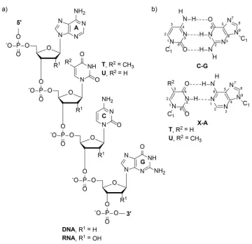
Figure 1.1. Chemical structure of nucleic acids ... 3
Figure 1.2. Comparison of the double helical structures adopted by DNA and RNA ... 5
Figure 1.3. Secondary structures adopted by RNA ... 7
Figure 1.4. Higher-order nucleic acid structures ... 8
Figure 1.5. Examples of important nucleic acid three-way junctions ... 9
Figure 1.6. Conformations of perfectly paired and bulged 3WJs ... 11
Figure 1.7. DNA binding small molecules ... 13
Figure 1.8. Structures of the aminoglycosides ... 20
Figure 1.9. RNA binding small molecules ... 22
Figure 1.10. DNA four-way junction binding small molecules ... 26
Figure 1.11. DNA three-way junction binding small molecules ... 28
Figure 1.12. Comparison of nucleic acid junctions in the present of metallohelicates .... 29
Figure 1.13. Structure of triptycene ... 30
Figure 2.1. Triptycene scaffold for targeting nucleic acid three-way junctions ... 47
Figure 2.2. Thermal stabilization data ... 50
Figure 2.3. Fluorescence quench assay, thermal stability data, and gel shift data ... 52
Figure 2.4. Cytotoxicity and cell-uptake studies using human ovarian carcinoma cell lines ... 53
Figure 2.6a. Solvent gradient for analytical HPLC chromatograms... 62
Figure 2.6b. Analytical HPLC chromatogram of Trip 1 ... 63
xiii
Figure 2.6d. Analytical HPLC chromatogram of Trip 2 ... 64
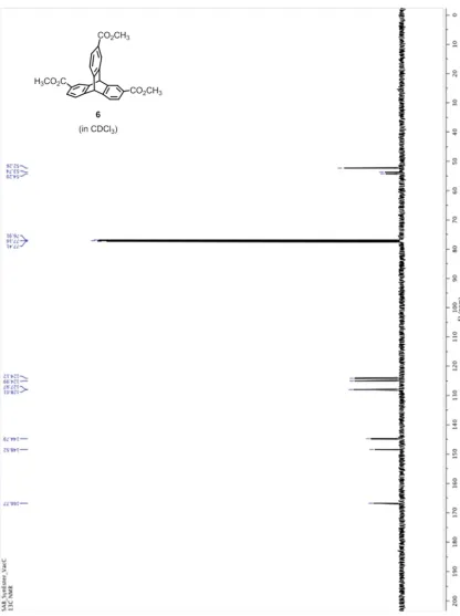
Figure 2.7. 1H NMR of triptycene 6 in CDCl3 ... 65
Figure 2.8. 13C NMR of triptycene 6 in CDCl3 ... 66
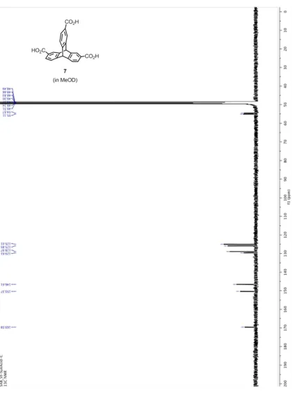
Figure 2.9. 1H NMR of triptycene 7 in MeOD ... 67
Figure 2.10. 13C NMR of triptycene 7 in MeOD ... 68
Figure 2.11. 1H NMR of Trip 1 in D2O ... 69
Figure 2.12. 13C NMR of Trip 1 in D2O ... 70
Figure 2.13.1H NMR of Trip 2 in D2O ... 71
Figure 2.14.13C NMR of Trip 2 in D2O ... 72
Figure 2.15.1H NMR of Trip 3 in D2O ... 73
Figure 2.16. 13C NMR of Trip 3 in D2O ... 74
Figure 2.17. 1H NMR of compound 4 in D2O ... 75
Figure 2.18. 13C NMR of compound 4 in D2O ... 76
Figure 2.19. Design of the DNA 3WJ sequence ... 77
Figure 2.20. UV thermal stabilization data for 1 in 10 mM CacoK, pH 7.2... 78
Figure 2.21. UV thermal stabilization data for 1 at different concentrations in 10 mM CacoK, pH 7.2... 78
Figure 2.22. UV thermal stabilization data for triptycene 2 ... 79
Figure 2.23. UV thermal stabilization data for triptycene 3 ... 80
Figure 2.24. UV thermal stabilization data for control 4 ... 81
xiv
Figure 2.26. Temperature-dependant CD spectra ... 83
Figure 2.27. CD thermal experiment ... 83
Figure 2.28. CD spectra of 20 µM DNA 3WJ2 in 10 mM CacoK, pH 7.2 at 4 °C ... 84
Figure 2.29. UV thermal stabilization data of 3WJ2 in the presence of triptycene 1 ... 84
Figure 2.30. UV thermal stabilization data of an RNA 3WJ in the presence of triptycenes 1, 2, and 3. ... 84
Figure 2.31: Cell uptake studies using MALDI-MS ... 87
Figure 3.1. Slipped DNA junctions formed by (CAG)·(CTG) repeats ... 97
Figure 3.2. Gel shift assay, titration of inhibitor 10 into TNR 3WJ ... 99
Figure 3.3. Gel shift assay, titration of triptycenes into TNR-I10 complex ... 100
Figure 3.4. Fluorescence-quenching assay and circular dichroism (CD) ... 102
Figure 3.5a. Analytical HPLC chromatogram of Trip 2 ... 108
Figure 3.5b. Analytical HPLC chromatogram of Trip 3 ... 109
Figure 3.5c. Analytical HPLC chromatogram of Trip 4 ... 109
Figure 3.6. 1H NMR of Trip 2 in D2O ... 110
Figure 3.7. 1H NMR of Trip 3 in D2O ... 111
Figure 3.8. 1H NMR of Trip 4 in D2O ... 112
Figure 3.9. 13C NMR of Trip 4 in D2O ... 113
Figure 4.1. The heat shock response in E. coli and strategy for small molecule modulation at the mRNA level ... 121
Figure 4.2. Stabilization of a model system RNA by triptycene derivatives 1 and 2 .... 123
xv
Figure 4.4. Modulation of σ32 mRNA (-19 to +229) by triptycene derivatives and
targeting σ32 in E. coli ... 125
Figure 4.5. Temperature-dependant circular dichroism CD of σ32 RNA (-19 to +229) 126 Figure 4.6. SHAPE reactivity data ... 127
Figure 4.7. Comparison of the structure of rpoH (-19 to +227) at 25 °C and the predicted structure from SHAPE experiments performed at 37 °C ... 129
Figure 5.1. Triptycene building blocks for the immobilization strategy ... 139
Figure 5.2. Representative disconnections for triptycene 6d ... 140
Figure 5.3. Synthesis of orthogonally protected building blocks and immobilization on resin ... 142
Figure 5.4. Fluorescence-quenching experiments using TNR junctions ... 144
Figure 5.5. 1H NMR of anthracene 2 in CDCl3 ... 152
Figure 5.6. 13C NMR of anthracene 2 in CDCl3 ... 153
Figure 5.7. 1H NMR of anthracene 3 in CDCl3 ... 154
Figure 5.8. 13C NMR of anthracene 3 in CDCl3 ... 155
Figure 5.9. 1H NMR of triptycene 4 in CDCl3 ... 156
Figure 5.10. 13C NMR of triptycene 4 in CDCl3 ... 157
Figure 5.11. 1H NMR of triptycene 5a in CDCl3 ... 158
Figure 5.12. 13C NMR of triptycene 5a in CDCl3 ... 159
Figure 5.13. HMBC spectrum of 5a in CDCl3 ... 160
Figure 5.14. Expanded HMBC spectrum of 5a in CDCl3 ... 161
xvi
Figure 5.16. Expanded HSQC spectrum of 5a in CDCl3 ... 163
Figure 5.17. 1H NMR of 5b in CDCl3... 164
Figure 5.18. 13C NMR of 5b in CDCl3 ... 165
Figure 5.19. HMBC spectrum of 5b in CDCl3 ... 166
Figure 5.20. Expanded HMBC spectrum of 5b in CDCl3 ... 167
Figure 5.21. HSQC spectrum of 5b in CDCl3... 168
Figure 5.22. Expanded HSQC spectrum of 5b in CDCl3... 169
Figure 5.23. 1H NMR of 5c in CDCl3 ... 170
Figure 5.24. 13C NMR of 5c in CDCl3 ... 171
Figure 5.25. HMBC spectrum of 5c in CDCl3 ... 172
Figure 5.26. Expanded HMBC spectrum of 5c in CDCl3 ... 173
Figure 5.27. HSQC spectrum of 5c in CDCl3 ... 174
Figure 5.28. Expanded HSQC spectrum of 5c in CDCl3 ... 175
Figure 5.29. 1H NMR of 5d in CDCl3... 176
Figure 5.30. 13C NMR of 5d in CDCl3 ... 177
Figure 5.31. HMBC spectrum of 5d in CDCl3 ... 178
Figure 5.32. Expanded HMBC spectrum of 5d in CDCl3 ... 179
Figure 5.33. 1H NMRof 6d in MeOD ... 180
Figure 5.34. 13C NMRof 6d in MeOD ... 181
Figure 5.35. 1H NMRof 7d in DMSO ... 182
Figure 5.36. 13C NMRof 7d in DMSO ... 183
xvii
Figure 5.37b. Analytical HPLC chromatogram of Trip-(Gly-His)3 (9d) ... 185
Figure 5.37c. Analytical HPLC chromatogram of Trip-(His-Lys-His)3 (10d) ... 186
Figure 5.37d. Analytical HPLC chromatogram of Trip-(His-Lys-Lys)3 (11d) ... 186
Figure 5.37e. Analytical HPLC chromatogram of Trip-(His-Lys-Asn)3 (12d) ... 187
Figure 5.38. Fluorescence-quenching experiment plots ... 188
1
2
1.1Background
The central dogma of molecular biology provides the flow of genetic information
within biological systems. In 1958, Francis Crick proposed that the transfer of genetic
information flows from DNA to RNA to protein.1 The DNA sequence for a particular
gene serves as a template for its corresponding mRNA by transcription. The mRNA then
serves as a template for the translation of individual amino acids in a protein. In normal
cells, the transfer of this information is tightly regulated to maintain balanced levels of
each component. Cancer cells and other disease-related processes have aberrant
regulation of the components leading to proliferation.
Classically, medicinal chemistry has focused on the design of small molecules
towards protein targets for potential therapeutic use. Nucleic acids play important roles in
the regulation of gene expression and gene products through the interaction with ligands,
such as proteins, enzymes, metabolites, small molecules, and other nucleic acids. The
ability to target the DNA or RNA encoding a protein involved in a disease state may be a
useful therapeutic. Targeting disease relevant pathways with small molecules at the DNA
and RNA level could lead to new therapeutic strategies and deeper biological insight,
impacting human health.
1.2Nucleic Acid Structure
1.2.1 Deoxyribonucleic acid (DNA) Structure
Deoxyribonucleic acid (DNA) is a double-helical biopolymer consisting of
3
phosphate group. Each monomer is connected through a phosphodiester linkage between
the 2’-deoxyribose rings at the 3’ and 5’ positions. The nitrogenous bases are composed
of purines [adenine (A) and guanine (G)] and pyrimidines [cytosine (C) and thymine (T)],
which provide the building blocks of the genetic code.2 The chemical structure of a
nucleic acid strand is shown in Figure 1.1. Nucleic acids also have two different grooves
due to helix formation. The DNA major groove is wide and shallow while the minor
groove is deep and narrow.
4
The modern history of nucleic acids began in 1951 when Chargaff reported that
different species contained different base compositions; however the ratio of A and T as
well as C and G were very similar.3 This is known as Chargaff’s rule and aided Watson
and Crick in the discovery of the structure of DNA. In 1952, x-ray diffraction data of a
fiber of DNA was obtained by Rosalind Franklin and Maurice Wilkins.4,5 The diffraction
data along with Chargaff’s rule led Watson and Crick to propose the base-paired
double-helical model of B-DNA in 1953.6 The model consists of two right-handed helical strands
oriented in an antiparallel direction with a sugar-phosphate backbone and nitrogenous
bases positioned inward. The bases occupy the core and are stacked and stabilized by
hydrogen bonds formed between two paired bases as well as hydrophobic interactions
(Figure 1.2). Adenine prefers pairing with thymine by two hydrogen bonds and C prefers
G by three hydrogen bonds. In order to maximize hydrophobic interactions, a helical
5
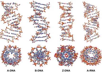
Figure 1.2. Comparison of the double helical structures adopted by DNA and RNA.
The most abundant form of DNA present in nature exists in the B-form, as
Watson and Crick had observed. However, DNA can adopt other helical conformations.
B-form DNA has the ability to undergo a conformational change to A-form DNA when
the relative humidity is reduced to 75%. The right-handed helix in A-DNA is wider and
flatter compared to B-DNA with a deep major groove and very shallow minor groove.7,8
In 1979, Andrew Wang and Alexander Rich observed a left-handed double helix formed
by d(CGCATGCG). This form of Z-DNA is formed in alternating pyrimidine-purine
sequences and has a deep minor groove and no noticeable major groove.9 A structural
6
Table 1.1. Structural features of DNA and RNA.
1.2.2 Ribonucleic acid (RNA) Structure
RNA differs from DNA structurally in two ways: the presence of a hydroxyl
group at the 2’ position of the sugar ring and the absence of a methyl group at the 5
position of thymidine resulting in uracil (Figure 1.1). Duplex RNA is predominantly
found in the A-form (Figure 1.2). However, due to the 2’-hydroxyl and that RNA is
single stranded and folds on itself to increase its stability, various, non-canonical,
complex three-dimensional structures can be formed. These include bulges and loops of
7
and three- and four-way junctions (Figure 1.3). The 2’-hydroxyl also presents another site
for hydrogen bonding with water or other regions of the RNA leading to these structures.
Figure 1.3. Secondary structures adopted by RNA.
1.2.3 Higher-Order Structures
Nucleic acids can adopt several other higher order structures such as triplexes,
G-quadruplexes, and junctions (Figure 1.4). Triplex structures form in homopyrimidine or
homopurine sequences in both DNA and RNA at specific salt concentrations and pH. The
third strand in a triple helix binds in the major groove of the duplex forming Hoogsteen
hydrogren bonds.10,11 G-quadruplexes have the propensity to form in guanine-rich
8
same strand (Figure 1.4a). Guanine tetrads are formed when four guanine bases
Hoogsteen hydrogen bond to form a square planar structure. These tetrads can stack on
one another forming a G-quadruplex, where strands can be parallel or antiparallel. These
structures are polymorphic and adopt structures dependent on concentration, sequence,
and cations.12,13
Figure 1.4. Higher-order nucleic acid structures. (a) Structure of a G-tetrad and an intramolecular antiparallel G-quadruplex. (b) Schematic and crystal structure of a four-way junction (PDB: 1DCW). (c) Schematic and crystal structure of a three-way junction (PDB: 1F44).
DNA junctions serve as important intermediates in several biological processes.
Four-way, or Holliday junctions, are present during homologous recombination, important for
double-strand break repair and in imparting genetic diversity (Figure 1.4b).14–20 Of
particular interest is the three-way junction (3WJ), which is formed when three
9
(Figure 1.4c). Nucleic acid 3WJs are very diverse existing in several different
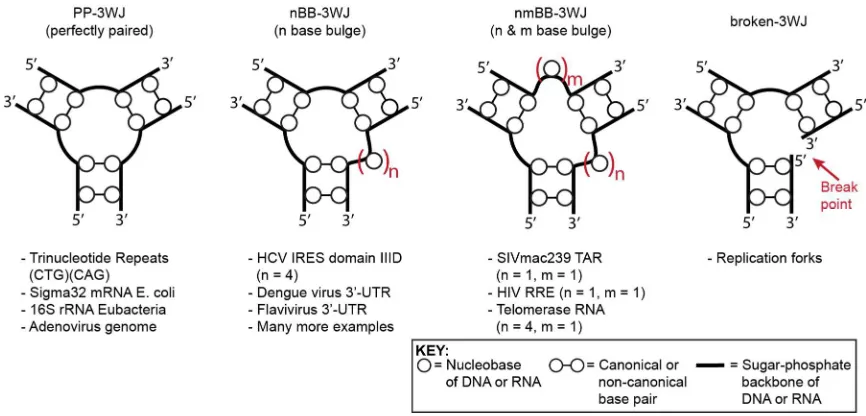
conformations with varying numbers of unpaired bases at the center (Figure 1.5).
Perfectly-paired 3WJs (3H) have been shown to adopt fully extended conformations
(Figure 1.6a) under a variety of conditions.21 This differs from what has been observed
for four-way junctions (4WJs) which form coaxial stacked conformations and ion
dependant folding.22 Nucleic acid 3WJs are ubiquitous structures found throughout
biological systems (Figure 1.5). The conformations of these junctions have been
well-studied.
10
1.3Nucleic Acid Junctions
1.3.1 DNA Three-Way Junctions
DNA three-way junctions occur in replication and recombination and are also
present in viral genomes and polymorphic triplet repeat expansions which are implicated
in neurodegenerative disease.23–27 DNA 3WJs are diverse, existing as perfectly paired
(3H) or with unpaired bases at one (3HSn) or more positions (2HSn HSm or HSn HSm HSp)
at the center of the junction. Using gel electrophoresis, Duckett and Lilley have shown
that perfectly paired (3H) junctions exist in a fully extended conformation where the
three angles between the arms are similar (Figure 1.6a).28 This was later confirmed by
Stuhmeier using FRET experiments, where the three distances measured from one end to
another of the junction were very similar.29 Shlyakhtenko et al. have carried out
cyclization experiments and demonstrated that the inter-helical angles ranged between 60
and 90°.30 Images of perfectly paired 3WJs using atomic force microscopy (AFM) have
also shown a large compositions of symmetric molecules.31 Additionally, thymine bases
at the junction are reactive towards osmium tetroxide and ethyl pyrocarbonate in the
presence and absence of metal cations. This further demonstrates that reactivity with
these reagents is not precluded by helix-helix stacking and that the junction is folded into
a more extended Y shape even in the presence of metal cations.28,32 Taken together, these
experiments demonstrate that there is no coaxial stacking of the helical regions in a
perfectly paired 3WJ. Some 3H junctions, however, have an asymmetric structure
11
Figure 1.6. Conformations of perfectly paired and bulged 3WJs. (a) A perfectly paired three-way junction adopting an extended structure. (b) A bulged 3WJ (3HS2) in the extended conformation
undergoes a conformational change where coaxial stacking is observed in the presence of magnesium cations. The angle of the third helix is dependent on the number of unpaired bases. Two stacking conformers can be formed.
Due to the unpaired, single-stranded region between the helical arms, 3HSn
junctions have a higher degree of flexibility compared to 3H junctions. Gel
electrophoresis studies by Leontis and coworkers, showed that these bulged junctions
with unpaired nucleotides at the interface form more stable structures.35 Lilley also
demonstrated that junctions containing unpaired nucleotides adopt a different
conformation in the presence of metal ions. In the absence of metal ions, the junctions
assumed an extended conformation in which the inter-helical angles were similar.
However, in the presence of metal ions, including magnesium and other multivalent ions,
the junctions underwent an ion-dependent folding transition. Thymine bases at the
junction were protected against OsO4 reactivity in these folded species. These results
were consistent with coaxial stacking of two helical arms in the presence of metal ions
(Figure 1.6b). The angle of the third arm is dependent on the number of unpaired bases.36
FRET studies later confirmed these observations. Bulge containing 3WJs were found to
be asymmetrical, where one inter-helical angle was significantly smaller than the other
Time-12
resolved FRET also demonstrated that the structure and flexibility of 3WJs are dependent
on the number of unpaired bases as well as the base composition of the bulge.37
Cyclization rates of DNA 3WJs containing unpaired bases were slower than those that
were perfectly paired.30 NMR studies of 3HS2 DNA junctions in the presence of
magnesium ions, revealed that two helical arms are coaxially stacked whereas the third
helix is extended away from the junction.38–41 Two stacking conformers can exist in these
junctions, depending on the stacking partner. The existence and stability of these
conformers is dependent on the sequence.42
1.3.2 RNA Three-Way Junctions
DNA junctions are important intermediates in biological processes. However,
RNA junctions occur much more frequently and are often important structural elements.
They occur in 5S rRNA, 16S rRNA, the hammerhead ribozyme, the group I intron
ribozyme, viral genomes, bacterial temperature sensors, and telomerase RNA.43–53
Although the global structure of RNA 3WJs has not been studied as thoroughly as DNA
junctions, it is thought that the same folding principles from DNA can be applied to
RNA.54
There are few examples of perfectly paired (3H) RNA 3WJs in nature. Folding of
these junctions is difficult while maintaining complete base pairing. This strain is
released by the addition of unpaired bases at the center of the junction to increase
conformational flexibility. The structure of several RNA 3WJs have been analyzed to
gain insight into their folding properties. The 2HS3HS2 junction in the 16S rRNA is the
13
junction adopts an extended conformation where no coaxial stacking is observed.
However, upon addition of magnesium, coaxial stacking was observed between two
arms.44–46 Another example of an RNA 3WJ is the hammerhead ribozyme containing an
HS1HS7HS3 junction. Coaxial stacking is observed between two stems in crystal
structures solved.55,56 Other structural studies, including FRET, gel electrophoresis, and
transient electric birefringence agree well with crystal structures.44,45,57–63 As with other
junctions, folding is observed upon addition of ions, including magnesium.
Figure 1.7. DNA binding small molecules.
Lescoute and Westhof have analyzed structures containing RNA 3WJs in order to
14
families depending on the length of the unpaired segments between the helices. These can
adopt different conformers just as is observed in DNA junctions.
1.4Nucleic Acid Recognition
1.4.1 Modes of DNA Recognition
DNA is an attractive target for the development of therapeutics. Mutations in
DNA sequences may lead to defects in protein function and misregulation leading to
increases or decreases in cellular processes, which may lead to disease. Many of the
chemotherapeutic drugs used today are DNA binding agents that cause damage and
inhibit cellular processes such as replication. These natural products and synthetic small
molecules generally bind to DNA by few traditional modes of binding.65,66 These include
intercalation, minor or major groove binding, covalent binding, and interactions with the
phosphate backbone. Some small molecules rely on more than one binding mode to
enhance their effectiveness. Examples of DNA binding small molecules are shown in
Figure 1.7.
1.4.1.1 Intercalation
DNA intercalation was first discovered in 1961 when Lerman was studying the
DNA binding properties of proflavine, a planar, aromatic chromophore.67 Intercalation is
defined as the full or partial insertion of planar aromatic rings between DNA base pairs in
either the major or minor groove. The interaction between the DNA and intercalator is
15
phosphate backbone. The rigidity of the backbone of DNA does not allow for
intercalation at every base pair. According to the neighbor-exclusion principle, a ligand
can insert at every other base pair.68 Upon intercalation the DNA helix is partially
unwound and causes elongation of the helix, which is thought to prevent DNA replication
and transcription resulting in cell death. Several intercalators have been discovered with
some examples shown in Figure 1.7. Several well-studied compounds such as acridines,
anthraquinones, and ethidium bromide are routinely used in molecular biology for the
staining of DNA. Natural products currently used in the clinic as anticancer drugs include
actinomycin D and anthracyclines such as doxorubicin. Actinomycin D was isolated in
1940 and was the first antibiotic to display activity against cancer cells by inhibiting
transcription.69,70 Doxorubicin was isolated in the 1950s and exhibits its anti-cancer
activity by intercalating and inhibiting topoisomerase II, thereby preventing relaxation of
DNA for transcription.71,72 Some of these molecules have the ability to recognize specific
base pair steps, however this still results in non-specific binding to several DNA
structures and sequences.
1.4.1.2 Covalent
Several alkylating agents have also been developed which covalently bind to
DNA. Chlorambucil, a nitrogen mustard, covalently modifies DNA by alkylation at the
N7 of G or A.73 Cisplatin is a very well-known platinum-based drug that covalently binds
to DNA and is used in the treatment of cancer. As in the case with chlorambucil, cisplatin
reacts with the N7 of neighboring purines causing a kink in the DNA.74 Other cisplatin
16
drugs display many side effects due to their non-specificity for cancer cells compared to
normal cells.
1.4.1.3 Groove Binding
Minor groove binders have similar structural features including positive charges
and aromatic or heteroaromatic rings that form a crescent shape.75 The natural product
distamycin A binds to the minor groove of DNA with a preference for A,T-rich
sequences (at least four base pairs of AT). The molecule binds in a 1:1 or 2:1 ratio of
ligand to DNA.76–78 Distamycin’s specificity and affinity is achieved through hydrogen
bonding in the minor groove, hydrophobic and electrostatic interactions, and shape
complementarity. Distamycin has been utilized as an inspiration for the design of minor
groove binders including polyamides developed by Dervan and coworkers in which a
combination of pyrrole, imidazole, and hydroxypyrrole can recognize specific DNA
sequences.79 Several natural products have also been found to interact with the minor
groove. Anthramycin, part of the pyrrolobenzodiazepine class of natural products, was
discovered in 1963. Several groups found that these molecules bound to DNA but not
RNA and that a covalent bond between the ligand and DNA is formed. 80,81 Other minor
groove binders include diarylamides, such as DAPI and pentamidine, and
bis-benzimidazoles, such as Hoechst 33258. These molecules also bind to A,T rich sequences
similar to distamycin A.
Due to the larger width and different hydrogen bonding pattern of the major
groove of DNA, this region is often targeted by proteins. Helix-turn-helix, zinc-binding,
17
oligonucleotides also have the ability to recognize the DNA major groove. These form a
triple helix by Hoogsteen base pairing. Peptide nucleic acids (PNAs), in which the sugar
phosphate backbone is replaced with a polyamide, have been utilized for binding as well.
The decrease in negative charge allows for enhanced binding to the DNA compared to
another DNA strand.83 Small molecule major groove binders typically exhibit other forms
of binding such as minor groove binding or intercalation. Nogalamycin is one such
example where major groove binding is driven by intercalation.
1.4.2 Targeting RNA with Small Molecules
RNA was initially considered the messenger between genetic information stored
in the form of DNA and proteins. However, with the advent of genomic high-throughput
techniques such as next-generation sequencing, sequencing of the human transcriptome
has allowed for the detection of thousands of noncoding RNA molecules with various
functions that are critical in living organisms. These include ribosomal RNA (rRNA) and
transfer RNA (tRNA) which play crucial roles in protein synthesis, small nuclear RNAs
(snRNAs) involved in splicing, telomerase-associated RNAs such as TERRA and TERC,
as well as smaller RNAs including microRNA (miRNA) that participate in RNA
interference. RNA is also a key component in the progression of infectious disease such
as HIV and hepatitis C, genetic disease such as Huntington’s or mytonic dystrophy
characterized by triplet repeat expansions, and metabolic disease including cancer.84–86
These noncoding RNAs and disease causing RNA motifs may provide new drug targets
for small molecules. Riboswitches, regulatory regions within an mRNA that bind a small
18
recently by a group at Merck.87 The druggability of RNA is often challenged; however
one may argue that it has been demonstrated by clinically approved antibiotics which
bind to ribosomal RNA. The targeting of a particular RNA would allow for the control
of specific cellular processes.
A plethora of natural products have been found to bind to DNA, however fewer
small molecules are known to bind to RNA. The major groove of A-form RNA is deeper
and narrower than that of B-DNA and the minor groove is shallower making it more
difficult to target. Due to RNA’s ability to form complex, three-dimensional structures
(Figure 1.3) similar to proteins, defined pockets are formed which have the ability to
interact with other nucleic acids, proteins, and small molecules. These structurally diverse
binding pockets provide regions for specifically targeting RNA by small molecules.
The aminoglycosides have been well-studied and used in understanding small
molecule-RNA binding interactions. (Figure 1.8) This class of molecules binds to cavities
or pockets in RNA formed from loops, bulges, etc; therefore they have the ability to
compete with RNA-RNA and RNA-protein interactions. However, they are promiscuous
binders and will bind to several RNA targets. Nevertheless, studies of
aminoglycoside-RNA interactions provide a platform for the design of new small molecules and are still
the subject of intense research efforts.
Electrostatic interactions are important for RNA binding. Generally, molecules
that interact with nucleic acids contain positive charges which favorably interact with the
negatively charged biomolecule, increasing the binding affinity. The number of amines
19
of the amines can also be tuned, thereby changing the overall charge. It has been
determined that half of the total binding energy in a number of aminoglycoside-RNA
interactions is due to electrostatics. Although these interactions are responsible for high
affinity, they often lead to nonspecific binding.
Nonionic interactions also contribute to RNA binding. Favorable interactions
between hydrophobic regions of small molecules and the bases occur, including
hydrophobic and π-stacking interactions. Hydrogen bonding between molecules and the
phosphate backbone or nucleobases contribute significantly to binding as well. This has
also been observed with the aminoglycosides, in which the amine and hydroxyl groups
act as hydrogen bond donors to the oxygen of the phosphate backbone.89,90 Different
substitution patterns on small molecules may have distinct interactions with RNA leading
to enhanced binding affinity.
Shape complementarity and conformational adaptation also play significant roles
in small molecule binding to RNA. Shape complementarity is defined as how well a
molecule and receptor fit both sterically and electrostatically. As discussed previously,
RNA adopts folds that lead to pockets available for binding. These pockets all differ in
size and shape as well as electronegativity. This allows for the development of small
molecules that interact with the RNA with high affinity and specificity. This is observed
in small molecule interactions with RNA aptamers.91,92 Comformational adaptation is
defined as conformational changes of the ligand or receptor that occur upon binding. Due
to the flexibility of RNA, rearrangements can occur upon ligand binding. This may lead
20
conformationally constrained aminoglycoside to selectively bind the 16S A site over
TAR RNA. They found that no selectivity was achieved which may be due to the ligand
adopting a different binding orientation.93
Figure 1.8. Structures of the aminoglycosides.
1.4.2.1 Aminoglycosides
The various antibiotics, including aminoglycosides94, tetracyclines95, and
oxazolidinones96, that target the ribosome, affecting protein synthesis, are some of the
best known cases of RNA binders. The aminoglycoside antibiotics contain a large
number of positive charges and bind to the A-site on the 16S rRNA of bacterial
21
open state which in turn allows for the recognition of noncognate tRNA resulting in
incorporation of the incorrect amino acid leading to cell death.97,98
Aminoglycosides also bind to nonribosomal RNA. They have been found to bind
to tRNA causing a conformational change by displacing a Mg2+ ion and inhibiting amino
acylation of the tRNA.99,100 Additionally aminoglycosides bind to ribozymes,101,102
ribonuclease P,103,104 and viral RNAs. In HIV, transcription of viral DNA is aided by the
trans-activator of transcription (Tat) protein. Tat recognizes a bulged RNA hairpin loop
known as the trans-activating response (TAR) element present in the beginning of the
transcript mediated by an arginine-rich area.105–107 Neomycin has been shown to be a
potent inhibitor of the interaction.108 Triazole-linked neomycin dimers consisting of
different length linkers were developed displaying increased affinity and specificity.109
The Rev protein in HIV-1 exports viral RNAs from the nucleus of the host to the
cytoplasm by binding to the Rev response element (RRE), a stem-loop RNA.105,110 To
achieve higher affinity and specificity towards RRE, Tor and coworkers have replaced
the amino groups with guanidinium groups. These ligands displayed a 10-fold increase in
potency at the inhibition of Rev-RRE compared to the unmodified aminoglycoside.
Linezolid is the first oxazolidinone to reach the clinic in 2000. Its mechanism of
action is not completely understood but is thought to act by binding to the 23S rRNA in
the 50S subunit near the binding site of other antibiotics including chloramphenicol and
preventing the formation of the initiation complex.96,111 The structural information from
22 1.4.2.2 Bulge Binding
Bulge regions occur in RNA which contain one or more unpaired bases on one
duplex strand. These can exist as single base bulges, containing one unpaired base, or
multiple base bulges, in which there is more than one unpaired base. Bulge regions within
a duplex have a destabilizing effect proportional to the number of unpaired bases.112,113
TAR RNA contains a bulge region that has been targeted with small molecules frequently
(Figure 1.9a).
Figure 1.9. RNA binding small molecules. (a) Small molecules that bind to the bulge region of TAR RNA. (b) Internal loop binders that bind to RRE RNA. (c) Small molecules targeting nucleotide repeat expansion.
Several DNA binding molecules including ethidium bromide, berenil, and
23
Hoechst 33285 prefers binding to the bulge region of TAR, demonstrating a higher
affinity for the native structure compared to a bulgeless structure.116 Modular ligands
have also been developed composed of a polyaromatic structure for intercalation, a
positively charged region to make contacts with the phosphate backbone, and a linker
connecting the two. One example of these compounds is compound 1 shown Figure 1.8,
consisting of a 6-chloro-2-methoxy acridine linked to spermidine.117 This molecule binds
to the bulge region of TAR as demonstrated by RNase footprinting and NMR.118 Other
conjugated molecules bind to TAR such as ethidium-arginine conjugates which
researchers have designed to bind to two sites on the RNA.119 Compound 2 has been
shown to bind to TAR with a Ki of 51 nM. The indole ring intercalates between two base
pairs while the amines make contacts with the backbone. All of these compounds have
used intercalation and electrostatic interactions as binding modes. Neocarzinostatin, 3, is
composed of two ring systems connected by a spirocycle causing a twist in the molecule
and stacking of the aromatic moieties. This compound was found to bind to various RNA
bulges including TAR.120
1.4.2.3 Internal Loop Binding
Internal loops occur in regions containing noncomplementary bases. These may
exist as symmetric loops in which the number of unpaired bases on either side of the
duplex is the same or unsymmetric when the number of unpaired bases is different. A
well studied target for internal loop binders is the Rev response element (RRE) (Figure
24
In a gel competition assay, several diphenylfuran derivatives were screened for
the disruption of the Rev-RRE interaction.121 Structure activity relationships (SAR)
studies were performed to determine the moieties required for binding. From this data
compound 4 showed the most potency with an IC50 = 0.3 μM. NMR studies demonstrated
that these ligands bind to the internal loop of the RNA as a dimer through the minor
groove.122 Marino and coworkers have evaluated several acridine-like molecules towards
the Rev-RRE interaction. Using a 2-aminopurine assay, they found that acridine orange
and proflavine had the highest affinity.123 These molecules have led to tight binders but
are not selective. To achieve selectivity, work has been done to recognize loop regions
with intercalators displaying the proper hydrogen bonding pattern for the mismatched
base. Compound 5 was developed to bind to guanine bases and azaquinoline 6 hydrogen
bonds with adenine mismatches.124,125 Dimers of these molecules have been used to target
sequence specific mismatches.
1.4.2.4 Hairpin Loops
Hairpin loops form when a sequence folds on itself into a duplex connected by
single stranded bases. This structural motif is one of the most predominant found in
RNA. In a gel shift assay, compound 7 was identified as a TAR RNA binder (Figure
1.9a). RNase footprinting experiments and ESI-MS confirmed loop binding to the
25 1.4.2.5 Targeting Expanded Repeats
Nucleotide repeat expansions have become targets for the design of small
molecules, since these have been associated with several diseases including Fragil X
Syndrome (SFX)128, Huntington’s disease129, and mytonic dystrophy type I (DM1)130 and
type II (DM2).131 These repeats [(CTG) or (CCTG)] form a hairpin structure and bind to
muscleblind-like 1 protein (MBNL1), a regulator of splicing, causing disregulation of
alternative splicing pre-mRNAs.132 Development of high affinity small molecules may
displace MBNL1, restoring its function (Figure 1.9c). Zimmerman and coworkers have
designed a molecule consisting of an acridine DNA intercalator and a triaminotriazine,
also a known binder. This compound, 8, does not discriminate between d(CTG) and
r(CUG) repeats, but shows some selectivity towards U-U mismatches.133 Pentamidine has
also been shown to disrupt the MBNL1 binding to r(CUG) repeats.134 Disney has
designed compounds in which an alkynyl kanamycin A is assembled on a peptoid
backbone, 9.135 Another series of these consist of Hoechst 33258, 10.136 This strategy
uses compounds previously shown to bind to nucleic acids which may lead to
nonspecificity.
1.5Recognition of Nucleic Acid Junctions
1.5.1 Four-Way Junctions
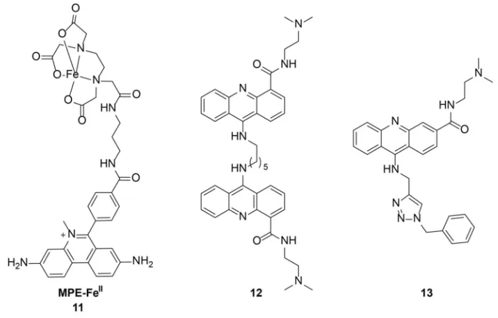
Work on targeting 4WJs had began in the late 1980s by Kallenbach when they
discovered that methidiumpropyl-EDTA-FeII [MPE-FeII], 11, could cleave a Holliday
26
bases from the branch point. In 2000, Segall and coworkers identified a peptide with the
sequence WKHYNY that stabilized 4WJs with protein bound. This peptide led to the
accumulation of 4WJs but did not inhibit recombination.137 A second generation peptide
with the sequence WRWYCR increased the potency by 50 times and acted as a dimer by
disulfide formation.138 Based on this dimer, several cyclic peptides were also tested.139
Searcey, Cardin, and coworkers have targeted 4WJs using bisintercalators with long
linkers to reach across the junction. Upon crystallization of C6-linked
bisacridine-4-carboxamide, 12, revealed that the acridine displaces an adenine forming a pseudo-base
pair.140 Recently, Searcey and coworkers have identified a compound, 13, that induces
the formation of a 4WJ (Figure 1.10).141
27
1.5.2 Three-Way Junctions
Hannon, Coll, and coworkers crystallized a metal complex binding to a DNA
three-way junction, revealing an alternative mode of binding.142 Hannon and coworkers
have worked on metallosupramolecular chemistry designing compounds in which three
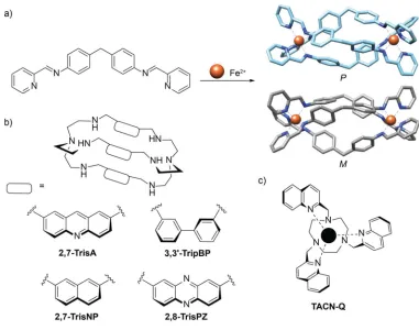
bis-pyridylimine ligands wrap around one another in the presence of two Fe2+ ions
forming two enantiomers, M and P (Figure 1.11a). These ligands have been shown to
bind to the major groove of DNA causing intramolecular coiling.143,144 Crystallization to
2.6 Å of the metal complex, [Fe2(C25H20N4)3]Cl4, with a palindromic sequence of DNA
resulted in the formation of a three-way junction with the complex in the center (Figure
1.12a). Crystals were only obtained with sequences composed of A-T at the center of the
junction. Interstingly, the M enantiomer was only observed in the crystal. The
predominant interactions observed were π-stacking between the central phenylene ring
and the bases at the junction as well as van der Waals interactions between the end
pyridine rings and the C5’ sugar. These studies were followed up by NMR145 and
analysis by gel electorphoresis,146 confirming the ability of the ligand to stabilize
different 3WJ sequences. Conjugation of arginine to the ends of the metallohelicates,
increased 3WJ stabilization and showed increased activity in cancer cells.147 However,
these ligands have also been shown to bind to G-quadruplex DNA as well as double
helical DNA.148,149
Monchaud and coworkers have used cationic azacryptands to stabilize DNA 3WJs
(Figure 1.11b). Several azacryptands were synthesized and tested in a FRET melting
28
previously studied small molecule, TACN-Q (Figure 1.11c), which had shown some
selectivity towards 3WJs.150 All compounds reported had the ability to stabilize the
junction to varying degrees. FRET melting experiments were also performed on duplex
DNA showing less stabilization compared to 3WJ DNA. Direct melting experiments in
the presence of G-quadruplex forming DNA, resulted in similar stabilization compared to
3WJ DNA.151 This is consistent with previous studies on these molecules that have
shown they interact with G-quadruplexes.152
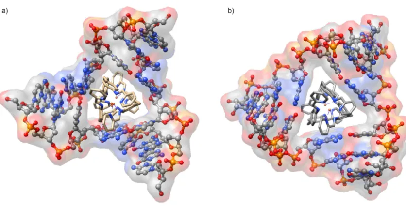
Figure 1.11. DNA three-way junction binding small molecules. (a) Assembly of Fe(II) triple helicate, with M- and P-enantiomers shown. (b) Azacryptands used in studies carried out by Monchaud and coworkers. (c) Chemical structure of TACN-Q.
Hannon, Coll, and coworkers have also crystallized the diiron(II) supramolecular
29
expected, the cylinder sits in the center of the junction and only the M enantiomer is
observed. The cylinder also makes contacts at the termini of the arms forming π
interactions between the bases and the phenyl rings.
Several differences were observed between the DNA and RNA 3WJs (Figure
1.12). The DNA junction was found to be cone shaped, where one face of the junction is
narrower than the other. The two faces of the RNA junction are more similar. Due to the
absence of a narrow opening, the cylinder may move more freely to maximize π-stacking
interactions thereby maximizing stabilization. Gel electrophoresis demonstrated that the
cylinders have the ability to stabilize RNA junctions in solution.153 Interestingly, the M
and P enantiomers show no difference in binding to the RNA, contrary to what is
observed with DNA.146
30
1.6Triptycene
The first reported synthesis of triptycene was by Bartlett and co-workers in
1942.154 This synthesis began with anthracene and p-benzoquinone to yield triptycene in
6 steps. Wittig and coworkers reported the synthesis of triptycene by a Diels-Alder
reaction using benzyne and anthracene in 1956.155 Standard routes have been employed
to access various substitution patterns.156–161 Triptycene belongs to a class of compounds
called iptycenes composed of phenyl rings fused together by a bicycle[2.2.2]octane
core.162 Triptycene is the simplest member of this family of compounds, consisting of
three phenyl rings attached through the bicyclic core.
Figure 1.13. Structure of triptycene.
The unique structure and electro and photochemical properties of triptycene have
led to its use in a variety of supramolecular and material applications. Triptycene-based
molecules have been used as ligands for catalysis, as molecular machines, host-guest
chemistry, gas adsorption, liquid crystals, building blocks in polymers, and many other
materials chemistry applications.156–158,163–177 However, triptycene has seldom been used
in biological applications and biomolecular recognition. Analogs have been shown
31
1.7Overview of Thesis
The design of DNA and RNA small molecule binders has focused on the
modification of natural products including the aminoglycosides or the conjugation to
known binding moieties such as intercalators which often lead to nonspecific binding.
The ability to target particular nucleic acids in a structure or sequence-dependent manner
remains a significant challenge.
As discussed previously nucleic acid junctions are important structural motifs
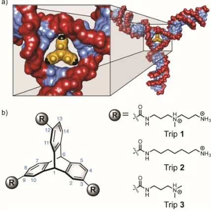
found in nature. We were interested in designing a small molecule that would be selective
towards three-way junctions. Previous 3WJ binders show modest, if any, selectivity
towards junctions. Additionally many of these molecules require the use of metals. We
proposed that triptycene provided a scaffold that was 3-fold symmetric with similar
dimensions to the junction interface. Additionally, triptycene lacks an extended, planar
π-surface area, which will avoid intercalation, and is amenable to diversification in which
two faces of the molecule can be modified to introduce specificity between junctions.
This targeting approach is based on shape complementarity.
Triptycenes were synthesized and the hypothesis was tested using an
oligonucleotide that formed a 3WJ. These studies demonstrated the triptycene’s ability to
discriminate DNA 3WJs from double-helical DNA. After establishing this, triptycenes
were evaluated for binding to d(CAG)·(CTG) trinucleotide repeats implicated in
neurodegenerative disease. Our studies show that these molecules have the ability to
modulate the structure of these junctions, which could lead to valuable probes in studying
32
3WJs. The mRNA from the rpoH gene in E. coli, involved in the heat shock response,
contains a perfectly paired 3WJ at the center, essential for its function. Our studies have
shown that these molecules can target this piece of RNA and show a decrease in the heat
shock response in bacteria. Work has also been done to synthesize bridge-head
substituted triptycenes as building blocks for solid phase synthesis for the generation of
large libraries.
1.8 Bibliography
1. Crick, F. Nature1970, 227, 561–563.
2. Bloomfield, V. A.; Crothers, D. M.; Tinoco, I. Nucleic Acids: Structure, Properties and Functions; University Science Books: Sausalito, CA, 2000.
3. Zamenhof, Stephen; Brawerman, George; Chargaff, E. Biochim. Biophys. Acta
1952, 9, 402–405.
4. Franklin, R. E.; Gosling, R. G. Nature1953, 171, 740–741.
5. Wilkins, M. H. F.; Stokes, A. R.; Wilson, H. R. Nature1953, 171, 738–740.
6. Crick, F.; Watson, J. Nature1953, 171, 737–738.
7. Shakked, Z.; Rabinovich, D.; Kennard, O.; Cruse, W. B.; Salisbury, S. A.; Viswamitra, M. A. J. Mol. Biol.1983, 166, 183–201.
8. Franklin, R. E.; Gosling, R. G. Acta Crystallogr.1953, 6, 673–677.
9. Wang, A. H. J.; Quigley, G. J.; Kolpak, F. J.; Crawford, J. L.; vanBoom, J. H.; vanderMarel G.; Rich, A. Nature1979, 282, 680–686.
10. Jain, A.; Wang, G.; Vasquez, K. M. Biochemie2008, 90, 1117–1130.
11. Frank-Kamenetskii, M. D.; Mirkin, S. M. Annu. Rev. Biochem.1995, 64, 65–95.
12. Agarwala, P.; Pandey, S.; Maiti, S. Org. Biomol. Chem.2015, 13, 5570–5585.
33 14. Holliday, R. Genet. Res.1964, 5, 282–304.
15. Broker, T. R.; Lehman, I. R. J. Mol. Biol.1971, 60, 131–149.
16. Sigal, N.; Alberts, B. J. Mol. Biol.1972, 71, 789–793.
17. Potter, H.; Dressler, D. Proc. Natl. Acad. Sci. U. S. A.1976, 73, 3000–3004.
18. Potter, H.; Dressler, D. Proc. Natl. Acad. Sci. U. S. A.1978, 75, 3698–3702.
19. Orr-Weaver, T. L.; Szostak, J. W.; Rothstein, R. J. Proc. Natl. Acad. Sci. U. S. A.
1981, 78, 6354–6358.
20. Schwacha, A.; Kleckner, N. Cell1995, 83, 783–791.
21. Im, K.; Jeong, D.; Hur, J.; Kim, S.-J.; Hwang, S.; Jin, K. S.; Park, N.; Kim, K. Sci. Rep.2013, 3, 3226.
22. Duckett, D. R.; Murchie, A. I. H.; Clegg, R. M.; Zechel, A.; von Kitzing, E.; Diekmann, S.; Lilley, D. M. J. Struct. Methods Hum. Genome Initiat. DNA Recomb.1990, 1, 157–181.
23. Singleton, M. R.; Scaife, S.; Wigley, D. B. Cell2015, 107, 79–89.
24. Woods, K. C.; Martin, S. S.; Chu, V. C.; Baldwin, E. P. J. Mol. Biol.2001, 313, 49–69.
25. Mirkin, S. M. Nature2007, 447, 932–940.
26. Liu, G.; Chen, X.; Bissler, J. J.; Sinden, R. R.; Leffak, M. Nat. Chem. Biol.2010, 6, 652–659.
27. Slean, M. M.; Reddy, K.; Wu, B.; Nichol Edamura, K.; Kekis, M.; Nelissen, F. H. T.; Aspers, R. L. E. G.; Tessari, M.; Schärer, O. D.; Wijmenga, S. S.; Pearson, C. E. Biochemistry2013, 52, 773–785.
28. Duckett, D. R.; Lilley, D. M. J. EMBO J1990, 9, 1659–1664.
29. Stühmeier, F.; Welch, J. B.; Murchie, a. I. H.; Lilley, D. M. J.; Clegg, R. M. Biochemistry1997, 36, 13530–13538.
34 1189.
31. Oussatcheva, E. A.; Shlyakhtenko, L. S.; Glass, R.; Sinden, R. R.; Lyubchenko, Y. L.; Potaman, V. N. J. Mol. Biol.1999, 292, 75–86.
32. Shlyakhtenko, L. S.; Appella, E.; Harrington, R. E.; Kutyavin, I.; Lyubchenko, Y. L. J. Biomol. Struct. Dynam.1994, 12, 131–143.
33. Guo, Q.; Lu, M.; Churchill, M. E.; Tullius, T. D.; Kallenbach, N. R. Biochemistry
1990, 29, 10927–10934.
34. Lu, M.; Guo, Q.; Kallenbach, N. R. Biochemistry1991, 30, 5815–5820.
35. Leontis, N. B.; Kwok, W.; Newman, J. S. Nucleic Acids Res.1991, 19, 759–766.
36. Welch, J. B.; Duckett, D. R.; Lilley, D. M. Nucleic Acids Res. 1993, 21, 4548– 4555.
37. Yang, M.; Millar, D. P. Biochemistry1996, 35, 7959–7967.
38. Leontis, N. B.; Hills, M. T.; Piotto, M.; Malhotra, A.; Nussbaum, J.; Gorenstein, D. G. J. Biomol. Struct. Dynam.1993, 11, 215–223.
39. Rosen, M. A.; Patel, D. J. Biochemistry1993, 32, 6576–6587.
40. Ouporov, I. V; Leontis, N. B. Biophys. J.1995, 68, 266–274.
41. Overmars, F. J.; Pikkemaat, J. A.; van den Elst, H.; van Boom, J. H.; Altona, C. J. Mol. Biol.1996, 255, 702–713.
42. Welch, J. B.; Walter, F.; Lilley, D. M. J. Mol. Biol.1995, 251, 507–519.
43. Shen, Zhu; Hagerman, Paul, J. J. Mol. Biol.1994, 241, 415–430.
44. Batey, R. T.; Williamson, J. R. J. Mol. Biol.1996, 261, 536–549.
45. Serganov, A. A.; Masquida, B.; Westhof, E.; Cachia, C.; Portier, C.; Garber, M.; Ehresmann, B.; Ehresmann, C. RNA1996, 2, 1124–1138.
46. Orr, J. W.; Hagerman, P. J.; Williamson, J. R. J. Mol. Biol.1998, 275, 453–464.
47. Forster, A. C.; Symons, R. H. Cell1987, 49, 211–220.
35
Cech, T. R.; Doudna, J. A. Science1996, 273, 1678–1685.
49. Watts, J. M.; Dang, K. K.; Gorelick, R. J.; Leonard, C. W.; Bess Jr, J. W.; Swanstrom, R.; Burch, C. L.; Weeks, K. M. Nature2009, 460, 711–716.
50. Mauger, D. M.; Golden, M.; Yamane, D.; Williford, S.; Lemon, S. M.; Martin, D. P.; Weeks, K. M. Proc. Natl. Acad. Sci.2015, 112, 3692-3697.
51. Ouellet, J.; Melcher, S.; Iqbal, A.; Ding, Y.; Lilley, D. M. J. RNA2010, 16, 1597– 1609.
52. Kortmann, J.; Narberhaus, F. Nat. Rev. Microbiol.2012, 10, 255–265.
53. Brown, Y.; Abraham, M.; Pearl, S.; Kabaha, M. M.; Elboher, E.; Tzfati, Y. Nucleic Acids Res.2007, 35, 6280–6289.
54. Lilley, D. M. J. Biopolymers1998, 48, 101–112.
55. Pley, Heinz W.; Flaherty, Kevin, M.; McKay, D. B. Nature1994, 372, 68–74.
56. Scott, W. G.; Scott, W. G.; Finch, J. T.; Finch, J. T.; Klug, A.; Klug, A. Cell1995, 81, 991–1002.
57. Amiri, K. M.; Hagerman, P. J. Biochemistry1994, 33, 13172–13177.
58. Bassi, G. S.; Murchie, a I.; Walter, F.; Clegg, R. M.; Lilley, D. M. EMBO J.
1997, 16, 7481–7489.
59. Bassi, G. S.; Mollegaard, Niels-Erik; Murchie, A. I. H.; von Kitzing, E.; Lilley, D. M. J. Nature1995, 2, 45–55.
60. Bassi, G. S.; Møllegaard, N. E.; Murchie, a. I. H.; Lilley, D. M. J. Biochemistry
1999, 38, 3345–3354.
61. Bassi, Gurminder S.; Murchie, A. I. H.; Lilley, D. M. J. RNA1996, 2, 756–768.
62. Ohlendorf, D. H.; Lipscomb, J. D.; Weber, P. C. Nature1988, 336, 403–405.
63. Tuschl, T.; Gohlke, C.; Jovin, T. M.; Westhof, E.; Eckstein, F. Science 1994, 266, 785–789.
64. Lescoute, A.; Westhof, E. RNA2006, 12, 83–93.