An Efficient Supervised Learning Technique for Tumour
Detection and Analysis from MR Image Data Set
Kshipra Singh
1, Prof. Umesh Kumar Lilhore
2, Prof. Nitin Agrawal
31M. Tech. Research Scholar, NRI Institute of Information Science & Technology Bhopal, Madhya Pradesh ,India
2Head PG, NRI Institute of Information Science & Technology Bhopal, Madhya Pradesh, India 3Associate Professor, NRI Institute of Information Science & Technology Bhopal, Madhya Pradesh, India
ABSTRACT
Image mining plays a vital role in image analysis. It is a sub field of data mining technique and mainly focuses on knowledge discovery from image data sets. An Image mining technique uses mainly three major steps, image segmentation, detection and finally extraction of of information. In medical field image analysis for a medical image set such as MRI image data set, are always challenging for the researchers because medical image mining needs more accuracy in the mining results. Existing image mining methods encounters with several issues such as poor accuracy, higher detection time, and inaccurate tumour growth rate. In this research work we are presenting an efficient supervised learning method for tumour detection and analysis from MR image dataset. Proposed supervised learning method uses hybrid method. Initial it uses existing BWT method for data pre-processing and segmentation than later apply SVM+ PCA with object labelling method to extract final results for tumour image such as tumour size, type, growth rate. Existing BWT method with SVM and proposed BWT with SVM+ PCA are implemented over simulator MATLAB and various performance measuring parameters are calculated and experimental results analysis clearly shows that proposed method performs outstanding over existing BWT with SVM tumour detection method.
Keywords : MRI, BWT, SVM, Object labeling, PCA, Brain Tumour
I.
INTRODUCTION
Image mining is a subfield of data mining process. It basically deals with knowledge discovery from image data set. Image segmentation holds a vital position in the region of clinical photograph processing. Segmentation may be used to discover a tumour from MRI photo [2].
A brain tumour can be broadly classified as number one brain tumour (a tumour originates in the mind) and secondary mind tumour (unfold to mind from elsewhere inside the body thru metastasis). Primary mind tumours do no longer spread to other body elements and can be malignant or benign and
II.
MR IMAGE MINING
Image mining may be an important technique that is employed to mine data squarely from the image. Image segmentation is that the primary introduces image mining. Image mining is solely associate degree expansion of information mining within the field of image process.
Figure 1. MR Images
Image mining handles with the hidden knowledge extraction, image knowledge association and extra patterns that don't seem to be clearly accumulated in the pictures [1]. Mining of medical MR images is called as MR image mining [3].
III.
EXISTING METHODS
Nilesh Bhaskarrao Bahadure et al. 2017, worked on “Image Analysis for MRI Based Brain Tumour Detection and Feature Extraction Using Biologically Inspired BWT and SVM”. As per researcher [1] the segmentation, detection, and extraction of infected tumour vicinity from magnetic resonance (MR) photos are a primary subject, however, a tedious and time taking mission achieved by way of radiologists or medical experts, and their accuracy depends on their experience handiest.
Vasupradha Vijay et al. 2016 worked on “Automated Brain Tumour Segmentation and Detection in MRI using Enhanced Darwinian Particle Swarm Optimization (EDPSO)”. As per Vasupradha [2], scientific image segmentation is the most difficult problems inside the research subject of MRI scan analysis.
Deepa et al. 2016 worked on “Review of Brain
Tumour Detection from MRI Images”. According to Deepa [3], these days photograph processing
performs an important role in the scientific area and scientific imaging is a developing and challenging subject.
Kamil Dimililer et al. 2016 worked on “Effect of image enhancement on MRI brain images with neural networks”. As per Kamil [4] given that human frame’s trendy control metabolism stops, vintage cells do no longer die and those bizarre cells form a mass of tissue called a tumour.
Asra Aslam et al. 2015 worked on “Improved Edge Detection Algorithm for Brain Tumour Segmentation”. As per Asra [5], photograph segmentation is used to split gadgets from the historical past, and for this reason, it has proved to be a powerful tool in biomedical imaging. In this paper, an advanced edge Detection algorithm for mind-tumor segmentation is offered.
Shweta Kharya et al. 2012 worked on “Using data mining techniques for diagnosis and prognosis of cancer disease”. As per Shweta [6] breast cancer is one in all the leading cancers for ladies in developed countries as well as India. It is the second most typical explanation for cancer death in girls. The high incidence of carcinoma in girls has increased considerably within the last years.
IV.
PROBLEM STATEMENT
The main difficulty in image segmentation method is that the selection of a correct technique for a specific quite image dataset. There’s not usually accepted technique for brain imaging image segmentation. Image mining techniques are widely used in medical image processing such as tumour detection from MR images.
A Brain tumour is associate degree abnormal growth of cells inside the bone. Ordinarily, the growth can grow from the cells of the brain, blood vessels, nerves that emerge from the brain. There are a unit 2 kinds of growth which are- benign (non-cancerous) and malignant (cancerous) tumors. Various image mining techniques are widely used for processing of MR images. Existing image mining techniques have following issues-
Accuracy in region and size detection
Total detection time
Detection rate % or growth rate %
V.
PROPOSED SOLUTION
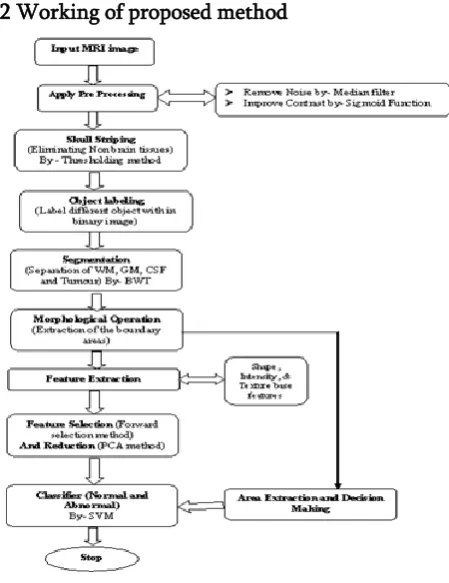
In this research work, we are presenting an efficient supervised learning method for tumour detection and analysis from MR image dataset. Proposed supervised learning method uses a hybrid method. Initially, it uses existing BWT method for data pre-processing and segmentation than later apply SVM+ PCA with object labelling method to extract final results for tumour image such as tumour size, type, growth rate. Existing BWT method with SVM and proposed BWT with SVM+ PCA are implemented over simulator MATLAB and various performance measuring parameters are calculated.
5.1 Proposed algorithm- Proposed Hybrid Brain tumour detection method for MR images
Input- Image set (Tumour image and normal image) Output- Tumour detected (Tumour area in pixel, detection time and accuracy %)
Step 1- Take in input image I1 from image data set I
Step 2- (Apply Pre-processing) 2.1 - Noise Removal (Median Filter)
2.2- Contrast Enhancement (Sigmoid Function) Step 3- Skull Striping (Elimination of non-brain tissues)
3.1 Convert Image into Gray Scale
3.2 Convert Image into Binary (By Thresholding) 3.3 Search number of connected objects
3.4 Find Mask by assigning 1 to inside and 0 to the outside (Tumour Region)
3.5 Find Mask by assigning 1 to inside and 0 to the outside (Tumour Region)
Step 4 Object labeling (label different objects within the image)
4.1 Take the Binary output of skull image 4.2 Label different object within the image Step 5 Segmentation (BWT)
5.1 Segmentation of abnormal brain tissues and normal tissues such as gray matter (GM), white matter (WM), and cerebrospinal fluid (CSF).
Step 6 Morphological Pha
6.1 Erosion operation of morphology is employed to eliminate white pixel
6.2 Eroded regions and the original image are both divided into two equal regions
6.3 The black pixel region extracted from the erode operation is counted as a brain MR image mask Step 7 Feature extractions-
7.1 Forward selection method apply
Step 8 Feature Selection and reduction (by PCA) 8.1 Apply PCA for feature selection reduction Step 9 Apply SVM Classification
5.2 Working of proposed method
Figure 2. Working of proposed method
VI.
RESULT ANALYSIS
Existing BWT method with SVM and proposed BWT with SVM+PCA methods are implemented over simulator MATLAB and various performance measuring parameters are calculated such as accuracy %, detection time, precision and detection rate %.
For performance analysis of existing method and proposed method are tested on total 100 MRI 3-D images in which 35 images are normal and 65 are abnormal images. These images are collected from open MRI online dataset (https://openfmri.org/dataset/). These all the Images are based on T-2 W MRI type.
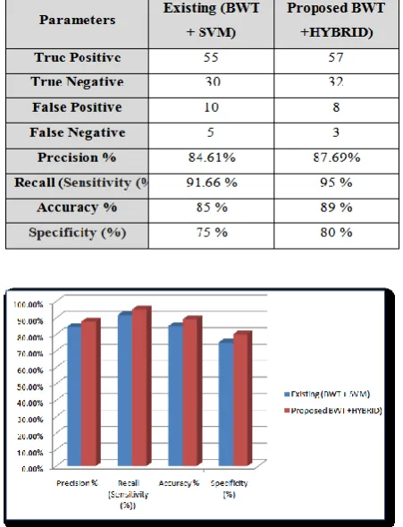
6.1 Confusion matrix-A confusion matrix has been created for an existing and proposed method. Confusion matrix shows all the possible prediction results. In confusion matrix, possible outcomes of a two-classes (normal and abnormal) prediction are represented as True Positive (or TP), True Negative (or TN), False Positive (or FP) and False Negative (or
FN). Here images have two category normal images and abnormal images. TP, TN, FP, and FN can be defined as –
TP= True positive shows the total number of abnormal images which has been correctly classified by the classifier.
TN= True negative shows the total number of normal images which has been correctly classified by the classifier.
FP= False positive shows the total number of normal images which has been classified as abnormal by the classifier.
FN= False negative shows the total number of abnormal images which has been classified as normal by the classifier.
Table 1. Confusion Matrix
6.2 Simulation parameters and Results- Following parameters are calculated for existing and proposed methods-
Figure 3. Simulation results
6.2.1 Precision-Precision can be defined as the ration of abnormal images with correct results.
Precision = (TP/ TP+FP
6.2.2 Accuracy-Accuracy can be defined as the ratio of test results which are accurate.
6.2.3 Specificity- Specificity can be defined as the ratio of the finding all the normal images from all the normal cases.
Specificity= (TN/ TN+FP)
6.2.4 Recall- Recall or sensitivity can be defined as the probability of the test finding the abnormal case among all the abnormal cases.
Recall= TP/ TP+ FN
Table 2. Comparisons of Accuracy % Results for existing and proposed Method
Graph 1. Comparisons of Accuracy % Results for existing and proposed Method
Result Influence- The above Table 6.2.1 and Graph 6.2.1 shows simulation results of accuracy % calculated for existing and proposed a method. Experimental results clearly show that proposed method has better precision %
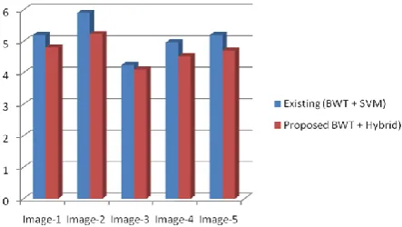
6.2.5 Detection area- For any image area or region can be calculated as the total number of vertical resolution, horizontal resolution and number of pixels of an image.
Detection area=Vr* Hr* Tp
Where Vr= Vertical resolution, Hr = Horizontal resolution, Tp= Number of a pixel in the infected area
Table 3. Detection area % for existing and proposed method
Graph 2. Detection area % for existing and proposed method
Result Influence-The above table 6.2.5 and Graph 6.2.5 shows tumour detection results in pixel for existing and proposed methods. The above results clearly show that proposed method has better detection area as compared to the existing method.
6.2.6 Detection time- This is the sum which requires detecting a tumour in MR images.
Graph 3. Detection time for existing & proposed
6.3 Classifications accuracy-Tumour area detection is calculated for three data sets. Dataset -1 contains total 100 images (35 images normal and 65 abnormal), data set- 2 contains 150 images (70 normal images and 80 abnormal images) and data set- 3 contains 200 images (69 normal images and 131 abnormal images. In first simulation results are calculated without applying feature extraction method for all three datasets (both methods existing and proposed). In the second phase of simulation again results are calculated after applying feature extraction method and following results calculated-
Table 5. Classifications accuracy
Result Influence- The above table and graph 5.5.4 show simulation results for tumour detection accuracy % without applying feature extraction method (PCA) and with feature extraction method for three datasets. An experimental results clearly influence, that’s proposed method have better results. Accuracy % increases after applying PCA feature extraction method.
VII.
CONCLUSIONS & FUTURE WORKS
This research works presented an efficient supervised learning method for tumour detection and analysis from MR image dataset. Proposed supervised learning method uses a hybrid method. Initial it uses existing BWT method for data pre-processing and segmentation than later apply SVM+ PCA with object labelling method to extract final results for tumour image such as tumour size, type, growth rate. Experimental results clearly show proposed method have 3.56 % better accuracy as compared to the existing method. The proposed method also detect better tumour area % (5-8 %) in less time (4-6 % efficient) over existing method.
In future work, we will implement it with the real-time environment. Also, proposed method will be tested with various other methods.
VIII.
REFERENCES
[1]. Nilesh Bhaskarrao Bahadur, Arun Kumar Ray, and Har Pal Thethi, "Image Analysis for MRI Based Brain Tumor Detection and Feature Extraction Using Biologically Inspired BWT and SVM", Hindawi International Journal of Biomedical Imaging Volume 2017, Article ID 9749108, PP 1-12
[2]. Vasupradha Vijaya Dr. A .R. Kavitha, S. Roselene Rebecca "Automated Brain Tumor Segmentation and Detection in MRI using Enhanced Darwinian Particle Swarm Optimization(PSO)", 2nd International Conference on Intelligent Computing, Communication & Convergence (ICCC-2016), PP 476-482
[3]. Deepa, Akansha Singh, "Review of Brain Tumor Detection from MRI Images", IEEE 2016, PP 3997-4001
Computing, ICAFS 2016, 29-30 August 2016, Vienna, Austria, PP 39-44
[5]. N. N. Gopal and M. Karnan, "Diagnose brain tumor through MRI using image processing clustering algorithms such as Fuzzy C-Means along with intelligent optimization techniques," 2010 IEEE International Conference on Computational Intelligence and Computing Research, 2010.
[6]. J. Selva Kumar, A. Lakshmi, and T.Arivoli, "Brain Tumor Segmentation and Its Area Calculation in Brain MR Images using K-Mean Clustering and Fuzzy C-Mean Algorithm", 2012 IEEE-International Conference On Advances In Engineering, Science And Management (ICAESM -2012) March 30- 31, 2012.
[7]. Rajesh C. Patil, Dr. A. S. Bhalchandra, "Brain Tumour Extraction from MRI Images Using MATLAB" International Journal of Electronics, Communication & Soft Computing Science and Engineering ISSN: 2277-9477, vol. 2, no. 1, April 2012.
[8]. M.-N. Wu, C.C. Lin, and C. C. Chang, "Brain Tumor Detection Using Color-Based K-Means Clustering Segmentation," Third International Conference on Intelligent Information Hiding and Multimedia Signal Processing (IIH-MSP 2007), 2007.
[9]. R. Dubey, M. Hanmandlu, and S. Vasikarla, "Evaluation of Three Methods for MRI Brain Tumor Segmentation," 2011 Eighth International Conference on Information Technology: New Generations, 2011.
[10]. T. S. D. Murthy and G. Sadashivappa, "Brain tumor segmentation using Thresholding, morphological operations, and extraction of features of the tumor," 2014 International Conference on Advances in Electronics Computers and Communications, 2014.
[11]. E. F. Badran, E. G. Mahmoud, and N. Hamdy, "An algorithm for detecting brain tumors in MRI images," The 2010 International
Conference on Computer Engineering & Systems, 2010. 11. Merlyn Mary Michael, "Survey on brain segmentation techniques," International Journal of Modern Trends in Engineering and Research, vol. 1, no. 6, pp, 187-192, December 2014.
[12]. Daizy Deb, Bahnishikha Dutta, and Sudipta Roy, "A noble approach for noise removal from brain image using Region filling," 2014 IEEE International Conference on Advanced Communications Control and Computing Technologies, 2014.
[13]. Azian Azamimi Abdullah, Bu Sze Chize, and Yoshifumi Nishio, "Implementation of an Improved Cellular Neural Network Algorithm For BrainTumor Detection," International Conference on Biomedical Engineering (ICoBE), Penang, pp. 27-28, February 2012. [14]. Ishita Maiti, Dr. Monisha Chakra borty, "A
New Method for Brain Tumor Segmentation Based on Watershed and Edge Detection Algorithms in HSV Color Model", National Conference on Computing and Communication Systems (NCCCS), Vol. 73, No. 3, pp. 329–345, March 2012.
[15]. J. Vijay, J. Subhashini, "An Efficient Brain Tumor Detection Methodology Using K-Means Clustering Algorithm," IEEE International Conference on Communication and Signal Processing, pp. 653-657, April 3-5, 2013. [16]. Bilotta. E., Cerasa. A., Pietro. P., Quattrone. A.,
Staino. A., Stramandinoli. F., "A CNN Based Algorithm for the Automated Segmentation of Multiple Sclerosis Lesions," Evo Applications, Part I, pp. 211-220, 2010.
[17]. K. S. Angel Viji, J. Jayakumar, "Performance evaluation of standard image segmentation methods and clustering algorithms for segmentation of MRI brain tumor images," European Journal of Scientific Research, Vol. 79, No. 2, pp. 166-179, 2012.
characterization of brain tumor from MR images," IEEE International Conference on Advances in Recent Technologies in Communication and Computing, 2009.
[19]. Arash Azim Zadeh Irani and Bahari Belton "A K-means Based Generic Segmentation System" Sixth International Conference on Computer Graphics, Imaging and Visualization, 2009. [20]. K. S. Tamilselvan, Dr.G.Murugesan, and B.
Gnanasekaran, "Brain Tumor Detection from Clinical CT and MRI Images using WT-FCM Algorithm," IEEE International Conference on Green Computing, Communication and Conservation of Energy (ICGCE), pp. 260-263, 2013.
[21]. Anam Mustaqeem, Ali Javed, Tehseen Fatima, "An Efficient Brain Tumor Detection Algorithm Using Watershed & Thresh Holding Based Segmentation," International Journal of Image, Graphics and Signal Processing, Vol. 10, pp. 34-39, 2012.
[22]. Janani M and Dr. Manicka Chezian. R, "A Survey On Content-Based Image Retrieval System", International Journal of Advanced Research in Computer Engineering & Technology, Volume 1, Issue 5, pp 266, July 2012.
[23]. Nikita Jain, Vishal Srivastava "DATA MINING TECHNIQUES: A SURVEY PAPER" IJRET: International Journal of Research in Engineering and Technology, Volume: 02 Issue: 11, Nov-2013.
[24]. Venkatadri.M and Lokanatha C. Reddy,"A comparative study on decision tree classification algorithm in data mining", International Journal Of Computer Applications In Engineering , Technology And Sciences (IJCAETS), Vol.- 2, no.- 2, pp. 24- 29, Sept 2010.
[25]. https://openfmri.org/dataset/
[26]. Mr. Dharmesh Patel, Mr.Umesh Lilhore, "A survey on image mining techniques for brain
tumor classification", 4(10): October -2015, PP 592-595.
[27]. Prashant Aher and Prof. Umesh Lilhore, "An improved CBMIR architecture, based on modified classifiers & feedback method for tumor image retrieval from MRI images", International Journal of Modern Trends in Engineering and Research (IJMTER) Volume 03, Issue 12, December – 2016, PP 156-160. [28]. Prashant Aher, Prof. Umesh Lilhore, "Survey of