Comparing Behavior of Chondrocyte Cells on Different
Polyhydroxybutyrate Scaffold Structure for Cartilage Tissue
Engineering
Reza Samanipour1*, Saeed Karbasi2, Batool Hashemibeni3
1. Department of Nuclear Engineering, Faculty of Nuclear Engineering and Basic Sciences, Najafabad Branch, Islamic Azad University, Najafabad, Iran. 2. Department of Medical Physics and Biomedical Engineering, School of Medicine, Isfahan University of Medical Sciences, Isfahan, Iran.
3. Department of Anatomical Sciences, School of Medicine, Isfahan University of Medical Sciences, Isfahan, Iran.
* Corresponding Author: Reza Samanipour, PhD Candidate
Address: Department of Nuclear Engineering, Faculty of Nuclear Engineering and Basic Sciences, Najafabad Branch, Islamic Azad University, Najafabad, Iran. Tel: +98 (938) 9294442
E-mail: [email protected]
A B S T R A C T
Article info: Received: 01 Jan. 2016
Accepted: 31 Mar. 2016
Available Online: 01 Jul. 2016
Key Words:
Cartilage tissue engineering, Polyhydroxybutyrate, Scaffolding, Casting solvent, Electro spinning
Introduction: As the ability to repair cartilage tissue in body is limited, finding a suitable
method for cartilage regeneration has gained the attention of many scholars. For this purpose, scaffold structure and morphology, along with cell culture on it, can be a novel method to treat cartilage injuries, osteoarthritis.
Methods: In this study, polyhydroxybutyrate (PHB) is selected as the scaffold. Firstly, PHB (6% w/v) solution was prepared using chloroform solvent by employing solvent and electrospinning methods. With regard to phase studies, morphology, and specifying agent groups, we used
specific characterization devices such as Fourier transform infrared spectroscopy (FTIR). To
compare the behaviour of cellular scaffolds, they were divided into 2 groups of scaffolds, and
the chondrocyte cells were cultured. To perform phase studies, analysis of MTT and trypan
blue were carried out for measuring the viability and attachment on the surface of the scaffold,
and the specification of scanning electron microscope (SEM) was employed to determine the
morphology of the cells.
Results: Through performing MTT test on the first, third and seventh days, it was found that these types of scaffolds are significantly different from those in the control group (P<0.05).
Scanning electron microscope (SEM) indicates good attachment of chondrocytes on all scaffolds. Results obtained from trypan blue exclusion test also indicated an increase in cell attachment on scaffolds.
Conclusion: Comparing cell behavior on two scaffolds indicates that cell attachment, cell growth and proliferation, and cell migration on the electrospun scaffold is better than the scaffold provided by using solvent casting approach.
Citation: Samanipour R, Karbasi S, Hashemibeni B. Comparing Behavior of Chondrocyte Cells on Different Polyhydroxybutyrate Scaffold Struc-ture for Cartilage Tissue Engineering. Anatomical Sciences. 2016; 13(2):105-116.
1. Introduction
artilage tissue has little ability for self-repair because it has no blood vessels and nerves. Injury to the cartilage, for any reason, leaves a permanent and chronic lesion, which can result in disability or deformation of damaged organ depending on the case. Currently, different types of cartilage implantations such as autograft, allograft, and xenograft are being used to treat massive cartilage damages. However, each method has some disadvantages, along with its advan-tages, that can make their use problematic. One of the treatment methods is using chondrocytes of the patient, but it has many limitations such as decline of prolif-eration capability of chondrocytes in old people. On the other hand, chondrocytes isolated from body more
toward fibroblast phenotype after monolayer culture
and expression of effective factors in cartilage-building would be changed in them.
Tissue engineering is a novel approach, which has
been considered over the years to surpass the limitations
and repair injured tissues [1,2]. It includes growing
spe-cial cells in an extracellular domain to create structures similar to natural organs or tissues. In this method, 3-D scaffold is usually applied to simulate natural extra-cellular matrix, initial cell attachment, and cell
prolif-eration [3]. The main purpose of tissue engineering is
making tissue structures that can reconstruct structure
and functions of injured tissues after implantation. To
achieve this goal, choosing a proper cell is an important issue as mentioned before. However, a cell cannot
cre-ate functional structures by itself. The reality is that a
cell can use its maximum potential to generate a tissue that is cultured on the scaffold. Applied scaffoldings in tissue engineering have been designed in a manner that provide the required 3-D environment to protect and conduct cell processes like migration, proliferation, and
differentiation to generate functional tissues [4].
A lot of biodegradable polymers and ceramics have been used as biocompatible scaffolds in tissue
engi-neering [5,6]. The mechanism of degradation of bio
-degradable polymers in the body is different based on their molecular structure, although in most cases, deg-radation products of the polymers are also biodegrad-able and cause no damage to living tissues. Among the degradations, one can refer to bulk degradation and surface degradation. In the former, polymer begins to be degraded from inside, but in surface degradation, it
is degraded layer by layer from the surface [7]. Among
biodegradable polymers, polyhydroxyalkanoates or
PHAs are considered because of their superior bio-degradability and biocompatibility as well as suitable
physical and mechanical properties [8,9].
In our study, we used solvent castings and electrospin-ning methods to make PHB scaffolds. Some studies are conducted on the cartilage restoration using
electrospin-ning scaffolds. Li et al. [10] provided a nanofiber scaf
-fold of polycaprolactone and examined the ability of the scaffold in cartilage generation using mesenchymal stem
cells. The scaffolds produced by them had uniform fi -bers with diameter of 700 nm running random directions in the scaffold. Results obtained from 21 days culture of mesenchymal stem cells on scaffolds in the presence of
growth factor TGF-β1 indicated differentiation of these
cells from chondrocyte phenotypes. In Thorvaldsson et
al. [11] study, scaffolds were made from microfibers of polylactic acid covered by nanofibers of polycaprolac
-tone. The study was conducted with the aim of control
-ling the size of porosity and cell infiltration and using advantages of nanofibers in cell growth. In this study, scaffolds with porosity of 95% and 97% were obtained.
To investigate cell behavior on scaffolds, human
chondrocyte cells are seeded on the scaffold. Results
obtained from the study indicate that pore size has a significant effect on cell infiltration inside the scaffold. Moreover, the presence of nanofibers in the structure
increases the bioactivity of scaffolds. Zheng et al. [12]
conducted a study on gelatin/polycaprolactone
electro-spinning scaffold in cartilage tissue engineering. Their
aim was assessing the effect of different ratios of gela-tin to caprolactone in the structure on the attachment and proliferation of chondrocytes.
The obtained results indicated that scaffolds with
more gelatin can increase attachment and proliferation of chondrocytes and formation of cartilage tissue in the
first 3 weeks of implantation. However, after 12 weeks,
new cartilage tissues were formed in all samples and
no significant difference was observed among samples.
In another study by Sombatmankhong et al. [13], 3-D
fiber scaffolds were made of polyhydroxybutyrate/
polyhydroxybutyrat-co-hydroxyvalerate (PHBV) alloy
using electrospinning method. In this study, fibers with diameter of 2.3 to 4 μm were obtained. Produced fiber scaffolds had more hydrophilicity than the films from
the same material prepared for comparison [14].
More-over, tensile resistance failure of alloy fibers compared to pure fibers of PHB and PHBV was obvious. Indirect toxicity evaluation using rat fibroblast cells (L929) indi
This study mainly focuses on a scaffold design for cell evaluation of the seeded scaffolds. To optimize the seed
-ing efficiency and observe the cell proliferation into the
inner structure, we developed a special design for the
scaffold. The objective of the design was to maximize its
surface and facilitate the seeding process to enhance cell adhesion and good supply of the scaffold interior with
medium. The cultivation of cells seeded onto the scaffolds
was carried out under static and dynamic conditions. Cell evaluation was carried out on days 3 and 7. Cell adhesion on the designed structure was also analysed. Additionally, cell growth inside the bulk material and cell morphology were examined. Comparing cell behavior on the two scaf-folds and using PHB alone (without combination with other materials as main scaffold to culture chondrocytes) can be regarded an innovation of our study. After making scaffolds, porosity percentage was determined. Finally, to investigate attachment of chondrocytes, cell culture and
SEM imaging were employed [16].
2. Materials and Methods
Materials
We used materials listed in Table 1. In addition, the
employed cell line was chondrocyte cell line
manufac-tured by Tehran Pasteur Institute.
Scaffold preparation
In this study, we used Haji Ali applied method to
pre-pare scaffold using solvent casting [17]. First, 6% w/v
polyhydroxybutyrate solution was prepared, and then it
was entirely solved in chloroform at 55°C-60°C for 12 h.
As the study has considered salt/polymer weight
propor-tion of 9 to 1 to create porosity, as per 0.6 g polymer, 5.4 g salt (particle size of 212-250 μm) was weighed and added to the solution. The salt in polymer solution was properly
mixed by vertex to distribute salt particles, and then it was poured immediately in 8-cm Petri dish (here as a frame).
The mixture was placed in outside environment for 48 h
to bring out the solution uniformly and slowly. Next, a white trapped disk formed of polymer and salt particles
remained. The sample was washed using deionized wa -ter. Washing samples and bringing out salt particles from
them lasted 5 days. For final drying, samples were placed
in vitro for 24 h, and then for 48 h in outside temperature under the pressure of 0.8 kPa. By the end of solvent cast-ing process, washcast-ing particles and drycast-ing samples, scaf-folds were prepared for evaluating and performing tests.
We used Heydar Khan Tehrani method to prepare
scaffold using electrospinning method [18]. In this
method, polymer solution was used with a similar mechanism to solvent casting method, but to provide PHB solution, in addition to chloroform solvent,
de-odorized methanol fraction (DEMEF) method (8:2)
was also used. Produced PHB solution was placed on
horizontal injection pump, and then attached to the tip
of a 21-gauge syringe.
Physical properties
Porosity measurement
In order to determine the percentage of porosity, water
replacement formula was applied as follows [19]:
where P, Mw, Md, and Va are “porosity percentage,”
“wet mass of scaffolding”, “dry mass of scaffolding”, and “apparent volume”, respectively.
Fourier transforms infrared spectrometer
Infrared spectrometry is carried out based on radia-tion absorpradia-tion and studying the vibrionic transiradia-tions
of molecules and multi-atomic ions. This method is
used as a powerful and developed method to determine the structure and amount of chemical compounds. It is mostly employed to identify organic compounds be-cause their spectra are usually complicated and have large number of maximum and minimum peaks, which
can be employed for comparative purposes. This test is
appropriate for polymer samples [19].
Cell culture
Sterilization of scaffold
For sterilization, each group was placed in a 9-cm plate. Scaffolds were sterilized in 75% ethanol for 3 h
under ultraviolet radiation for 1 h, and then they were
immersed in DMEM (Dubecco’s Modified Eagle Me -dium, Invitrogen, California, USA) overnight.
Cell seeding
The human osteoblast cell lines C28/I2 (Species: human; Tissue: cartilage) were purchased from Pas
-teur Institute of Iran (IPI) (Tehran, Iran). C28/I2 cells
were cultured in RPMI1640 (Roswell Park Memorial Institute) supplemented with 10% fetal bovine serum (FBS), and 100 mg/mL streptomycin (Jinke Biotech).
Cells were incubated in a 5% CO2 incubator at 37˚C,
and were harvested by trypsinization followed by addi
-P(%)= Mw-Md ×100
tion of fresh culture medium to create a new single cell suspension. When the cells reached the plateau phase of
growth, they were harvested by trypsinization, followed
by addition of fresh culture medium to create a new single cell suspension with desired seeding cell number
per 100 mg [20]. Inoculation was performed in
polysty-rene 24-well flat-bottom culture plates. Scaffolds were
placed in the center of the wells added with 1 mL cell suspension to allow full attachment of cells to scaffolds. Cultivation was conducted for 3, 7, 14, and 28 days.
Culture media were changed every 4 days [21].
Cell viability
For attachment study, C28/I2 was allowed to attach on scaffold and all of the scaffold specimens for 8 and
24 h, respectively. At each specified seeding time, the viability of the attached cells was quantified by MTT
assay [22]. Each sample was rinsed with phosphate
buffer saline (PBS; Sigma–Aldrich, USA) to remove unattached cells prior to MTT assay. Since no studies
were carried out on the expression of attachment pro-teins or the strength of the attached cells, this evalu-ation only served as a qualitative measure of the cell
attachment study. The viability of the cells was again quantified by MTT assay [23].
Quantification of viable cells (MTT assay)
The MTT assay is based on the reduction of the yellow tetrazolium salt to purple formazan crystals by dehydroge
-nase enzymes secreted from the mitochondria of metaboli
-cally active cells. The amount of the formed purple forma
-zan crystals is proportional to the number of viable cells. First, each sample was incubated at 37° C for 1 h with 250(10) µL/well of MTT solution at 0.5(5) mg/mL with -out phenol red for a 24-well (or 96-well) scaffold. After
incubation, MTT solution was removed. A buffer solution containing dimethyl sulfoxide (DMSO; Carlo Erba, Italy) of about 900(100) µL/well and glycine buffer (pH=10) of about 125(5) µL/well was added into the wells to dissolve the formazan crystals. After 10 min of rotary agitation, the
solutions were transferred into a cuvette and were placed
in a thermo Spectronic Genesis10 UV–vis spectrophotom
-eter, from which the absorbance at 540 nm representing
the number of viable cells was measured [24].
Cell attachment
Trypan blue
Chondrocyte cells from line of C28/I2 were used for
the assays. Briefly, osteoblast cells were released from
nearly confluent cultures with trypsin (Sigma-Aldrich Chemie GmbH, Germany), and were washed with a
solution containing trypsin inhibitor (Sigma-Aldrich
Chemie GmbH, Germany). After 30 min, non-bound cells were washed away, and attached cells were fixed,
stained, and counted.
Scanning electron microscopy examination
It was tested in accordance with the ISO 10993-5 stan
-dards and carried out directly. To study the cell morphol -ogy and adhesion, a cell suspension containing 20000 cells with UV light was seeded on the surface of sterile
scaffold. The specimens were washed twice by phosphate
buffered saline (PBS) and immersed in PBS containing
3% glutaraldehyde (pH=7.4) for 4 h. Then, they were de -hydrated in increasing concentrations of ethanol (from
30%, 50%, 70%, 90%, and 95% to 100%), followed by lyophilization. They were then mounted on aluminum stumps, coated with gold in a sputtering device for 1.5 min at 15 mA and examined under a scanning electron mi
-croscope (KYKY-2800, Tehran University-Iran) [25-26].
Statistical analysis
Significant differences were determined with ANO -VA test (1-way analysis of variance) to compare the
means of different data sets. Statistical significance was accepted at 0.05 confidence level. The results were reported as mean ±MTT and trypan blue.
3. Results
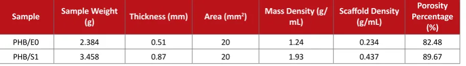
Assessment of density and porosity of scaffolds
As mentioned before, porosity is an important
param-eter for cell scaffolds. To measure density and porosity of
scaffolds in solvent casting method, thickness of samples
was determined using a thickness gauge device (Table 2).
Mass density for PHB polymer was considered to be 1.24
g/mL [27]. It should be mentioned that two iterations
were considered for each sample and measured values
were reported as average. According to Table 2, porosity
of PHB samples made by both methods is higher than 80%, indicating that samples have high and suitable po-rosity for cell attachment and proliferation. Popo-rosity per-centage of produced scaffolds through electrospinning
method was measured using MATLAB software.
FTIR of PHB
the C-O bond in PHB; and 1128cm-1 and 1176-1 peaks are related to the asynchronous and synchronous stretch-ing vibration of C-O bond, respectively. CH3 groups
create an acute peak at 1377cm-1, which is the result of
the stretching vibration of this bond. At wave number of
1724cm-1, a very clear and acute peak, which is the result
of the stretching vibration of carbonyl groups, is noticed.
Since the nature of the scaffold and the method of its cre-ation, and on the whole, its morphology are effective on cel-lular behaviour, the selection of polyhydroxybutyrate polymer
using two manufacturing methods; namely, electrospinning
and solvent casting in this research shows very good results with regard to cell culture and chondrocyte cells.
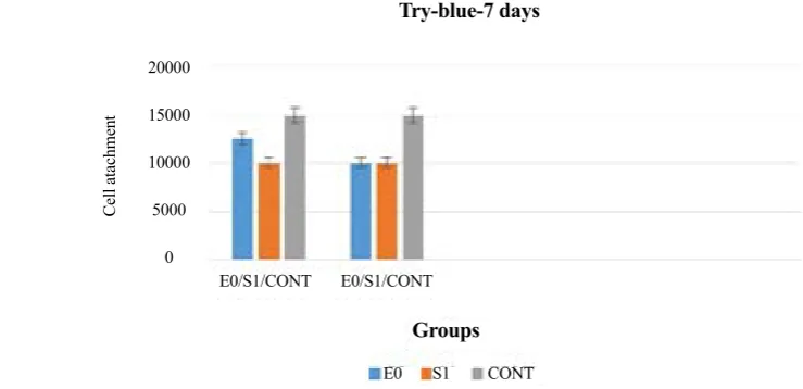
Trypan blue test results for cell attachment
Total number of attached cells to surface of scaffolds was estimated using Equation 2 and then analyzed in Ex
-cel software. As illustrated in Figures 1and 2, the
num-ber of cells attached to scaffolds is approximately 70% to
80% of total cells poured on scaffold surface. This indi -cates that the scaffolds are good beds for cell attachment
(CONT=control sample without the presence of scaf
-fold). By comparing Figures 1and 2, we can say that cell
attachment on day 7 is more than that on day 3 for both
scaffolds. This indicates suitability of the scaffolds for
cell attachment and performing cell culture tests.
By comparing Figures 1and 2, we can say that cell
attachment on day 7 is more than that on day 3 for both
scaffolds. This indicates suitability of the scaffolds for
cell attachment and performing cell culture tests.
MTT test results for cell proliferation
Metabolic activity of cells on scaffolds was
deter-mined using MTT analysis. Obtained results have been Figure 1. FTIR absorption spectrum of PHB.
Figure 2. Trypan blue diagram for day 3.
E0: cell+scaffolds made by electrospinning, S1: cell+scaffold made by solvent casting. E0/S1/CONT
Cell atachment
E0/S1/CONT
E0 S1 CONT
Groups Try-blue-3 days
20000
15000
10000
5000
Figure 4. MTT diagram for day 3.
E0: cell+scaffolds made by electrospinning, S1: cell+scaffold made by solvent casting.
Figure 5. MTT diagram for day 7.
E0: cell+scaffolds made by electrospinning, S1: cell+scaffold made by solvent casting.
OD (optical density
OD (optical density
E0/S1/CONT
E0/S1/CONT E0/S1/CONT
E0/S1/CONT
E0
E0 S1
S1
CONT
CONT
Groups
Groups MTT-3days
MTT-3days
0.3 0.25 0.2 0.15 0.1 0.05 0
0.3 0.25 0.2 0.15 0.1 0.05 0 Figure 3. TB diagram for day 7.
E0: cell+scaffolds made by electrospinning, S1: cell+scaffold made by solvent casting. E0/S1/CONT
Cell atachment
E0/S1/CONT
E0 S1 CONT
Groups Try-blue-7 days
20000
15000
10000
5000
presented in Figures 3and 4. As it is obvious, increase in culture time has resulted in increased chondrocytes proliferation on scaffolds. Moreover, percentage of sur-vived cells on the electrospinned scaffolds is more than scaffold made by solvent casting. In addition, obtained
values have been analysed using SPSS. The results in -dicate that the relationship between survival of cells on
scaffold surface has correlation coefficient of 75% with P value under 0.05, i.e. this correlation is not random.
Certainly, P-valueE0 for samples made by
electrospin-ning method is 0.023 and P-values1 for samples made
by solvent casting is 0.033. Through comparing P-val -ues obtained from samples with control group, it could
be found that there was a significant difference between the groups and control group (P-value<0.05).
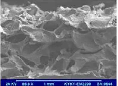
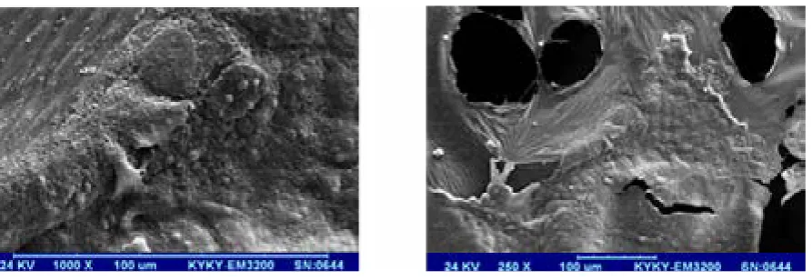
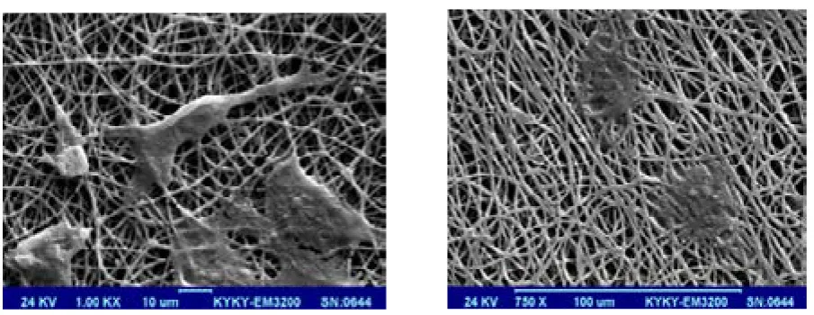
Morphology of scaffolds
As it is obvious in SEM images, all scaffolds have had
good biocompatibility for cell culture (Figures 5-12).
Cells have a tendency for colon formation in day 1, and as time goes by, they began to discharge redundancies.
Therefore, PHB creates a good interaction between
scaffold and cells with better attachment and growth
of cell redundancies, and finally more migration and
expansion of cells. As mentioned before, lack of cell penetration and migration deep inside the scaffolds are
basic problems with tissue engineering. Through com -paring obtained SEM images, it could be found that cell attachment, migration, and penetration on electrospun scaffold is better than solvent casting-based scaffold
Table 1. The materials used in the study.
Company Country Material
Sigma Aldrich Germany Natural origin polyhydroxybutyrate (molecular weight of 300000)
Scharlau Spain Chloroform (CHCl3) (Purity: 99.47%)
Merck Germany Sodium chloride (NaCl) (Purity: 99.5%)
Merck Germany 96% ethanol (C2H5OH)
Gibco USA RPMI 1640 Medium
Gibco USA Phosphate buffered saline (PBS)
Gibco USA Fetal bovine serum (FBS)
Gibco USA Trypsin/EDTA
Gibco USA Penicillin/Streptomycin
Promega, Madison USA 3-(4,5-Dimethylthiazol-2-yl)-2,5-diphenyltetrazolium bromide (MTT)
Merck Germany Glutaraldehyde
Merck Germany Dimethyl sulfoxide (DMSO)
Sigma Aldrich USA Trypan Blue
Sina Gen Iran Distilled water
Sina Gen Iran Deionized water
Table 2. Density and porosity of scaffolds made by electrospinning (E0) and solvent casting (S1).
Porosity Percentage
(%)
Scaffold Density
(g/mL)
Mass Density (g/
mL) Area (mm2)
Thickness (mm) Sample Weight
(g) Sample
82.48 0.234
1.24 20
0.51 2.384
PHB/E0
89.67 0.437
1.93 20
0.87 3.458
Figure 6. SEM images of scaffold surface made by solvent casting.
Figure 7. SEM images of cell attachment to scaffold surface made by solvent casting.
Figure 9. SEM images of cell penetration inside structure of scaffold surface made by solvent casting.
Figure 10. SEM images of scaffold surface made by electrospinning method.
because of suitable surface topography, its nanoscale, open porosity, pore continuity, and link to each other, that makes the surface to volume ratio high.
4. Discussion
The study results were obtained from two fields of
construction and assessment of PHB using two meth-ods of solvent casting and electrospinning and through
optimization of factors affecting the process. The scaf -folds have been evaluated with regard to porosity and attachment behavior of chondrocytes on them by
try-pan blue, MTT, and SEM tests. By analyzing the re -sults, it was found out that attachment of chondrocytes on scaffold made by electrospinning method is better than solvent casting method because of its nanoscale, higher surface energy, and good topography. Moreover, because of continuity of porosities in electrospun scaf-fold, migration and proliferation of chondrocytes on this scaffold are more than that on the scaffold made by solvent casting method. Because of high percentage of porosities created on scaffold by solvent casting and good growth and proliferation of chondrocytes on the scaffold, it can be considered as a good bed for extra-cellular matrix of cartilage. In addition, chondrocytes show good attachment on the scaffolds.
Acknowledgements
I would like to express my deep gratitude to Dr Sale-hi, faculty member of Isfahan University of Medical Sciences for the service and generous support to
con-duct this research. This article is extracted from the thesis
of Nuclear Engineering faculty of Islamic Azad University of Najafabad. This paper had not any finanacial support.
Conflicts of Interests
The authors declared no conflict of interest.
References
[1] Hunter W. Of the structure and disease of articulating car-tilages 1743. Clinical Orthopaedics & Related Research. 1995; 317:3-6. PMID: 7671493
[2] Tuli R, Li WJ, Tuan RS. Current state of cartilage tissue en-gineering. Arthritis Research & Therapy. 2003; 5(5):235– 38. doi: 10.1186/ar991
[3] Portocarrero G., Collins G., Livingston Arinzeh T. Chal-lenges in cartilage tissue engineering. Journal of Tissue Science & Engineering. 2013; 4:e120. doi: 10.4172/2157-7552.1000e120
[4] Lanza R, Langer R, Vacanti JP. Principles of tissue engi-neering. New York: Academic Press; 2011.
[5] Smith IO, Liu Xh, Smith LA, Ma PX. Nanostructured polymer scaffolds for tissue engineering and regenera-tive medicine. Wiley Interdisciplinary Reviews: Nano-medicine and Nanobiotechnology. 2009; 1(2):226-36. doi: 10.1002/wnan.26
[6] O’Brien FJ. Biomaterials & scaffolds for tissue engineer-ing. Mater Today. 2011; 14(3):88-95. doi: 10.1016/S1369-7021(11)70058-X
[7] Gardian C, Ferroni L, Favero L, Stellini E, Stomaci D, Siv-olella S, et al. Nanostructured biomaterials for tissue en-gineered bone tissue restoration. International Journal of Molecular Sciences. 2012; 13(1):737-57. doi: 10.3390/ ijms13010737
[8] Tsuji H, Suzuyoshi K. Environmental degradation of
bio-degradable polyesters 1. Poly (ε-caprolactone), poly
[(R)-3-hydroxybutyrate], and poly (L-lactide) films in con
trolled static seawater. Polymer Degradation & Stability. 2002; 75(2):347-55. doi: 10.1016/s0141-3910(01)00240-3
[9] Chen GQ, Wu Q. The application of polyhydroxyal-kanoates as tissue engineering materials. Biomateri-als. 2005; 26(33):6565-578. doi: 10.1016/j.biomateri-als.2005.04.036
[10] Williams SF, Martin DP, Horowitz DM, Peoples OP. PHA applications: addressing the price performance issue: I. Tissue engineering. International Journal of Biological Macromolecules. 1999; 25(1):111-21. doi: 10.1016/s0141-8130(99)00022-7
[11] Li WJ, Tuli R, Okafor C, Derfoul A, Danielson KG, Hall
DJ, et al. A three-dimensional nanofibrous scaffold for
cartilage tissue engineering using human mesenchy-mal stem cells. Biomaterials. 2005; 26(6):599-609. doi: 10.1016/j.biomaterials.2004.03.005
[12] Thorvaldsson A, Stenhamre H, Gatenholm P, Walk-enström P. Electrospinning of highly porous scaffolds for cartilage regeneration. Biomacromolecules. 2008; 9(3):1044-49. doi: 10.1021/bm701225a
[13] Zheng R, Duan H, Xue J, Liu Y, Feng B, Zhao S, et al. The
influence of Gelatin/PCL ratio and 3-D construct shape
of electrospun membranes on cartilage regeneration. Biomaterials. 2014; 35(1):152-64. doi: 10.1016/j.biomateri-als.2013.09.082
[14] Sombatmankhong K, Suwantong O,
Waleetorncheep-sawat S, Supaphol P. Electrospun fiber mats of poly
(3-hydroxybutyrate), poly (3-hydroxybutyrate-co-3-hy-droxyvalerate), and their blends. Journal of Polymer Sci-ence Part B: Polymer Physics. 2006; 44(19):2923-933. doi: 10.1002/polb.20915
[15] Choi YS, Cha SM, Lee YY, Kwon SW, Park CJ, Kim M. Adipogenic differentiation of adipose tissue derived adult stem cells in nude mouse. Biochemical & Biophysi-cal Research Communications. 2006; 345(2):631-37. doi: 10.1016/j.bbrc.2006.04.128
[16] Blitterswijk CA, Moroni L, Rouwkema J, Siddappa R, Sohier J. Tissue engineering–an introduction. New York: Academic Press; 2008.
[17] Haji Ali H. [Synthesis and evaluation of mechanical prop-erties, degradation, and bioactivity of composite scaffold of bioactive glass/polyhydroxybutyrate particles for bone tissue engineering (Persian)] [MSc. thesis]. Tehran: Iran University of Science and Technology; 2011.
[18] Heydar Khan Tehrani A. [Manufacturing nanocomposite hydroxyapatite-polyhydroxybutyrate (nHA-PHB) scaf-fold using electrospinning method for tissue engineering (Persian)] [MSc. thesis]. Isfahan: Isfahan University of Technology; 2010.
[19] Subia B, Kundu J, Kundu S. Biomaterial scaffold fabrica-tion techniques for potential tissue engineering applica-tions. In: Eberli D, editor. Tissue Engineering. New York: Intech Open Access Publisher; 2010, p. 142-57.
[20] Mandl EW, van der Veen SW, Verhaar JA, van Osch GJ. Serum-free medium supplemented with high-concentration FGF2 for cell expansion culture of hu-man ear chondrocytes promotes redifferentiation capacity. Tissue Engineering. 2002; 8(4):573-80. doi: 10.1089/107632702760240490
[21] Gan L, Kandel RA. In vitro cartilage tissue formation by co-culture of primary and passaged chondrocytes. Tissue Engineering. 2007; 13(4):831-42. doi: 10.1089/ ten.2006.0231
[22] Panossian A, Ashiku S, Kirchhoff CH, Randolph MA, Yaremchuk MJ. Effects of cell concentration and growth period on articular and ear chondrocyte transplants for tissue engineering. Plastic & Reconstructive Surgery. 2001; 108(2):392-402. doi: 10.1097/00006534-200108000-00018
[23] Naumann A, Dennis JE, Aigner J, Coticchia J, Arnold J, Berghaus A, et al. Tissue engineering of autologous carti-lage grafts in three-dimensional in vitro macroaggregate culture system. Tissue Engineering. 2004; 10(11-12):1695-706. doi: 10.1089/ten.2004.10.1695
[24] Aigner J, Tegeler J, Hutzler P, Campoccia D, Pavesio A, Hammer C, et al. Cartilage tissue engineering with novel nonwoven structured biomaterial based on hyaluronic acid benzyl ester. Journal of Biomedical Materials Re-search. 1998; 42(2):172-81. PMID: 9773813
[25] Isogai N, Kusuhara H, Ikada Y, Ohtani H, Jacquet R, Hillyer J, et al. Comparison of different chondrocytes for use in tissue engineering of cartilage model structures. Tissue Engineering. 2006; 12(4):691-703. doi: 10.1089/ ten.2006.12.691
[26] Hidaka C, Cheng C, Alexandre D, Bhargava M, Torzilli
PA. Maturational differences in superficial and deep
zone articular chondrocytes. Cell Tissue Research. 2006; 323(1):127-35. doi: 10.1007/s00441-005-0050-y