Essential role of
AWP1
in neural crest specification
in
Xenopus
JEONG-HAN SEO
1, DONG-SEOK PARK
1, MINA HONG
1, EUN-JU CHANG
1and SUN-CHEOL CHOI
*,1,21Department of Biomedical Sciences, Cell Dysfunction Research Center (CDRC) and 2Department of Biochemistry and Molecular Biology. University of Ulsan College of Medicine, Pungnap-Dong, Songpa-Gu, Seoul, Republic of Korea
ABSTRACT The neural crest (NC) comprises a transient and multipotent embryonic cell population, which gives rise to a wide variety of cell types, including craniofacial cartilage, melanocytes, and neurons and glia of the peripheral nervous system. The NC is induced by the integrated action of Wnt, FGF, and BMP signaling, and its cell fates are subsequently specified by a genetic cascade of specific transcription factors. Here we describe a critical role of AWP1 in NC induction during Xeno-pus early development. XenopusAWP1 (XAWP1) was found to be expressed in the presumptive preplacodal ectoderm, neural tissue, and posterior dorsal mesoderm, but was absent in the neural fold along the anterior-posterior axis of the neurulae. Notably, XAWP1 was induced by FGF8a in naïve ectodermal tissue. XAWP1-depleted embryos exhibited defects in pigmentation, craniofacial cartilage, and in the dorsal fin. A knockdown of XAWP1 impaired both endogenous and the FGF8a or Wnt8-induced expression of NC markers without affecting mesoderm formation. Furthermore, NC induction inhibited by XAWP1 depletion was rescued by co-expression of activating forms of `-catenin or TCF3. In addition, overexpression of XAWP1, in concert with BMP inhibition, induced the expression of neural plate border specifiers, Pax3 and Msx1, and these regulatory factors recovered NC induction in the XAWP1-depleted embryos. `-catenin stability and Wnt-responsive reporter activity were also impaired in AWP1-depleted cells. Taken together, these results suggest that XAWP1 functions as a mediator of Wnt signaling to regulate NC specification.
KEY WORDS:
AWP1, neural crest induction, Wnt, Xenopus
Introduction
The neural crest (NC) is a vertebrate multipotent embryonic cell population, which arises at the border between the neural plate and epidermis along the antero-posterior (AP) axis posterior to the diencephalon and migrates to several parts of the embryo to dif-ferentiate into a large variety of cell types, including melanocytes, craniofacial cartilage and neurons and glia of the peripheral ner-vous system (Le Douarin and Dupin, 2003; Sauka-Spengler and Bronner-Fraser, 2008). During gastrulation, NC cells are induced E\ WKH FRPELQHG DFWLRQ RI :QW ÀEUREODVW JURZWK IDFWRU )*) bone morphogenetic protein (BMP), and Notch signals that origi-nate from the epidermis, neural plate, and underlying mesoderm (Sauka-Spengler and Bronner-Fraser, 2008; Steventon et al., 2005). These NC-inducing signals are integrated at the neural plate border (NPB) to activate a genetic cascade of transcription IDFWRUVÀUVWLQGXFLQJWKHH[SUHVVLRQRI13%VSHFLÀHUVMsx1, Pax3, Zic1, Ap2, Dlx3) that, in turn and in concert with the same signaling
www.intjdevbiol.com
*Address correspondence to: Sun-Cheol Choi. Department of Biomedical Sciences, Cell Dysfunction Research Center(CDRC), University of Ulsan College of Medicine, Pungnap-Dong, Songpa-Gu, Seoul 138-736, Republic of Korea. Fax: 82-2-3010-5307. e-mail: [email protected]
Accepted: 22 August 2013. Final, author-corrected PDF published online: 22 January 2014. Edited by: Sally Dunwoodie
ISSN: Online 1696-3547, Print 0214-6282
© 2014 UBC Press Printed in Spain
Abbreviations used in this paper: *58JWVMUWZXPWOMVM\QKXZW\MQV#./.ÅJZWJTI[\ OZW_\PNIK\WZ#57UWZXPWTQVWWTQOW#6+VM]ZITKZM[\#68*VM]ZITXTI\MJWZLMZ# 7,+WZVQ\PQVMLMKIZJW`aTI[M#@)?8Xenopus)?8
SDWKZD\VWULJJHUWKHH[SUHVVLRQRI1&VSHFLÀHUVSox9, Sox10, Slug, FoxD3) (Heeg-Truesdell and LaBonne, 2004; Prasad et al.,
6DXND6SHQJOHUDQG%URQQHU)UDVHU1&VSHFLÀHUV ultimately control NC behaviors such as the epithelial to mesen-chymal transition (EMT), delamination, migration and differentiation by regulating the adhesive properties, shape, motility, cell-cycle, cell survival and multipotency of NC progenitors (Sauka-Spengler and Bronner-Fraser, 2008; Theveneau and Mayor, 2012). However, our current understanding of the complex gene regulatory network IRU 1& VSHFLÀFDWLRQ PLJUDWLRQ DQG GLIIHUHQWLDWLRQ LQWR GLYHUVH derivatives appears to be preliminary. More connections and the hierarchical organization of the transcription factors involved in NC formation remain to be elucidated.
prolifera-tion through `-catenin/TCF complex-dependent gene transcripprolifera-tion (MacDonald et al., 2009). A large body of evidence suggests that canonical Wnt/`-catenin signaling is implicated in NC induction and VXEVHTXHQWGHYHORSPHQW6WXKOPLOOHUDQG*DUFLD&DVWUR Double-homozygous null mutant mice for Wnt1/Wnt3a show severe abnormalities in NC derivatives (Ikeya et al., 1997). In Xenopus DQG]HEUDÀVKWKHPRUSKROLQRPHGLDWHGNQRFNGRZQRIWnt8 or Wnt3a inhibits the expression of NPB and NC markers (Elkouby et al., 2010; Hong et al., 2008; Lewis et al., 2004). In addition, Wnt6, which is secreted from the chick non-neural ectoderm adjacent to WKH13%LVQHFHVVDU\DQGVXIÀFLHQWWRLQGXFH1&FHOOVLQDYLDQ HPEU\RV*DUFLD&DVWUR et al.*DLQDQGORVVRIIXQFWLRQ analyses of other Wnt signaling components, including `-catenin, Frizzled7, LRP6, Dishevelled, and Kremen2, further support the model that NC formation involves the canonical Wnt pathway (Abu-Elmagd et al., 2006; Hassler et al., 2007; Tamai et al., 2000).
,QWKLVVWXG\ZHKDYHLGHQWLÀHGAWP1 as a critical regulator
RI 1& VSHFLÀFDWLRQ LQXenopus early embryogenesis. AWP1
DOVRNQRZQDV]LQFÀQJHU$1W\SH=)$1'ZDVRULJLQDOO\ LGHQWLÀHGDVDSURWHLQDVVRFLDWHGZLWKWKHVHULQHWKUHRQLQHNLQDVH PRK1, a lipid-activated kinase belonging to the protein kinase C superfamily (Duan et al., 2000). AWP1 contains A20 and AN1 ]LQFÀQJHUGRPDLQVDWLWV1DQG&WHUPLQDOUHVSHFWLYHO\DQGLV NQRZQDVXELTXLWLQELQGLQJQHJDWLYHPRGXODWRURI1)ѣ%DFWLYLW\ (Fenner et al., 2009). Recently, AWP1 has also been shown to DIIHFWDSRSWRWLFFHOOGHDWKDVDUHJXODWRURI1)ѣ%DFWLYLW\&KDQJ
et al., 2011) and protein export during biogenesis of the peroxi-some, a subcellular organelle (Miyata et al., 2012). AWP1 is expressed in several human tissues and also in the early stages of mammalian development (Duan et al., 2000), but its physi-ological function remains unknown. We have found that AWP1 affects the expression of NPB and NC markers in the neural fold by mediating Wnt/`-catenin signaling. Notably, it is a target gene
RI)*)VLJQDOLQJDQGLVDOVRHVVHQWLDOIRU1&LQGXFWLRQYLDWKLV pathway. Our present results thus suggest that AWP1 functions to PHGLDWHWKHFURVVWDONRI:QWDQG)*)VLJQDOLQJLQ1&LQGXFWLRQ
Results
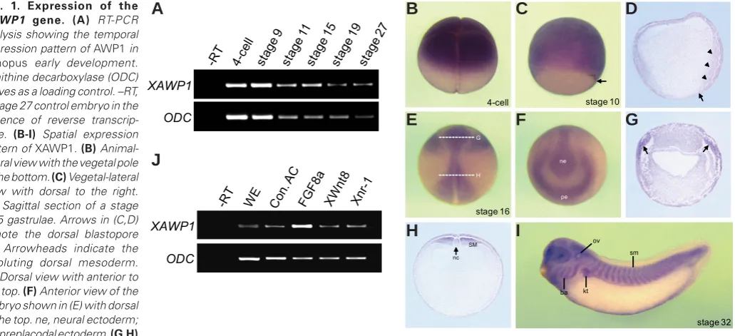
Expression pattern of AWP1 in early Xenopus embryogenesis To investigate the biological function of AWP1 in vertebrates, ZHÀUVWREVHUYHGLWVGHYHORSPHQWDOH[SUHVVLRQSDWWHUQLQXenopus laevis embryos. Temporally, XAWP1 shows both maternal and zygotic transcription in early embryogenesis as revealed by RT-PCR analysis (Fig. 1A). We also performed in situ hybridization to examine its spatial expression pattern. At the early cleavage stages, XAWP1 maternal transcripts are localized to the animal hemisphere of the embryo (Fig. 1B), and this pattern persists into the blastula stages (data not shown). As gastrulation proceeds, its expression is observed in the mesoderm and overlying ectoderm (Fig. 1 C,D). During neurulation, its zygotic message is detectable in the presumptive preplacodal ectoderm and neural tissue but QRWLQWKHDQWHULRUDQGODWHUDOQHXUDOIROGV)LJ(*2IQRWH
XAWP1 is strongly expressed in the posterior dorsal mesoderm
LQQHXUXODH)LJ(+$WWKHWDGSROHVWDJHVLWVVSHFLÀFH[
-pression is found in the eyes, as well as in NC derivatives such as branchial arches and otic vesicle in the head region, and in mesodermal derivatives such as somites and kidney tubules along the trunk region of embryo (Fig. 1I). In addition, we examined whether XAWP1 expression could be induced by signals critical for XenopusGHYHORSPHQWLQFOXGLQJ)*):QWDQG1RGDO.LPHO -man, 2006). Intriguingly, RT-PCR analysis showed that injection of FGF8a RNA, but not Wnt8 and Xnr-1, could enhance XAWP1 expression in the naïve ectodermal tissue (Fig. 1J), suggesting a possible role of XAWP1 in developmental events involving )*)VLJQDOLQJ
Fig. 1. Expression of the
XAWP1 gene. (A) RT-PCR
analysis showing the temporal expression pattern of AWP1 in
Xenopus early development. Ornithine decarboxylase (ODC) serves as a loading control. –RT, a stage 27 control embryo in the absence of reverse transcrip-tase. (B-I) Spatial expression pattern of XAWP1. (B) Animal-lateral view with the vegetal pole to the bottom. (C) Vegetal-lateral view with dorsal to the right.
(D) Sagittal section of a stage 10.5 gastrulae. Arrows in (C,D) denote the dorsal blastopore lip. Arrowheads indicate the involuting dorsal mesoderm.
(E) Dorsal view with anterior to the top. (F) Anterior view of the embryo shown in (E) with dorsal to the top. ne, neural ectoderm; pe, preplacodal ectoderm. (G,H)
Transverse sections of the embryo in (E) at the levels indicated by the dashed lines. Arrows in (G) denote the preplacodal ectoderm. SM, somitic me-soderm; nc, notochord; ov, otic vesicle; ba, branchial arch; kt, kidney tubule; sm, somites. (J) Four-cell stage embryos were injected in the animal pole region as indicated with FGF8a (1 ng), XWnt8 (400 pg) and Xnr-1 (100 pg) and then animal caps were excised at stage 9 and cultured to stage 10.5 for RT-PCR analysis. WE, a stage 10.5 whole embryo. Con. AC, uninjected control animal caps.
Knockdown of XAWP1 causes defective neural crest deriva-tives
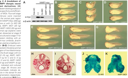
We employed an anti-sense morpholino oligo (MO)-mediated knockdown approach for loss-of-function analysis of XAWP1. For this, we designed two different MOs (MO1 and MO2), which target distinct sequences around the translation initiation site of XAWP1 mRNA. BLAST searches against Xenopus EST database revealed that several genes have internal nucleotide substitutions, possibly due to the allotetraploidy, displaying over 93% identity with XAWP1. $VWKH·UHJLRQVDURXQGWKHWUDQVFULSWLRQLQLWLDWLRQVLWHVRIWKHVH genes are highly conserved (data not shown), most of them appear WREHWDUJHWHGHIÀFLHQWO\E\WKHWZR02V:HVWHUQEORWWLQJDOVR demonstrated that both AWP1 MO1 and MO2, but not control (Co) 02FRXOGHIÀFLHQWO\LQKLELWWKHSURGXFWLRQRI;$:3SURWHLQ)LJ 2A). To examine the morphological phenotypes of XAWP1-depleted embryos, we targeted the injection toward the dorsal region of em-bryos. As shown in Fig. 2C, AWP1 MO1-injected embryos exhibited severe defects in craniofacial structures such as the eyes, and in SLJPHQWDWLRQDQGÀQIRUPDWLRQDORQJWKHWUXQNUHJLRQRIHPEU\RV (89%, n=68), and this malformation could be partially rescued by co-injection of human AWP1 (hAWP1) RNA (Fig. 1D, 58%, n=73), which has no the target sequence of the MO and appears to be UHVLVWDQWWRLWVLQKLELWRU\HIIHFW7RIXUWKHUFRQÀUPWKHVSHFLÀFLW\ of these MO phenotypes, we also investigated whether the two MOs could synergize to impair the formation of craniofacial
struc-tures. Thus, we titrated the MOs down to levels that did not yield the phenotypes on their own. Targeted injection of either MO1 or MO2 at such suboptimal levels did not elicit any defects (Fig. 2E, F), whereas co-injection of the MOs at the same reduced doses interfered with head formation and pigmentation (74%, n=72; Fig. *SKHQRFRS\LQJWKHGHIHFWLYHHPEU\RVWKDWZHUHLQMHFWHGZLWK a high level of MO1 alone. These results thus corroborate the VSHFLÀFHIIHFWVRIWKH02VLQGLVUXSWLQJWKHFUDQLRIDFLDOVWUXFWXUHV In addition, histological analysis clearly showed that injection of AWP1 MO1, but not Co MO, could inhibit formation of the lens and multilayered retina in the eye and of head mesenchyme in the injected side of the embryo (Fig. 2 H,I). We further examined the morphology of craniofacial cartilage, a NC derivative, in XAWP1-depleted embryos. As visualized by alcian blue staining, unilateral injection of AWP1 MO1 resulted in a severe loss of craniofacial VNHOHWDOHOHPHQWVLQFOXGLQJ0HFNHO·VFHUDWREUDQFKLDODQGFHUD -tohyal cartilage in the injected side compared with the uninjected control side of embryo (Fig. 2K). In contrast, injection of Co MO had no effect on the size or morphology of the craniofacial cartilage (Fig. 2J). Taken together, these data suggest that XAWP1 plays a critical role in the formation of NC-derived structures.
;$:3LVUHTXLUHGIRUQHXUDOFUHVWVSHFLÀFDWLRQ
We next investigated the effects of depleting XAWP1 on the initial steps of NC formation. Targeted injection of AWP1 MO1, but Fig. 2. A knockdown of
XAWP1 disrupts neural
crest derivatives. (A)
Western blotting analysis of the specificity and efficien-cy of MOs. Four-cell stage embryos were injected in the animal pole region with XAWP1-Myc (200 pg) alone or with Co MO (30 ng), AWP1 MO1 (20 ng) or AWP1 MO2 (30 ng) and animal cap explants were then dissected at stage 9 and cultured to stage 11 for western blotting with an anti-myc antibody. `-actin was used as a loading con-trol. (B-G) Embryos were injected in the dorso-animal region of two blastomeres at the 8-cell stage with Co MO (10 ng), AWP1 MO1 (10 ng for C and D, 5 ng for E and G), AWP1 MO2 (30 ng) and hAWP1 (5 pg) as indicated and cultured to stage 38. Arrows and arrowheads indicate de-fects in the dorsal fin and pigmentation, respectively. sub, suboptimal. (H,I)
Four-cell stage embryos were injected in the dorso-animal region of one blastomere with AWP1 MO1 (5 ng) or Co MO (10 ng) and then subjected to paraffin sectioning and eosin staining at stage 40. An arrow indicates the defective eye in the injected side. (J,K) One blastomere of a four-cell stage embryo was injected in the dorso-animal region with AWP1 MO1 (5 ng) or Co MO (10 ng) and then stained with alcian blue at stage 45. Arrows indicate se-verely disrupted ceratobranchial and Meckel’s cartilage in the injected side. cb, ceratobranchial cartilage; mc, Meckel’s cartilage; ch, ceratohyal cartilage.
Į-myc
AWP1 MO1(sub) AWP1 MO2(sub) MO1(sub) + MO2(sub)
Slug
AWP1 MO1(sub) AWP1 MO2(sub) MO1(sub)+MO2(sub)
Co MO AWP1 MO
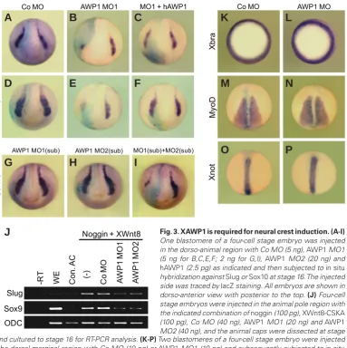
not Co MO, abrogated the expression of the NC markers, Sox10 and Slug in the injected side, compared with the uninjected side of embryos (86%, n=50 for Sox10; 65%, n=65 for Slug; Fig. 3 A,B, D,E). These repressive effects of the MO were rescued to some degree by co-expression of hAWP1 (54%, n=58 for Sox10; 67%, n=70 for Slug; Fig. 3 C,F). Moreover, while single injection of MO1 or MO2 at below threshold levels did not affect the expression of Sox10)LJ*+WKHFRLQMHFWLRQRIWKH02VDWWKHVDPHUHGXFHG FRQFHQWUDWLRQVFRXOGHIÀFLHQWO\UHSUHVVLWVH[SUHVVLRQLQWKHLQMHFWHG VLGHQ )LJ,7KLVFRQÀUPHGWKHVSHFLÀFLW\RIWKH02V LQLQKLELWLQJ1&VSHFLÀFDWLRQ1&PDUNHUVDUHLQGXFHGHFWRSLFDOO\LQ animal cap tissue by co-expressing Wnts and BMP inhibitors such as noggin and chordin (Chang and Hemmati-Brivanlou, 1998). We also used this animal cap assay to test whether XAWP1 is required for ectopic NC induction. Co-injection of Wnt8 and noggin RNAs induced the expression of Slug and Sox9 in the animal cap tissues (Fig. 3J). MO1 or MO2-mediated knockdown of XAWP1 could down-regulate this ectopic expression, whereas the co-injection of Co MO had no effect (Fig. 3J). These results thus suggest that XAWP1LVHVVHQWLDOIRUWKHHDUO\VSHFLÀFDWLRQRI1&FHOOV$VXAWP1
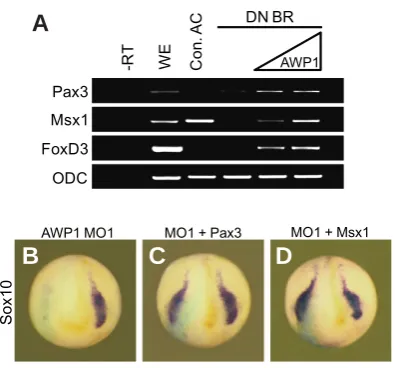
the presence of a dominant negative (DN) BMP4 receptor, which inhibits BMP signaling, suggesting a possible role for AWP1 as a PHGLDWRURI:QWDQGRU)*)VLJQDOLQJ7KXVZHIXUWKHUH[DPLQHG whether Msx1 and Pax3 might function downstream of XAWP1 to UHJXODWH1&VSHFLÀFDWLRQ7KHWDUJHWHGLQMHFWLRQRIAWP1 MO1 impeded the expression of the marker Sox10 in the injected side of WKHHPEU\RQ )LJ%ZKLFKFRXOGEHHIÀFLHQWO\UHVFXHG by co-injection of Msx1 or Pax3 RNA (77%, n=52 for Msx1; 85%, n=50 for Pax3; Fig. 4 C,D). Taken together, these data indicate that XAWP1 acts upstream of NPB regulators to specify NC cells.
XAWP1 mediates neural crest induction by Wnt and FGF signaling
Wnt/`FDWHQLQDQG)*)VLJQDOVKDYHEHHQVKRZQWREHFUXFLDO IRU1&LQGXFWLRQ6WXKOPLOOHUDQG*DUFLD&DVWUR:HH[DP -ined whether NC induction by these signals depends on XAWP1 activity. The unilateral injection of FGF8a RNA strongly enhanced the expression of Sox10 along the neural fold in the injected side of the embryo (74%, n=60; Fig. 5A), and co-injection of AWP1 MO1, but not Co MO, could impair this expansion (93%, n=45; Fig. 5B,
Fig. 3. XAWP1 is required for neural crest induction.(A-I)
One blastomere of a four-cell stage embryo was injected in the dorso-animal region with Co MO (5 ng), AWP1 MO1 (5 ng for B,C,E,F; 2 ng for G,I), AWP1 MO2 (20 ng) and
hAWP1 (2.5 pg) as indicated and then subjected to in situ hybridization against Slug or Sox10 at stage 16. The injected side was traced by lacZ staining. All embryos are shown in dorso-anterior view with posterior to the top. (J) Four-cell stage embryos were injected in the animal pole region with the indicated combination of noggin (100 pg), XWnt8-CSKA (100 pg), Co MO (40 ng), AWP1 MO1 (20 ng) and AWP1 MO2 (40 ng), and the animal caps were dissected at stage 9 and cultured to stage 16 for RT-PCR analysis. (K-P) Two blastomeres of a four-cell stage embryo were injected in the dorsal-marginal region with Co MO (10 ng) or AWP1 MO1 (10 ng) and subsequently subjected to in situ hybridization against Xbra at stage 10.5 or MyoD or Xnot at stage 14. Embryos are shown in vegetal view with dorsal to the top (K,L) or in dorsal view with anterior to the top (M-P).
is expressed in the mesoderm at the gastrula and neurula stages as described above, we next examined whether it might be involved in mesoderm induction and/or maintenance. Although the XAWP1-depleted embryos exhibited the seemingly dis-rupted trunk structure as shown above, its knockdown had no effect on the expression of an early pan-mesodermal marker, Xbra and late mesodermal mark-ers, MyoD and Xnot, suggesting that it is not critical for mesoderm formation.
XAWP1 acts upstream of QHXUDOSODWHERUGHUVSHFLÀHUV
C), suggesting a critical role of XAWP1 in FGF8a induction of the NC. In addition, injection of XWnt8-expressing plasmid resulted in ectopic rostral expansion of Sox10 in the injected side (62%, n=71; Fig. 5D), which could be potently inhibited by co-injection of AWP1 MO1 (89%, n=60) but not by Co MO (Fig. 5 E,F). Similarly, targeted expression of a DN XWnt8 RNA strongly abrogated the expression of Sox10DORQJWKHODWHUDOQHXUDOULGJHQ )LJ*DQG this suppression could be rescued by co-expressing XAWP1 RNA (55%, n=87; Fig. 5H). Furthermore, the AWP1 MO1-mediated re-pression of Sox10 exre-pression (61%, n=83; Fig. 5I) could be reverted to normal levels by co-injecting an activating form of `-catenin (pt `-catenin) or of Tcf3 (VP16-Tcf3) RNA (67%, n=87 for pt `-catenin; 65%, n=83 for VP16-Tcf3; Fig. 5 J,K). pt `-catenin is a stabilized mutant of `-catenin that lacks the phosphorylation sites required for its degradation, and expression of pt `-catenin or VP16-Tcf3 is able to induce strongly the transcription of Wnt-responsive genes in the nucleus (MacDonald et al., 2009). Thus, these data suggest that XAWP1 controls NC induction downstream of Wnt8 ligand and upstream of the `-catenin/Tcf3 complex in the Wnt signaling pathway. We conclude from this evidence that XAWP1 functions WRPHGLDWHWKHDFWLYLWLHVRI:QWDQG)*)VLJQDOLQJLQ1&LQGXFWLRQ
XAWP1 affects the stability of `-catenin
Wnt/`-catenin signaling regulates gene transcription by con-trolling the stability of `-catenin protein (MacDonald et al., 2009). Since XAWP1UHJXODWHV1&VSHFLÀFDWLRQE\DFWLQJLQDSRVLWLRQ between a Wnt ligand and the `-catenin/Tcf3 complex in the Wnt signaling cascade as shown above, we tested whether it could affect the stability of `-catenin. Interestingly, injection of AWP1 MO1, but not Co MO, reduced the level of `-catenin-myc protein, which could be reversed by co-expression of hAWP1 RNA (Fig. 6A). Furthermore, co-injection of XAWP1 up-regulated slightly its level (Fig. 6A). These results suggest that XAWP1 plays a critical role in the stabilization of `-catenin. Next, we performed reporter
DVVD\V WR FRQÀUP WKH UHTXLUHPHQW RIAWP1 for Wnt/`-catenin signaling. The expression of a constitutively active form of Wnt receptor, LRP6 (6N LRP6) or Dishevelled increased the luciferase activity driven by Wnt-responsive elements in a TOPFlash reporter gene (Fig. 6B). However, the siRNA-mediated knockdown of AWP1 could interfere with the full activation of this reporter by the Wnt signaling components, although a control siRNA had a marginal effect on reporter activation (Fig. 6B). Overall, these results sug-gest that AWP1 can affect Wnt signaling by modulating the stability of `-catenin.
Discussion
Although AWP1 has been shown to be expressed in mouse and human early embryos as well as in several human tissues (Duan et al., 2000), its physiological function remains unknown. ,QWKLVVWXG\ZHKDYHLGHQWLÀHGWKHSLYRWDOUROHRIAWP1 in NC formation during vertebrate early development. In Xenopus neu-rulae, XAWP1 is absent in the neural fold, a neural crest-forming region but is detectable in its surrounding tissues, including the Fig. 4. XAWP1 regulates neural crest induction upstream of Msx1 and
Pax3.(A) Four-cell stage embryos were injected in the animal pole region
with DN BMP4 receptor (1 ng) with or without XAWP1 (0.5, 2 ng), and the animal cap explants were excised at stage 9 and cultured to stage 11 for RT-PCR analysis. (B-D) Four-cell stage embryos were injected in the dorso-animal region of one blastomere with AWP1 MO1 (5 ng) with or without Msx1 (100 pg) or Pax3 (70 pg) as indicated and then subjected to
in situ hybridization against Sox10 at stage 16.
Sox
10
AWP1 MO1 MO1 + Pax3 MO1 + Msx1
Pax3 Msx1
ODC
-R
T
WE Con.
AC
AWP1
DN BR
FoxD3
B
C
D
A
Fig. 5. Neural crest induction by FGF or Wnt signal requires XAWP1
function.(A-K) One blastomere of a eight-cell stage embryo was injected
in the dorso-animal region as indicated with FGF8a (20 pg), XWnt8-CSKA (100 pg), DN XWnt8 (400 pg), XAWP1 (300 pg), pt `-catenin (100 pg),
VP16-Tcf3 (50 pg), Co MO (5 ng) and AWP1 MO1 (5 ng) and then subjected to in
situ hybridization against Sox10 at stage 16. Arrows indicate the expanded expression of Sox10.
XWnt8 XWnt8 + Co MO XWnt8 + AWP1 MO1
DN XWnt8 DN XWnt8 + XAWP1
Sox
10
AWP1 MO1 MO1 + pt ȕ-catenin MO1 + VP16-Tcf3
Sox
10
Sox
10
FGF8a FGF8a + Co MO FGF8a + AWP1 MO1
G
B
C
D
E
F
H
I
J
K
preplacodal ectoderm, neural ectoderm, and axial and paraxial mesoderm (Fig. 1). This expression pattern suggests that XAWP1 affects neural crest induction non-autonomously in the adjacent tissues but not in the neural crest progenitors. In support of this, the paraxial somitic mesoderm where the strong expression of XAWP1 is visible, has been shown to be a key source of neural
FUHVWLQGXFLQJVLJQDOVVXFKDV:QWDQG)*)LQXenopus (Hong et al., 2008; Monsoro-Burq et al., 2003). Furthermore, XAWP1 acts as
DPHGLDWRURI:QWDQG)*)VLJQDOLQJWRVSHFLI\QHXUDOFUHVWFHOOVDV shown in this work. In contrast, XAWP1 appears not to be critical IRUPHVRGHUPDOVSHFLÀFDWLRQDVHYLGHQFHGE\QRFKDQJHVLQWKH expression of early and late mesodermal markers in the XAWP1-depleted embryos (Fig. 3), which display the seemingly defective trunk structure. In addition to the marked expression of XAWP1 in the preplacodal ectodermal region, the craniofacial defects caused by its knockdown, in particular, the malformed eyes, suggest that preplacodal development as well as neural crest formation is also disrupted in the XAWP1-depleted embryos. The neural crest cells can organize the vertebrate eye by contributing to the alignment
Fig. 6.AWP1 is involved in Wnt/`-catenin signaling.(A) Four-cell stage
embryos were injected in the animal pole region with the indicated com-binations of `-catenin-myc (100 pg), Co MO (40 ng), AWP1 MO1 (40 ng),
hAWP1 (400 pg) and XAWP1 (400 pg), and the animal caps were excised at stage 9 and cultured to stage 11 for western blotting. (B) HEK 293T cell were transfected as indicated with 6N LRP6 (450 ng), XDsh (450 ng),
AWP1 siRNA (50 nM) and Co siRNA (50 nM). Three independent experi-ments were performed and a single representative result is shown. Error bars denote standard deviations.
RI UHWLQD DQG OHQV YLD 7*)` DQG :QW VLJQDOLQJ *URFRWW et al., 2011). Moreover, it has been shown that lens develop from the preplacodal ectoderm (PE), requiring the sequential activation of a variety of transcription factors (Ogino et al., 2012). A combination RI)*)VLJQDOZLWK:QWLQKLELWLRQSURPRWHVSUHSODFRGDOGHYHORS -ment (Streit, 2007). Xenopus FGF8 is expressed in the placodal region and anterior neural ridge at the neurula stages (Fletcher et al., 2006). Our analysis revealed that FGF8a can induce the expression of XAWP1 in naïve ectodermal tissue (Fig. 1). Taken WRJHWKHUWKHVHUHVXOWVVXJJHVWWKDW)*)VLJQDOPD\EHUHVSRQVLEOH for the expression of XAWP1 in the preplacodal ectoderm. XAWP1 is also detected in developing neural ectoderm. This expression pattern of XAWP1, in concert with its assumed mediation of Wnt signaling in the underlying mesoderm, suggests a possibility that it might affect neural induction and/or patterning. Indeed, depletion of XAWP1 was found to expand the expression of a pan-neural marker, Sox2 and a forebrain marker, Otx2 but to repress that of a hindbrain marker, Krox20 (data not shown). Neural crest induction is independent of the anterior-posterior patterning of the neural tissue (Wu et al., 2005). Therefore, XAWP1 is likely to affect these two processes separately.
XAWP1DFWLYLW\LVUHTXLUHGIRU1&LQGXFWLRQE\:QWRU)*)D ligand (Fig. 5). Notably, it acts upstream of the `-catenin/Tcf3 FRPSOH[WRFRQWURO1&LQGXFWLRQ*LYHQWKDWFGF8a induces the NC indirectly by activating the expression of Wnt8 (Hong et al., 2008), it seems likely that XAWP1PHGLDWHVERWK:QWDQG)*) induction of the NC by affecting the Wnt signaling pathway. Sup-portingly, the depletion of AWP1 impairs the stability of `-catenin and the activity of a Wnt-responsive reporter gene, indicating its possible role as a Wnt signaling component. However, there is also HYLGHQFHWKDW:QWDQG)*)VLJQDOVDFWLQSDUDOOHOWRUHJXODWH1& VSHFLÀFDWLRQ0RQVRUR%XUT et al., 2005). Thus, it is possible that these two signals converge on AWP1 activity during NC induction. 0RUHRYHU)*)LQGXFWLRQRIAWP1 expression (Fig. 1) suggests that it might have a role in mediating the crosstalk between the :QWDQG)*)VLJQDOVLQ1&IRUPDWLRQ
It appears that the mechanism underlying the activity of AWP1 in the Wnt signaling pathway involves the control of `-catenin stability DVGHVFULEHGDERYH,WLVZHOONQRZQWKDW*6.DQG&.SKRV -phorylated `-catenin undergoes `-Trcp-dependent ubiquitination for proteasome degradation (MacDonald et al., 2009). Since AWP1 has been shown to mediate polyubiquitination of proteins (Fenner et al., 2009), it is tempting to speculate that it could somehow affect the ubiquitination-mediated degradation of the `-catenin protein. In addition, AWP1LVLPSOLFDWHGLQWKHUHJXODWLRQRI1)ѣ%DFWLYLW\ (Chang et al., 2011). A recent study has shown that `-catenin acts DVDWUDQVFULSWLRQDOFRDFWLYDWRURI1)ѣ%GHSHQGHQWWUDQVFULSWLRQ (Armstrong et al.0RUHRYHU1)ѣ%VLJQDOLQJLVDOVRLQYROYHG LQWKHVSHFLÀFDWLRQRIPRXVHPHVHQFHSKDOLF1&FHOOV)XMLWD et al.,
*LYHQWKHVHÀQGLQJVLWLVSRVVLEOHWKDWWKHORVVRIIXQFWLRQ of AWP1LQWHUIHUHVZLWKWKHFRRSHUDWLRQEHWZHHQ:QWDQG1)ѣ% signaling, which could lead to the inhibition of NC induction by Wnt signaling. In addition, it has been shown that a balance between the anti-apoptotic and apoptotic activities in the neural fold plays a critical role in the proper formation of the NC (Tribulo et al., 2004). Interestingly, the level of AWP1 expression affects the incidence of DSRSWRWLFFHOOGHDWKE\UHJXODWLQJWKHDFWLYLW\RI1)ѣ%&KDQJ et al., 2011). We also observed that both gain- and loss-of-function of XAWP1 could increase the rate of apoptotic cell death in neural
tissue (data not shown). Thus, the effects of XAWP1 on apoptotic FHOOGHDWKFRXOGEHUHOHYDQWWRLWVUHJXODWLRQRI1&VSHFLÀFDWLRQ Further experiments will be necessary to test these hypotheses and to elucidate the precise mechanism underlying the activity of AWP1.
Materials and Methods
Embryo manipulation and lineage tracing
In vitro fertilization, embryo culture and microinjection were carried out
as described previously (Sive et al., 1989). The developmental stages of WKHHPEU\RVZHUHGHWHUPLQHGDFFRUGLQJWRWKH1LHXZNRRSDQG)DEHU·V normal table of development (Nieuwkoop and Faber, 1994). For lineage
tracing, `JDODFWRVLGDVHP51$/DF=SJZDVFRLQMHFWHGZLWK02V
DQGRWKHU51$VDQGLWVDFWLYLW\ZDVYLVXDOL]HGZLWKWKH;*DOVXEVWUDWH (Duchefa Biochemie).
Plasmid constructs, MOs and RNA synthesis
The complete coding region of Xenopus AWP1*HQH%DQN$FFHVVLRQ
1R%&ZDVDPSOLÀHGE\3&5ZLWKWKHSULPHUV·$7*$77$7*
-*&*&$**$*·IRUZDUGDQG·77$7$7&77&7*$$7&7777&7&&·
UHYHUVH DQG WKHQ VXEFORQHG LQWR D S*(0 7 YHFWRU XAWP1-T). The
DNA fragment, which was produced by digesting the XAWP1-T construct with EcoRI and SpeI restriction enzymes, was inserted into the EcoRI/
XbaI sites of a pCS2 vector (XAWP1-CS2+). The PCR product
encom-passing the coding region and MO target sites was generated with the SULPHUV ·&***$7&&&**&7$&**77&&&*77*&$7*· IRUZDUG DQG · &*&**$7&&*&*7$7&77&7*$$7&7777&7&· UHYHUVH and then inserted into the BamHI site of a pCS2+-Myc vector to produce a XAWP1-Myc construct. For sense mRNA synthesis, XAWP1-CS2+ and XAWP1-Myc constructs were linearized with NotI and transcribed with Sp6 RNA polymerase. The coding region of human AWP1*HQH -%DQN$FFHVVLRQ1R$-ZDVDPSOLÀHGE\3&5ZLWKWKHSULPHUV · **$$77&&$7**&7&$$*$$$&7$$7· IRUZDUG DQG ·7*&7& -7$*$*&7&$$$7&7777**$7&77· UHYHUVH DQG LQVHUWHG LQWR WKH
EcoRI/XbaI sites of a pCS2+-Myc vector to generate the hAWP1-Myc
construct. Capped mRNAs were in vitro synthesized with the mMessage P0DFKLQHNLW$PELRQ$QWLVHQVH02VZHUHSXUFKDVHGIURP*HQH7RROV
Whole-mount in situ hybridization was performed as described previ-ously (Harland, 1991). Anti-sense RNA probes were in vitro synthesized with digoxigenin-labeled nucleotides. An anti-sense XAWP1 probe was generated by transcribing the SpeI-linearized XAWP1-T construct with T7 RNA polymerase. BM purple (Roche) was used as a substrate for the alkaline phosphatase.
Western blotting
$QLPDOFDSWLVVXHVZHUHKRPRJHQL]HGLQ7ULWRQ;O\VLVEXIIHU P07ULV+&O7ULWRQ;P01D&OJO\FHUROP0(*7$ 1.5 mM MgCl2, 1 mM DTT, 1 mM sodium orthovanadate, 50 mM NaF, 10 +g/ml aprotinin, 10 +g/ml leupeptin). Equal amounts of protein were VHSDUDWHGE\6'63$*(:HVWHUQEORWWLQJZDVFDUULHGRXWDFFRUGLQJ to a standard protocol with anti-Myc (1:1000, Santa Cruz) and anti-`-actin (1:1000, Santa Cruz) antibodies.
RT-PCR
For RT-PCR analysis, total RNA was extracted from whole embryos and animal cap explants with TRI Reagent (Molecular Research Center) and treated with RNase-free DNase I (Roche Molecular Biochemicals) to remove genomic DNA. RNA was transcribed with M-MLV reverse transcriptase
3URPHJDDWʝIRUKRXU3&5SURGXFWVZHUHDQDO\]HGRQDJDURVH gels. The numbers of PCR cycles for each primer set were determined HPSLULFDOO\WRPDLQWDLQDPSOLÀFDWLRQLQWKHOLQHDUUDQJH7KHVHTXHQFHVRI the PCR primers used are as follows: XAWP1·7$&$&$**&&$*&7& -*77*&&·IRUZDUG·$$&**7*$*7&&&*&$*$$7$&*·UHYHUVH
Slug (Mizuseki et al., 1998); Sox9 (Monsoro-Burq et al., 2003); Pax3 (de
Croze et al., 2011); and Msx1 (Suzuki et al., 1997).
Histology and cartilage staining
)RUKLVWRORJ\HPEU\RVZHUHÀ[HGLQ0(0)$DWVWDJHDQGHPEHGGHG in Paraplast (Leica). Sections were cut at a thickness of 10 +m on a rotary microtome and stained with eosin (Sigma). For cartilage staining, embryos ZHUHÀ[HGLQ0(0)$DWVWDJHGHK\GUDWHGLQHWKDQRODQGVWDLQHGIRU three nights in 0.04% alcian blue/30% acetic acid in ethanol. Embryos were then washed extensively with ethanol and rehydrated in 2% KOH solution. Finally, the embryos were washed in 20% glycerol/2% KOH for at least 1 hour and dehydrated through a glycerol series into 80% glycerol. Cranial cartilage was manually dissected out and photographed.
Luciferase reporter assay
)RUWKH723ÁDVKUHSRUWHUDVVD\+(.7FHOOVZHUHWUDQVIHFWHGLQ 12-well dishes with Lipofectamine (Invitrogen). Approximately 48 hours after VL51$WUDQVIHFWLRQHDFKZHOOUHFHLYHGQJRI723ÁDVKUHSRUWHUSODVPLG (Upstate) and 1 ng of pRL-TK Renilla vector (as an internal control). Cells were lysed 24 hours after plasmid transfection and luciferase activity was measured with a dual luciferase assay system (Promega).
Acknowledgements
This work was supported by the National Research Foundation of Korea(NRF) MRC grant funded by the Korea government (MSIP)(2008-0062286).
References
$%8(/0$*'0*$5&,$025$/(6&DQG:+((/(5*1)UL]]OHG
mediates canonical Wnt signaling in neural crest induction. Dev Biol 298: 285-298.
$5067521*1-)$*2772)3527+0$11&DQG58335$0D -ternal Wnt/beta-catenin signaling coactivates transcription through NF-kappaB binding sites during Xenopus axis formation. PLoS One 7: e36136.
&+$1*&DQG+(00$7,%5,9$1/28$1HXUDOFUHVWLQGXFWLRQE\;ZQW%
in Xenopus. Dev Biol 194: 129-134.
&+$1*(-+$-.$1*66/((=+DQG.,0++$:3ELQGVWR
tumor necrosis factor receptor-associated factor 2 (TRAF2) and is involved in TRAF2-mediated nuclear factor-kappaB signaling. Int J Biochem Cell Biol 43: 1612-1620.
'(&52=(10$&=.2:,$.)DQG0216252%854$+5HLWHUDWLYH
AP2a activity controls sequential steps in the neural crest gene regulatory network.
Proc Natl Acad Sci USA 108: 155-160.
DUAN, W., SUN, B., LI, T.W., TAN, B.J., LEE, M.K. and TEO, T.S. (2000). Cloning and characterization of AWP1, a novel protein that associates with serine/threonine kinase PRK1 in vivo. Gene 256: 113-121.
ELKOUBY, Y.M., ELIAS, S., CASEY, E.S., BLYTHE, S.A., TSABAR, N., KLEIN, P.S., ROOT, H., LIU, K.J. and FRANK, D. (2010). Mesodermal Wnt signaling organizes the neural plate via Meis3. Development 137: 1531-1541.
)(11(5%-6&$11(//0DQG35(+1-+,GHQWLÀFDWLRQRISRO\XELTXLWLQ
binding proteins involved in NF-kappaB signaling using protein arrays. Biochim
Biophys Acta 1794: 1010-1016.
)/(7&+(5 5% %$.(5 -& DQG +$5/$1' 50 )*) VSOLFHIRUPV
mediate early mesoderm and posterior neural tissue formation in Xenopus.
Development 133: 1703-1714.
FUJITA, K., YASUI, S., SHINOHARA, T. and ITO, K. (2011). Interaction between NF-kappaB signaling and Notch signaling in gliogenesis of mouse mesencephalic neural crest cells. Mech Dev 128: 496-509.
-dermal Wnt function as a neural crest inducer. Science 297: 848-851.
*52&2777-2+16216%$,/(<$3DQG675(,7$1HXUDOFUHVWFHOOV RUJDQL]HWKHH\HYLD7*)EHWDDQGFDQRQLFDO:QWVLJQDOOLQJNat Commun 2: 265.
HARLAND, R.M. (1991). In situ hybridization: an improved whole-mount method for
Xenopus embryos. Methods Cell Biol 36: 685-695.
+$66/(5 & &58&,$7 &0 +8$1* </ .85,<$0$ 6 0$<25 5 DQG
NIEHRS, C. (2007). Kremen is required for neural crest induction in Xenopus and promotes LRP6-mediated Wnt signaling. Development 134: 4255-4263.
+((*758(6'(// ( DQG /$%211( & $ VOXJ D IR[ D SDLU RI VR[
transcriptional responses to neural crest inducing signals. Birth Defects Res C
Embryo Today 72: 124-139.
+21*&63$5.%<DQG6$,17-($11(7-3)JIDLQGXFHVQHXUDO
crest indirectly through the activation of Wnt8 in the paraxial mesoderm.
Devel-opment 135: 3903-3910.
IKEYA, M., LEE, S.M., JOHNSON, J.E., MCMAHON, A.P. and TAKADA, S. (1997). Wnt signalling required for expansion of neural crest and CNS progenitors.
Nature 389: 966-970.
KIMELMAN, D. (2006). Mesoderm induction: from caps to chips. Nat Rev Genet 7: 360-372.
LE DOUARIN, N.M. and DUPIN, E. (2003). Multipotentiality of the neural crest. Curr
Opin Genet Dev 13: 529-536.
/(:,6-/%211(5-02'5(//05$*/$1'-:022157'256.< 5,DQG5$,%/(':5HLWHUDWHG:QWVLJQDOLQJGXULQJ]HEUDÀVKQHXUDO
crest development. Development 131: 1299-1308.
0$&'21$/' %7 7$0$, . DQG +( ; :QWEHWDFDWHQLQ VLJQDOLQJ
components, mechanisms, and diseases. Dev Cell 17: 9-26.
0,<$7$12.80272.08.$,612*8&+,0DQG)8-,.,<$:3 =)$1'IXQFWLRQVLQ3H[H[SRUWE\LQWHUDFWLQJZLWKF\VPRQRXELTXLWLQDWHG3H[
and Pex6 AAA ATPase. 7UDIÀF 13: 168-183.
0,=86(.,..,6+,00$768,01$.$1,6+,6DQG6$6$,<Xenopus
=LFUHODWHGDQG6R[WZRIDFWRUVLQGXFHGE\FKRUGLQKDYHGLVWLQFWDFWLYLWLHV
in the initiation of neural induction. Development 125: 579-587.
0216252%854$+ )/(7&+(5 5% DQG +$5/$1' 50 1HXUDO
crest induction by paraxial mesoderm in XenopusHPEU\RVUHTXLUHV)*)VLJQDOV
Development 130: 3111-3124.
0216252%854$+ :$1* ( DQG +$5/$1' 5 0V[ DQG 3D[
FRRSHUDWHWRPHGLDWH)*)DQG:17VLJQDOVGXULQJXenopus neural crest
induc-tion. Dev Cell 8: 167-178.
NIEUWKOOP, P.D. and FABER, J. (1994). Normal table of Xenopus laevis (Daudin):
a systematical and chronological survey of the development from the fertilized egg till the end of metamorphosis*DUODQG3XE1HZ<RUN
2*,12+2&+,+5(=$+0DQG<$68'$.7UDQVFULSWLRQIDFWRUV
involved in lens development from the preplacodal ectoderm. Dev Biol 363: 333-347.
35$6$' 06 6$8.$63(1*/(5 7 DQG /$%211( & ,QGXFWLRQ RI
the neural crest state: control of stem cell attributes by gene regulatory, post-transcriptional and epigenetic interactions. Dev Biol 366: 10-21.
6$8.$63(1*/(5 7 DQG %5211(5)5$6(5 0 $ JHQH UHJXODWRU\
network orchestrates neural crest formation. Nat Rev Mol Cell Biol 9: 557-568.
SIVE, H.L., HATTORI, K. and WEINTRAUB, H. (1989). Progressive determination during formation of the anteroposterior axis in Xenopus laevis. Cell 58: 171-180.
67(9(1721%&$5021$)217$,1(&DQG0$<255*HQHWLFQHW
-ZRUNGXULQJQHXUDOFUHVWLQGXFWLRQIURPFHOOVSHFLÀFDWLRQWRFHOOVXUYLYDOSemin Cell Dev Biol 16: 647-654.
STREIT, A. (2007). The preplacodal region: an ectodermal domain with multipotential progenitors that contribute to sense organs and cranial sensory ganglia. Int J
Dev Biol 51: 447-461.
678+/0,//(57-DQG*$5&,$&$67520,&XUUHQWSHUVSHFWLYHVRIWKH
signaling pathways directing neural crest induction. Cell Mol Life Sci 69: 3715-3737.
68=8.,$8(121DQG+(00$7,%5,9$1/28$Xenopus msx1 mediates
epidermal induction and neural inhibition by BMP4. Development 124: 3037-3044.
TAMAI, K., SEMENOV, M., KATO, Y., SPOKONY, R., LIU, C., KATSUYAMA, Y., HESS,
)6$,17-($11(7-3DQG+(;/'/UHFHSWRUUHODWHGSURWHLQVLQ:QW
signal transduction. Nature 407: 530-535.
THEVENEAU, E. and MAYOR, R. (2012). Neural crest delamination and migration: from epithelium-to-mesenchyme transition to collective cell migration. Dev Biol 366: 34-54.
75,%8/2 &$<%$5 0- 6$1&+(= 66 DQG 0$<25 5 $ EDODQFH
between the anti-apoptotic activity of Slug and the apoptotic activity of msx1 is required for the proper development of the neural crest. Dev Biol 275: 325-342.
:8-<$1*-DQG./(,1361HXUDOFUHVWLQGXFWLRQE\WKHFDQRQLFDO
Wnt pathway can be dissociated from anterior-posterior neural patterning in
Byung-Yong Park, Chang-Soo Hong, Faraz A. Sohail and Jean-Pierre Saint-Jeannet Int. J. Dev. Biol. (2009) 53: 535-540
http://dx.doi.org/10.1387/ijdb.092855bp
5 yr ISI Impact Factor (2011) = 2.959
Msx1 and Msx2 have shared essential functions in neural crest but may be dispensable in epidermis and axis formation in Xenopus
Deepak Khadka, Ting Luo and Thomas D. Sargent Int. J. Dev. Biol. (2006) 50: 499-502
http://dx.doi.org/10.1387/ijdb.052115dk
Early stages of neural crest ontogeny: formation and regulation of cell delamination
Chaya Kalcheim and Tal Burstyn-Cohen Int. J. Dev. Biol. (2005) 49: 105-116 http://dx.doi.org/10.1387/ijdb.041949ck
Origins and plasticity of neural crest cells and their roles in jaw and craniofacial evolution
Paul A Trainor, Kristin R Melton and Miguel Manzanares Int. J. Dev. Biol. (2003) 47: 541-553
http://www.intjdevbiol.com/web/paper/14756330
;HUODQRYHO&16VSHFLÀFVHFUHWRU\SURWHLQHVWDEOLVKHVWKHERXQGDU\EHWZHHQQHXUDO
plate and neural crest
S Kuriyama and T Kinoshita Int. J. Dev. Biol. (2001) 45: 845-852