International Journal of New Innovations in Engineering and Technology
Stress responses of olive flounder,
Paralichthys olivaceus
by sudden rise of
temperature in low and high water
temperature conditions
Jun Wook Hur
Bio-Monitoring Center, Sejeong 30121, Republic of Korea
Abstract- We examined effects of sudden change in water temperature (WT) on the stress responses of the cultured olive flounder, Paralichthys olivaceus in large size (FL, 30.2 cm in TL, 295.6 g in BW) and in small size (FS, 13.7 cm, 22.6 g). In Exp.Ⅰ, the WT was increased from 14 to 17℃ during a 6 h period (0.5℃/hour) and maintained at 17℃ for 3 h. The temperature was returned to 14℃ for 6 h. In Exp.Ⅰ, the temperature was increased from 14 to 20 (12 h) and 23℃ (18 h). After increasing the temperature, the fish were maintained at 23℃ for 3 h, and then returned to the original temperature for 12 and 18 h after 3 h, respectively. In Exp.Ⅱ, the WT increased from 23 to 26, 29 and 32℃ by the same method of Exp.Ⅰ. During the increase from 14 to 23℃, plasma cortisol levels of FS showed no significant differences in the group of temperature increase of 6 and 9℃, but FL showed significant differences of physiological change. In Exp.Ⅱ, plasma cortisol levels at 23℃ were 2.4 ng/mL in FL and 0.1 ng/mL in FS. In the increase from 23 to 26℃, the cortisol levels were 20.7 ng/mL in FL and 16.2 ng/mL in FS at 6 h. In the 6℃ increase group, the levels increased to 14.9 ng/mL in FL and 5.5 ng/mL in FS at 12 h. In the 9℃ increase group, the levels increased to 48.5 ng/mL in FL and 10.6 ng/mL in FS at 18 h. The glucose levels of FL in the 9℃ increase group increased from 30.0 (23℃) to 59.0 mg/mL at 18 h and 48.5 mg/mL at 21 h. Lactic acid levels of FL in the 9℃ increase group were increased from 0.7 to 2.6 mmol/L at 18 h to 3.6 mmol/L at 21 h and to 1.7 mmol/L at 51 h. The survival rate was also very high during times of temperature increases and the stress response also occurred for maintenance of homeostasis following, but it was recovered in a short period of time. But there was a significant change in respiration rate by increasing the temperature from 29 to 32℃. In this study, a single stress event of a rise and drop of WT was given. It is necessary to study stress responses to repeated WT changes in future study.
Keywords- olive flounder, Paralichthys olivaceus, water temperature, stress, cortisol
I. INTRODUCTION
Physiological stresses of fish in fish farming are mainly induced from artificial factors [1-2] such as changes of water temperature [3-6], salinity [7-8], rearing density, handling, confinement and transport [9].
reported that fish showed primary, secondary, and tertiary responses against the stress [13]. The primary response showed rapid exchange of plasma catecholamine and corticosteroid. When this response to stressful condition exceeded normal level, harmful secondary and tertiary responses occurred. Therefore, stress induces decreased energy metabolism, decreased growth rate and distracted reproduction.
WT change, one environmental stress factors, directly affects the metabolism of rearing fish. Barton and Iwama suggested that a rapid change of WT resulted in the changes of in vivo metabolism and hematology [10]. Rapid drop of WT such as cold-water mass, often occurring along the coast of eastern waters of Korea in summer, affects the growth and survival of farm fish [3-6]. On the other hand, the increasing of in WT due to heated effluent water from power plant, and high WT during the summer season affects also the health of fish in culture farm. Water at about 7℃ higher than natural seawater is made by the cooling system in power plant. This heated effluent water caused rapid change in WT at an adjacent coast. The rearing fish cannot easily adapt to such temperature changes and the growth and survival of farm fish will be directly influenced by these changes. To obtain basic data on the stress response of farm fish to heated effluent water, we studied the stress responses of olive flounder, Paralichthys olivaceus to sudden changes in WT.
II. MATERIALS AND METHODS
2.1 Preparation of the experimental fish and conditions
Olive flounder (large size, FL, mean length: 30.2±0.5 cm, weight: 295.6±13.0; small size, FS, 13.7±0.1 cm, 22.6±0.6 g) were purchased from Finfish Research Center, National Fisheries Research and Development Institute, Korea, and kept for 3 weeks under a natural photoperiod in the tank supplied with a seawater-running system. Before the initiation of the feeding trial, fish were acclimated to experimental conditions for 2 weeks. The WT and salinity of the seawater during the period ranged from 19 to 21℃ and from 34 to 35 g/L, respectively. During the acclimation and experimental period, fish were fed a commercial extruded pellet (Ewha Oil and Fat Industry Co. Ltd., Korea) containing 56.0% crude protein and 12.0% crude lipid twice a day. Fish in the each group were hand-fed to apparent satiation, 100% of satiation, twice a day at 09:00 and 18:00 h. Uneaten feed was removed 30 min after feeding and deducted from feed consumption calculations.
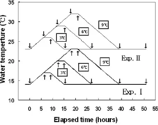
An experiment tank of 270 L flow-through (water volume: 250 L) was used, and the experiments were conducted by manipulation WT (two types, Fig. 1 in a running seawater culture system. Control of WT was automatically regulated by seawater temperature control system (Hana Com., Korea).
International Journal of New Innovations in Engineering and Technology
Figure 1. WT changes designed for the Exp. Ⅰ and Ⅱ.
2.2 Blood collection and analysis
The bloods of experimental fish were sampled from different tanks just before and after, 3 h after the WT increase and at the time returning to the original WT, and at the end of the experiment. Blood samples were collected from the caudal blood vessel complex using heparinized syringes within 1 min without anesthesia. Hematocrit, red blood cells, and hemoglobin were analyzed immediately using an automatic blood analyzer (Excell 500, USA). Blood samples were kept in 2-mL vacuum containers treated with sodium fluoride/potassium oxalate (Vacutainer, UK) and in 1.5-mL polypropylene microcentrifuge tubes held on ice for less than 5 min before centrifugation at 5,600 g for 5 min. Plasma was then collected and stored in a deep freeze (CLN-500 UW Nihon Freezer; Nihon Co., Japan) at –80℃ until analysis.
2.3 Statistical analysis
The experiment was performed in triplicate and results are reported as means ± SD unless otherwise stated. Data were analyzed by one-way ANOVA with the SPSS statistical package (SPSS Inc., USA). Means were separated by using Duncan's multiple range test and were considered significantly different at P<0.05.
III. Results
3.1 Plasma cortisol, glucose and lactic acid
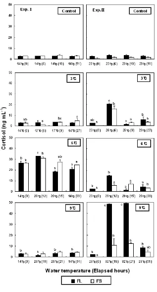
In the control group of low-WT condition, plasma cortisol levels ranged 2.7±0.1-3.1±0.1 ng/mL (FL) and 3.0±0.3-3.6±0.8 ng/mL (FS) during the experimental period (Fig. 2). The levels of the control group showed no significant differences to 3.0±0.4 ng/mL in FL and 2.8±0.8 ng/mLin FS at the beginning of the experiments of the experiment group. In the increase of 3℃, in FS showed no significant differences at 6 (1.3±0.2 ng/mL) and 9 h (3.7±0.2 ng/mL), but in the decrease from 17 to 14℃, it was significantly increased to 5.2±1.8 ng/mL at 15 h. In high-WT conditions, the cortisol levels from increasing from 23 to 26℃ were significantly increased in all FL (20.7±0.6 ng/mL) and FS (16.2±2.6 ng/mL) at 6 h. In the increase of 6℃, the cortisol levels of FL and FS was significantly increased to 14.9±0.5 and 5.5±1.5 ng/mL at 12 h, respectively. In the increase of 9℃ the cortisol levels were significantly increased to 48.5±2.5 ng/mL in FL and 10.6±6.2 ng/mL in FS at 18 h.
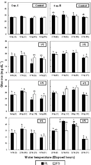
Plasma glucose levels were shown in Fig. 3. In increase of 6 and 9℃ in low-WT conditions, the levels of FS was significantly increased from 26.5±0.7 to 31.0±1.4 mg/mL at 12 h and to 32.0±0.0 mg/mL at 18 h. In the case of FL, the levels were significantly increased from 30.0±0.0 (at the beginning of experiments) to 59.0±1.4 mg/mL at 18 h and 48.5±5.0 mg/mL at 21 h by increasing 9℃.
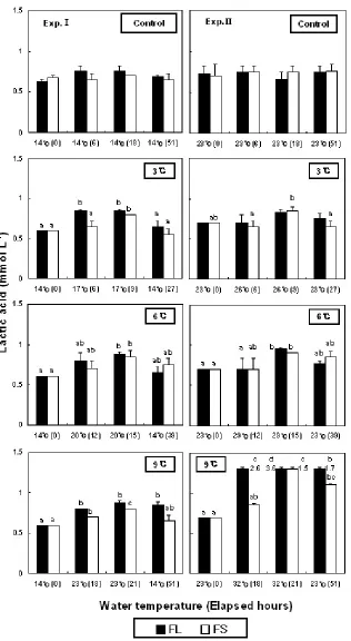
In low-WT conditions, the lactic acid levels from increasing 3℃ were significantly increased from 0.6±0.0 (FL and FS) to 0.9±0.0 (FL), 0.9±0.1 mmol/L (FS) at 15 h, respectively (Fig. 4). In high-WT condition, the levels of FL from an increase of 9℃ were significantly increased from 0.7±0.0 to 2.6±0.5 (18 h), 3.6±0.1 (21 h) and 1.7±0.1 mmol/L (51 h).
3.2 Hematocrit, hemoglobin and osmolality
International Journal of New Innovations in Engineering and Technology
International Journal of New Innovations in Engineering and Technology
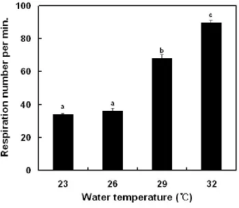
Figure 5. Variations of respiration number in olive flounder, Paralichthys olivaceus after WT changes.
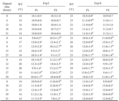
Hemoglobin levels in low and high-WT conditions were shown in Table 3. Hemoglobin levels in low-WT during the experimental period showed no significant differences from the control groups. However, by increasing WT from 23 to 26℃ of FL, hemoglobin was significantly increased from 10.0±1.0 g/dL at the beginning of experiments to 12.0±0.9 g/dL at 6 h. In the increase of 9℃ of FL, hemoglobin was increased from 11.0±1.6 to 15.0±1.1 g/dL at 21 h. In the increase of 9℃ of FS, it was increased from 8.9±0.8 to 13.0±0.5 g/dL at 21 h.
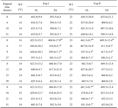
Plasma osmolality levels in low-WT and high-WT conditions are shown in Table 4. The levels of FL and FS in low-WT during experimental period showed no significant differences, ranging from 433.5±7.8-443.0±9.9 mOsm/kg (FL) and 390.0±7.1-394.5±7.8 mOsm/kg (FS), respectively. The levels showed no differences in increasing from 14 to 17, 20 and 23℃, but the levels from increasing by 9℃ in high-WT were increased from 441.5±0.7 mOsm/kg at the beginning of experiments to 474.0±2.8 mOsm/kg at 18 h.
3.3 Respiration number and survival
International Journal of New Innovations in Engineering and Technology until the end of the experiment in low-WT condition. In high-WT condition, they were 100% (FL and FS) by increasing 3 and 6℃, but in increasing 9℃, survival was 93.3% in FL (no data).
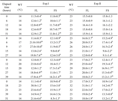
Table 1. Variations of blood hematocrit (%) in olive flounder, Paralichthys olivaceus after WT changes
Values are means ± SD (FL; n=6, FS; n=8) for experiments run on two occasions. Means sharing the same superscripted letter are not significantly different (Duncan’s multiple range test P>0.05). FL: olive flounder in large group, FS: olive flounder in small group.
Elapsed time (hours)
WT (℃)
Exp.Ⅰ WT
(℃)
Exp.Ⅱ
FL FS FL FS
0 14 11.5±0.4a 11.8±0.5b 23 15.5±0.8 15.8±1.3
6 14 12.6±1.2b 10.6±1.1a 23 15.4±0.9 16.1±1.2
12 14 12.0±0.9ab 11.7±0.8ab 23 16.4±1.0 16.1±2.2
18 14 12.6±0.9b 10.7±0.7ab 23 15.8±0.8 15.0±1.6
51 14 12.9±1.2b 11.0±1.2ab 23 15.9±1.4 15.9±1.3
0 14 11.6±0.3a 12.1±0.9b 23 16.0±3.7b 15.2±4.9b
6 17 21.0±10.0ab 13.2±3.5b 26 20.0±5.3c 17.4±1.4b
9 17 27.0±10.0b 11.9±0.5b 26 24.0±1.3c 16.5±2.4b
15 14 13.0±2.6a 9.0±0.8a 23 21.0±1.1c 9.6±2.2a
27 14 19.0±7.6ab 12.6±3.0b 23 12.0±0.4a 9.1±1.4a
0 14 12.0±0.3a 12.3±4.0a 23 17.0±2.7a 12.4±1.1a
12 20 23.0±8.6b 18.4±5.1c 29 25.0±4.6b 19.3±4.3d
15 20 12.0±1.2a 17.3±3.6bc 29 20.0±1.5a 16.9±1.7cd
27 14 18.0±6.9ab 11.0±1.7a 23 20.0±1.5a 15.3±0.4bc
39 14 17.0±4.5ab 14.2±1.4ab 23 18.0±2.1a 13.2±1.3ab
0 14 11.1±0.4a 10.9±0.4b 23 17.0±3.3a 10.9±1.6a
18 23 30.8±1.2c 15.6±3.0c 32 31.0±1.1c 14.2±0.7b
21 23 23.6±8.6b 15.9±1.5c 32 22.0±3.6b 17.0±2.3c
39 14 14.9±4.2a 14.4±3.5c 23 19.0±2.4a 13.3±0.8b
Table 2. Variations of blood hemoglobin (gdL-1) in olive flounder, Paralichthys olivaceus after WT changes
Values are means ± SD (FL; n=6, FS; n=8) for experiments run on two occasions. Means sharing the same superscripted letter are not significantly different (Duncan’s multiple range test P>0.05). FL: olive flounder in large group, FS: olive flounder in small group.
Elapsed time (hours)
WT (℃)
Exp.Ⅰ WT
(℃)
Exp.Ⅱ
FL FS FL FS
0 14 10.1±0.5 10.3±1.0 23 10.5±0.8a 10.9±0.7
6 14 10.9±0.8 10.8±0.7 23 11.3±0.9ab 11.0±1.1
12 14 10.8±1.0 10.4±1.4 23 11.9±0.6b 11.5±1.0
18 14 10.9±0.8 10.7±2.7 23 11.6±1.1b 11.2±0.7
51 14 10.8±0.9 10.4±0.6 23 11.8±1.0b 11.3±1.1
0 14 9.8±0.5a 10.5±1.2ab 23 10.0±1.0a 11.0±0.8ab
6 17 12.0±2.6a 12.4±2.1b 26 12.0±0.9b 12.0±1.2b
9 17 12.9±2.8a 10.2±2.7ab 26 12.0±1.0b 11.0±1.3ab
15 14 10.6±3.0a 9.5±3.3a 23 12.0±2.8b 10.0±1.7a
27 14 10.3±1.4a 9.1±1.3a 23 12.0±1.1ab 12.0±1.4ab
0 14 10.1±0.5a 11.2±1.4ab 23 12.0±1.4ab 10.0±2.0a
12 20 12.3±2.0b 14.6±2.5c 29 12.0±2.0a 9.9±1.8a
15 20 9.9±1.6a 13.2±2.7bc 29 14.0±1.7bc 12.0±1.3b
27 14 11.4±2.4ab 12.0±2.3ab 23 13.0±2.5abc 9.4±1.1a
39 14 10.4±1.3ab 10.6±0.8a 23 14.0±1.0c 11.0±1.4ab
0 14 10.9±0.6a 11.0±0.4bc 23 11.0±1.6a 8.9±0.8a
18 23 11.5±0.4a 12.0±0.3cd 32 14.0±0.5b 13.0±0.5c
21 23 12.4±1.9a 13.0±0.7d 32 15.0±1.1c 12.0±0.5c
39 14 11.2±1.2a 11.0±1.1b 23 12.0±1.0a 10.0±0.9b
International Journal of New Innovations in Engineering and Technology
Table 3. Variations of plasma osmolality (mOsm/kg) in olive flounder, Paralichthys olivaceus after WT changes
Values are means ± SD (FL; n=6, FS; n=8) for experiments run on two occasions. Means sharing the same superscripted letter are not significantly different (Duncan’s multiple range test P>0.05). FL: olive flounder in large group, FS: olive flounder in small group.
IV. DISCUSSION
Recently, many studies have been conducted out on stress response, reproduction ability, and metabolic physiology of fish through the hypothalamus-pituitary-interrnal (HPI) axis response. Barton and Iwama suggested that fish could adequately cope with stress from external environment changes but the stress above a threshold decreased physiological activity of fish and harmed health [10]. Fish with frequent stress events have difficulty in maintaining homeostasis and demands energy to overcome it. Because the energy that should be used for body growth and vital maintenance is consumed, growth retardation occurs and mortality rate increase [6,10,14].
The reports of Chang et al. and Park et al. are the only studies related to the stress response of fish by rapid WT change in Korea [4-5]. In those reports, flounder are known to cope with single stress event very well and have good adaptation ability. Wedemeyer et al. reported that the plasma cortisol level of salmonids in rest and
non-Elapsed time (hours)
WT (℃)
Exp.Ⅰ WT
(℃)
Exp.Ⅱ
FL FS FL FS
0 14 443.0±9.9 393.5±6.4 23 420.5±29.0 425.0±21.2
6 14 434.5±7.8 394.5±7.8 23 437.0±24.0 408.0±4.2
18 14 433.5±7.8 390.0±7.1 23 425.5±13.4 407.5±10.6
51 14 435.0±5.7 393.0±5.7 23 420.0±14.1 399.5±14.8
0 14 423.5±33.2 404.0±17.0ab 23 441.5±0.7bc 449.5±13.4ab
6 17 444.0±38.2 418.0±5.7b 26 467.0±18.4c 411.5±0.7c
9 14 420.0±28.3 395.0±7.1ab 23 437.5±3.5b 417.5±3.5b
27 14 397.5±3.5 382.5±3.5a 23 405.0±7.1a 396.5±2.1a
0 14 423.5±33.2 404.0±17.0 23 441.5±0.7 449.5±13.4
12 20 449.0±5.7 417.5±21.9 29 457.0±29.7 477.5±3.5
15 14 444.5±0.7 433.0±4.2 23 454.5±6.4 468.0±4.2
39 14 425.5±6.4 421.0±1.4 23 445.5±7.8 460.0±2.8
0 14 423.5±33.2 404.0±17.0 23 441.5±0.7ab 449.5±13.4
18 23 429.0±12.7 418.0±25.5 32 474.0±2.8c 433.5±13.4
21 14 433.5±3.5 423.0±2.8 23 446.0±7.1b 432.5±3.5
stress conditions was 30-40 ng/mL [15] and Pickering and Pottinger reported less than 5 ng/mL in the best condition [16]. Cortisol levels at the beginning of this study were 2.8-3.0 (14℃), 0.1-2.4 ng/mL (23℃) similar to two other report values. Chang et al. reported that cortisol levels of the same species were 3.6 ng/mL at 18℃, 1.5 ng/mL at 20℃ and 2.0 ng/mL at 23℃ [4]. Park et al. reported that the levels were 2.5 (18℃) and 2.6 ng/mL (20℃), respectively that are very similar to our reports [5]. The reason for the lower cortisol level in rest state than other salmon families are considered to be species specific to flounder as its activity against stress, living habit, and low motor activity, as suggesting in the reports of Chang et al. and Park et al. [4-5].
No change was observed in cortisol level from WT increase in Exp.Ⅰ of this study. But glucose and lactic acid levels increased with rising WT and decreasing WT at the end of the experiment. In Exp.Ⅱ, cortisol, glucose, and lactic acid levels increased with increased WT. In Exp.Ⅰ, cortisol levels did not increase, but glucose and lactic acid levels increased. It would be the appropriate temperature for the best growth in flounder. The WT for the ideal growth of flounder is around 18℃. When WT increased from 14℃, the temperature was similar to WT for the ideal growth of flounder causing cortisol levels to recover to beginning levels (from increase of WT to first blood collect). But in Exp.Ⅱ, the increased cortisol levels due to rising WT remained at the level it was that just after increasing the WT. And the increases in glucose and lactic acid levels were shown from typical stress response.
Barton and Iwama reported that the increased rate and time of cortisol level differed by fish species in a stressed condition [10]. For example, a peak level of cortisol in Atlantic salmon, Salmo salar due to shallow water depth was shown within less than 1 h. After 2 h, the measured value was shown to be similar to the values at the beginning of the experiment. Robertson et al. reported that red drum, Sciaenops ocellatus showed a peak in cortisol levels within 1 h by the handling the stress and the value recovered to stable 3 h later [17]. Like these studies, cortisol levels increased to a peak level in 1-3 h and it recovered in 6 h when acute stress was given [16,18]. But Waring et al. reported that cortisol levels of flounder, P. flesus and Atlantic salmon increased after 1 h following the handling of the stress and recovered to the starting value after 48 h following to the handling of the stress [19]. And cortisol levels of rainbow trout, Oncorhynchus mykiss [20], sea raven, Hemitripterus americanus [21] and turbot, Scophthalamus maximus [19] recovered after 24 h following stress. And the cortisol level of brown trout, S. trutta [16] recovered in 8 h. This suggests that recovery time of cortisol levels show differences according to fish species. It is possible to explain why cortisol did not increase in Exp.Ⅰ in this study. Barton and Iwama reported that the time of plasma cortisol levels, returning back to the starting value, showed difference according to fish species, and amount and type of stress [10]. And the reason that the peak reaching time of cortisol levels differed to the type of stress in this study can be explained by above report.
International Journal of New Innovations in Engineering and Technology REFERENCES
[1] E.M. Donaldson, “The pituitary-interrenal axis as an indicator of stress in fish. In: A.D. Pickering (Editor). Stress and Fish”, Academic Press, London, pp. 11-47, 1981.
[2] G.A Wedemeyer and D.J. McLeay, “Methods for determining the tolerance of fishes to environmental stressors. In Stress and Fish (Ed. by A.D. Pickering)”, Academic Press, London, pp. 247-275, 1981.
[3] Y.J. Chang, J.W. Hur, H.K. Lim and J.K. Lee, “Stress in olive flounder (Paralichthys olivaceus) and fat cod (Hexagrammos otakii) by the sudden drop and rise of water temperature”, Journal of the Korean Fisheries Society, 34, 91-97, 2001.
[4] Y.J. Chang, M.R. Park, D.Y. Kang and B.K. Lee, “Physiological responses of cultured olive flounder (Paralichthys olivaceus) on series of lowering seawater sharply and continuously”, Journal of the Korean Fisheries Society, 1999, 32, 601-606.
[5] M.R. Park, Y.J. Chang and D.Y. Kang, “Physiological response of the cultured olive flounder (Paralichthys olivaceus) to the sharp changes of water temperature”, Journal of Aquaculture, 12, 221-228, 1999.
[6] J.W. Hur, H.K. Lim and Y.J. Chang, “Effects of repetitive temperature changes on the stress response and growth of olive flounder,
Paralichthys olivaceus”,Journal of applied animal research, 33, 49-54, 2008.
[7] C.Y. Choi, “Environmental stress-related gene expression and blood physiological responses in olive flounder (Paralichthys olivaceus) exposed to osmotic and thermal stress”, Animal Cells and Systems, 14, 17-23, 2010.
[8] M. Yuan, Q. Jia, T. Wang, L. Tang, Y. Wang and W. Lu, “Dynamic responses of prolactin, growth hormone and their receptors to hyposmotic acclimation in the olive flounder Paralichthys olivaceus”,General and comparative endocrinology, 254, 8-13, 2017.
[9] J.W. Hur, I.S. Park and Y.J. Chang, “Physiological responses of olive flounder, Paralichthys olivaceus, to a series stress during transportation process” Ichthyological Research, 54, 32-37, 2007.
[10] B.A. Barton and G.K. Iwama, “Physiological changes in fish from stress in aquaculture with emphasis on the response and effects of corticosteroids”, Annual Review Fish Diseases, 1, 3-26, 1991.
[11] A.D. Pickering, “Growth and stress in fish production”, Aquaculture, 111, 51-63, 1993.
[12] H.K. Lim and J.W. Hur, “Effects of acute and chronic air exposure on growth and stress response of juvenile olive flounder,
Paralichthys olivaceus”,Turkish journal of fisheries and aquatic sciences, 18, 143-151, 2018.
[13] M. Mazeaud, F. Mazeaud, E.M. Donaldson, “Primary and secondary effects of sterss in fish: some new data with a general review”,
Transactions of the American Fisheries Society, 106, 201-212, 1977.
[14] C.B. Schreck, “Stress and rearing of salmonids”, Aquaculture, 28, 241-249, 1982.
[15] G.A. Wedemeyer, B.A. Barton and D.J. McLeay, “Stress and acclimation. In: Schreck, C.B., Moyle, P.B (eds.) Methods for fish biology”, American Fisheries Society, Bethesda, MD, pp. 451-489, 1990.
[16] A.D. Pickering and T.G. Pottinger, “Stress responses and disease resistance in salmonid fish: Effects of chronic elevtion of plasma cortisol”, Fish physiology and biochemistry,7, 253-258, 1989.
[17] L. Robertson, P. Thomas, C.R. Arnold and J.M. Trant, “Plasma cortisol and secondary stress responses of red drum to handling, transport, rearing density, and disease outbreak”. Progress of Fish-Culture, 49, 1-12, 1987.
[18] B.A. Barton, R.E. Peter and C.R. Paulence, “Plasma cortisol levels of fingerling rainbow trout (Salmo gairdneri) at rest, and subjected to handling, confinement, transport and stocking”, Canadian journal of fisheries and aquatic sciences,37, 805-811, 1980.
[19] C.P. Waring, R.M. Stagg and M.G. Powton, “Physiological response to handling in the turbot”, Journal of Fish Biology, 48, 161-173, 1996.
[20] N.W. Pankhurst and M. Dedual, “Effects of capture and recovery on plasma levels of cortisol, latate and gonadal steroids in a natural population of rainbow trout”. Journal of Fish Biology, 45, 1013-1025, 1994.
[21] M.M. Vijayan and T.W. Moon, “The stress response and plasma disappearance of corticosteroid and glucose in a marine teleost, the sea ravan”, Canadian Journal of Zoology, 72, 379-386, 1994.
[22] J.W. Hur, Y.J. Chang, H.K. Lim and B.K Lee, “Stress responses of cultured fishes elicited by water level reduction in rearing tank and fish transference during selection process”. Journal of the Korean Fisheries Society, 34, 465-472, 2001.
[23] I.S. Park, J.W. Hur and J.W. Choi, “Hematological responses, survival, and respiratory exchange in the olive flounder, Paralichthys olivaceus, during starvation”, Asian-Australasian journal of animal sciences, 25, 1276-1284, 2012.
[24] J.W. Hur, S.R. Woo, J.H. Jo and I.S. Park, “Effects of starvation on kidney melano -macrophage centre in olive flounder, Paralichthys olivaceus (Temminck et Schlegel)”, Aquaculture Research, 37, 821-825, 2006.