25
BH
Combining Radiographic, Urodynamic and Ultrasonographic Techniques for
the Evaluation of Urinary Bladder Structure and Function in an Animal
Model
Salah AS ()
1,2, Angela Ng MH
2, Christopher Ho CK
1,2, Ismail S
1, Ruszymah BHI
2,3, Zulkifili
MDZ
1,2Department of
1Surgery and
3Physiology, Universiti Kebangsaan Malaysia Medical Centre, Jalan Yaacob
Latif, Bandar Tun Razak, 56000 Cheras, Kuala Lumpur, Malaysia
2
Tissue Engineering Centre, Universiti Kebangsaan Malaysia Medical Centre, Jalan Yaacob Latif, Bandar
Tun Razak, 56000 Cheras, Kuala Lumpur, Malaysia
Abstract
The urinary bladder can be affected by either congenital or acquired disease leading to small, noncompliant, hypertonic urinary bladder with subsequent transfer of high pressure to upper tract leading to renal function deterioration and renal failure. The aim of urinary bladder reconstruction was to restore normal structure and functions of the urinary bladder. Successful reconstructions should be confirmed by measurement and evaluation of bladder structure and functions. In this study, different modalities such as radiography (CT scan), ultrasonography, and urodynamic were used to assess urinary bladder structure and functions in an animal model. The radiographic (CT scan) and ultrasonography have mostly structural data, while urodynamic studies produce functional parameters. Using these combined modalities we could measure and determine the normal urinary bladder volume, bladder filling pressure, voiding pressure, bladder shape, outline border, three dimension (3D) configurations, locations, and bladder dimensions. Radiography showed bladder as oval, contrast filled hollow organ, localized centrally. Mean bladder volume was 1.42 ml±0.03. Ultrasonography showed bladder as oval, elongated hypo echoic urine filled organ with, wall thickness 1mm at bladder dome and 2mm at bladder base. Mean volume was 1.44 ml±0.05. Urodynamic study showed low intravesical filling pressure with a mean value of - cm H2O, while the mean voiding pressure was 18-19cmH2O. Mean bladder volume was 1.40 ml±0.02. The acquired data for the normal (control) animals may be used as a reference for further evaluations of our future research on urinary bladder reconstructions using tissue engineering technology.
Keywords:
Urinary Bladder, Radiography, Urodynamic, Ultrasound, Evaluation.
Correspondence:
Associated Prof. Dr Zulkifili MD Zainuddin, Head of Urology Unit, Department of Surgery, Universiti Kebangsaan Malaysia Medical Centre, Jalan Yaakob Latiff, 56000 Cheras, Kuala Lumpur, Malaysia. Tel: 03-91733333 ext 2315 Email: [email protected].
Date of submission: 2nd June 2011 Date of acceptance: 23rd Sept 2011 Date of publication: 3rd Oct 2011
I
ntroductionThe urinary bladder is affected by several kinds of diseases which either congenitally inherited or by acquired degenerative, inflammatory, traumatic and neoplastic disorders developed during life, which leads
to dramatic deterioration in bladder structure and functions and requiring interference for bladder damage repair. Different methods for urinary bladder reconstruction include using the native autologous tissue of urinary tract or intestinal segment for bladder augmentation and reconstruction which is considered
26 as a gold standard for bladder repair. These procedures
are limited by the shortage of the native tissue and the associated harvest morbidity. Urinary bladder reconstruction with a congenitally different functional tissue as that of colon usually associated with complications as infections, mucus productions and malignancy transformation. Reconstructions with a functionally equivalent tissue are usually associated with satisfactory results which can be achieved by tissue engineering and regenerative medicine (1,2).
The aim of urinary bladder reconstruction was to restore normal structure and functions which is strongly affected by either congenital or acquired disease leading to small, noncompliant, hypertonic urinary bladder with subsequent transfer of high pressure to upper tract leading to renal functions deterioration and renal failure (3, 4). Our successful reconstructions should be confirmed by measurement and evaluations of returned bladder structure and functions to normal value.
With the advancement in radiology, the urinary tract imaging as a result, has become more accurate. The development of new equipment offering a great selection of options, and new imaging techniques are developed. Ultrasonography, computed tomography (CT) scan, and magnetic resonance imaging (MRI) provide higher soft tissue contrast resolutions than conventional radiography. The approach in any particular case also depends greatly on the equipment and professional skills available (5).
Radiography depends on different tissue absorbance to X-ray radiations. Contrast medium is frequently employed to enhance soft tissue visualizations. Direct installation of contrast media into the urinary bladder (cystograghy) is preferred over excretory urography for better examination of the bladder. Contrast is usually installed via a transurethral catheter. Cystography and cystourethrography are important radiologic techniques for evaluation of urinary bladder and detection of vesicoureteral reflux. CT cystography (CT of the pelvis following the installation of dilute contrast medium in to the bladder) has been shown to be useful in the evaluation of urinary bladder.
In CT scanning, a thin, parallel beam of X-rays is passed through the different body tissues with different absorbance capacity and detected by an array of solid or gas detectors.
The X-ray source and the detector are rapidly rotated in the gantry around the patient. The collected X-ray transmission data digitally interpreted to reconstruct a cross-sectional image (computed tomography) (6).
Sound is the mechanical propagation of pressure changes, or waves, through a deformable medium.
Medical sonography uses ultrasound to produce images. The frequencies range used in medical practice are between 3.5 and 15 MHz. Ultrasound images formed when part of the sound that was emitted by the transducer reflected back from tissue interfaces to the transducer. Reflected sound received by the transducer is converted into electrical signals that are analyzed by computer algorithms, and rapidly converted into video images viewed directly on a real-time display. Images are rapidly updated on the display, giving an integrated cross-sectional anatomic depiction of the site studied. Applications of bladder sonography include assessment of bladder volume and wall thickness, and detection of bladder calculi and tumors. The suprapubic transabdominal approach is the most commonly used procedure (6,7).
Urodynamic study represent an important part of the evaluation of non obstructive urinary bladder voiding dysfunction. Hydrodynamics principles were used for explanation of lower urinary tract physiology. Urodynamics studies now used for evaluating voiding problems resulting from lower urinary tract disease. The basic factors of normal bladder function are bladder capacity, accommodation, sensation, contractility, voluntary control, and response to drugs. All of them can be evaluated by cystometry. Cystometry can be performed by either of two basic methods: (1) allowing physiologic filling of the bladder with secreted urine and continuously recording the intravesical pressure throughout a voiding cycle (2) by filling the bladder with water and recording the intravesical pressure against the volume of water introduced into the bladder. With regard to the first (physiologic filling) method, the assessment of bladder function is based on voided volume. The second method permits accurate determination of the volume distending the bladder and of the pressures at each level of filling. If all the basic factors are within the normal range, bladder physiology can be assumed to be normal. Evaluation of every factor has its own implication, and before a definitive conclusion is reached, must be examined in the light of associated manifestations and findings (8).
Materials and Methods
Preparation of animals and materials
27 All experiments in this study were performed in
accordance with animal ethics approval.
Three groups were prepared with six animals in each group. Animals were anaesthetized using 0.1ml KTX (Ketamine, Zoletil, Xylazine) mixture via intravenous injections. For radiography the bladder was filled with a 50% Omnipaque (Ireland) 350mg/ml contrast agent using 3Fr urethral catheters. While 6 Fr urodynamic catheters inserted via suprapubic incisions were used for direct intravesical pressure measurement in urodynamic assessment.
Radiological evaluations of urinary bladder
SkyScan Tomography (CT-Scan) with 2D and 3D image was used for radiological assessment of urinary bladder. Scout film and 3D radiography performed. Rats were anesthetized by 0.1 ml (KTX) mixture by IV injections of previously hydrated animals. Lower abdominal area were palpated to detect full urinary bladder. With mild pressure on palpable rounded bladder a limited amount of urine was expelled (around 1 ml) and replaced with contrast agent without change in urine volume. Installation of 1ml 50% contrast agent into urinary bladder through external urethral orifice via 3 Fr urethral catheters. K-Y lubricant gel was used to avoid traumatic insertions. Catheter was longitudinally aligned with the course of the urethra and gentle pressure applied by the thumb and index finger to hold the catheter within urethral lumen and to prevent contrast leakage. One milliliter of 50% contrast agent was installed into urinary bladder for retrograde cystography. A radiological instrument (SKyscan 1076 high resolution in vivo scanner) was used to perform radiological evaluation in our study with a preinstalled software program. A 65mm composite object bed was used, with a length of 400mm and a maximum scanning length of 200mm. The animal was positioned with the urinary bladder located at central position at the scanning bed and with proper fixation. Initial radiological scout film (plain film) was taken and displayed on the screen. The selected site of the scout film, (urinary bladder) lower abdominal site was selected and a full format X-ray image of the site was noted. Scanning was started by selecting scanning width 68mm and pixel size 35um, while voltage and electrical current will set automatically corresponding to current pixel size and filter chosen. This was followed by reconstruction of the image. Preliminary plain abdominal X-ray was taken for detecting the presence of any calcification or osseous abnormality in pelvic area and it’s relation with the urinary bladder image after contrast administration. Cystography X-ray film for urinary bladder visualization both pre and post voiding and for
evaluations of bladder shape, location, outline border integrity and to exclude the presence of extravasations or any other pathology. Computed tomography (CT) film with its cross sectional images and its soft tissue resolution in relation to adjacent structures provided further information’s associated with the bladder wall, its lumen and the surrounding structures. The three dimension (3D) films were obtained for evaluation of urinary bladder configuration and volume.
Ultrasonic evaluation of urinary bladder
Ultrasonography was performed using Sonosite Bothell ultrasound instrument (USA). The probe (transducer) used was a linear multifrequency probe at 10 MHz. The animals were anesthetized with 0.1 ml KTX mixture intravenous injection of previously hydrated animals. Lower abdominal area was palpated for detection a full urinary bladder. Shaving of the area for hair removal before ultrasound performance was done. Water- based gel (K-Y) gel was used to secure contact with the body and eliminate air pocket between the transducer and the skin. Scanning of the urinary bladder was performed with multifrequency probe at frequency 10 MHz as a real time imagining. Ultrasonography examinations were directed to evaluate urinary bladder by assessing the full bladder volume and shape, the presence of any pathological foreign body as bladder stones, and assessment of post voiding residual urine. Examination of bladder wall aimed to determine its normal thickness or presence of any pathological change in the bladder wall. Surrounding tissue examinations was done to make comparisons after reconstructions, such as urine leakage and extravasations.
Urodynamic evaluation of urinary bladder
28 syringe was filled by normal saline connected to the
infusion catheter and allowed to fill urinary bladder under gravity. Having the syringe elevated around 20-30 cm, urinary bladder filling rate was gradual, stepwise and satisfactory with filling rate 0,05ml/sec. The aim of our study (cystometry) was the measurement of intravesical filling pressure which normally low and does not changed (increased) during bladder filling in normal urinary bladder with normal compliance and normal accommodation. The voiding pressure which is the intravesical recorded pressure during micturation, in bladder with normal voiding pressure urine should be expelled completely without pressure transfer to upper tract. Bladder capacity (volume) measurement as baseline data for the subsequent reconstructions studies.
Formula definition for the study was explained accordingly.
However, the bladder has a power of accommodation, that is, it can maintain an almost constant intraluminal pressure throughout its filling phase regardless of the volume of fluid present, and this directly influences compliance. As the bladder progressively accommodates larger volumes with no change in intra luminal pressure, the compliance values become higher.
Compliance = Volume/Pressure
Results
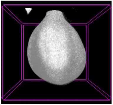
Radiography of urinary bladder using Sky-scan tomography initiated by plain abdominal X-ray scout film was taken as a preliminary film before contrast agent administration for visualization of soft tissue (urinary bladder) area (Figure 1 and 2). This film showed no pathological calcification nor radiopaque shadow, in animals with normal pelvic area. After 1ml of radio contrast agent administration, the normal urinary bladder shape wasvisualized as rounded, oval, hollow organ with smooth regular outline border, with bladder neck and proximal urethra directed caudally and bladder dome directed cranially. Urinary bladder localized centrally at lower abdominal (pelvic) area, midway between pelvic bones laterally. Urinary bladder dimensions (longitudinal, transverse, oblique and antero-posterior) were measured with mean longitudinal, transverse, oblique and antero-posterior dimension determination. Capacity of urinary bladder volume were measured while bladder full of urine using preinstalled software program with a mean urinary bladder volume of 1.42±0.03 ml (Table1). Post voiding film showed no residual urine (no more radiopaque bladder shadow)
Figure 1: Lower abdominal (Pelvic) and the urinary bladder X-ray. (A) Plain antero-posterior view. Show urinary bladder and pelvic soft tissue area as a preliminary standard X-ray view for detection of any pathological calcification at urinary bladder or the surrounding structure. No radiopaqe shadow can be detected and the bony structure was normal. (B) Cystography, lateral view. Represent urinary bladder visualization (after contrast agent administration). Showed normal full urinary bladder location, shape, boundaries (anterior and posterior) and size. (C) Cystography, anterior-posterior view. It shows normal full urinary bladder location, shape, boundaries (lateral), and size. This can be considered as a basic radiological image for further radiological evaluation of urinary bladder.
29 Table 1: Represent urinary bladder volume measurement with different methods with mean value measurement of normal urinary bladder volumeby urodynamic, radiological (CT scan) and by ultrasound means. The mean value for each method considered as a basic reference for reconstructed bladder evaluation.
Method Volume (ml) Mean volume
1 2 3 4 5 6
Urodynamic 1.42 1.38 1.39 1.43 1.41 1.40 1.40± .02
Radiography 1.39 1.44 1.46 1.38 1.41 1.42 1.42±0.03
Ultrasonography 1.40 1.39 1.47 1.53 1.41 1.43 1.44±0.05
Table 2: Represent urinary bladder dimensions measurement. Normal urinary bladder dimensions measurement (longitudinal, transverse, oblique and antero-posterior) was taken by ultrasound as a basic bladder dimensions measurement for reconstructed bladder dimensions evaluation with the normal value.
Directions of dimension Dimension measurement(cm) Mean value
1 2 3 4 5 6
Transverse 1.01 0.99 1.07 1.05 1.04 10.9 10.4±0.04
A-P 0.81 0.79 0.80 0.82 0.79 0.83 0.81±0.02
Oblique 0.86 0.89 0.86 0.89 0.86 0.92 0.88±0.02
Longitudinal 2.29 2.39 2.36 2.34 2.33 2.27 2.33±0.04
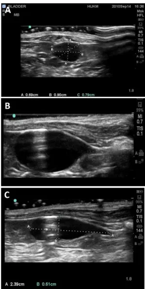
By utrasonography the full urinary bladder shape was seen as oval, elongated hollow organ with hypo echoic urine filled lumen, with bladder dome oriented cranially bladder base (neck) directed caudally (Figure 3). The urinary bladder was located centrally at the lower abdomen. The urinary bladder wall was seen as smooth, regular, border outline the urinary bladder. The mean wall thickness was 1mm at bladder dome and mean wall thickness of 2mm at bladder base. Urinary bladder dimension measurement of the full urinary bladder with mean longitudinal, transverse, oblique and the mean antero-posterior dimension (Table 2). Volume (capacity) of full urinary bladder measurement was essential part of our assessment, with a mean full urinary bladder volume was 1.44±0.05 ml. No post voiding residual urine was detected.
Urodynamic evaluation was started by connecting the outer urodynamic catheter end to pressure transducer (Figure 4). The increase in intravesical pressure was monitored continuously on a rectigraph. The inner catheter was connected to an infusion pump and the rate of infusion was set at 0.05ml/sec by allowing normal saline to fill the bladder under gravity.
Difficulties encountered during urodynamic performance were due to machine rapid infusion rate, which was substituted by filling the bladder under gravity.
Intravesical filling pressure recording showed pressure elevation to certain value then remained constant during all the time of bladder filling until full capacity of bladder was reached with a mean value of 5-6 cm H2O. Voiding pressure occurred when the bladder filling reached maximum capacity, followed by sudden rise of bladder pressure and voiding. The mean voiding pressure was -19cmH2O which sustained until bladder was completely evacuated. The mean bladder capacity (volume) was 1.40±0.02 ml.
Discussion
30 Figure 3: Ultrasound image, urinary bladder. (A) Cross
section image. (B) longitudinal section image. (C) longitudinal section with dimension. A, B and C images Showed normal urinary bladder lumen which normally seen as hypo echoic urine filled lumen. No foreign bodies and no intra luminal stones. Bladder wall seen with normal appearance and thickness. Bladder surrounding structures is free of fluid collection. With different bladder dimension recording for post reconstruction comparison.
Figure 4: Urodynamic study representative diagram, of urinary bladder urodynamic evaluation (cystometry), vertical (pressure), horizontal (volume), showing urinary bladder filling pressure curve with a mean value of (5-6 cm H2O) followed by sharp elevation of the curve which represent the voiding pressure with a mean value of (18-19 cm H2O). Normal bladder volume 1.40±0.02 ml.
ultrasonography, and urodynamic studies can be utilized.
Aiming to determine the basic data information of a normal urinary bladder structure and functions radiography (X-ray) applications using ( Sky scan 1076 scanner) computed tomography scan was carried out for the evaluation of normal urinary bladder. The plain film showed normal soft and bony tissue structure, no radiopaque shadow nor pathological calcifications was detected in normal pelvic region of non operated animals. While any radiopaque shadow may indicate urinary bladder stone formation or abnormal calcification within the bladder wall or surrounding tissue. Stone formation during reconstruction procedure may occur due to urine stagnation, at the bladder regeneration period and the usage of synthetic biomaterial (PLGA) and the stitch which is considered as foreign bodies that can represent a nidus for stone formation.
Radiography of that region after radiopaque contrast agent administration showed normal urinary bladder image (shape) as an oval rounded hollow organ filled by urine with smooth regular rounded border outline. The bladder shape maintenance after reconstructions indicate scaffold stability against surrounding tissue and intra abdominal pressure during regeneration in vivo. While bladder shape deformity may indicate that the scaffold physical properties is inadequate which lead to scaffold collapse or shrinkage. The bladder seen localized centrally at lower abdominal area between pelvic bony structures, normal urinary bladder dome oriented cranially while bladder base and neck oriented caudally. Abnormal deviation of the bladder from it’s normal position may indicate excessive fibrotic change and adhesion surrounding the bladder during regeneration.
31 The second evaluation method incorporated in our
study was the ultrasound evaluation of normal urinary bladder. It was a real time examination which complements the radiological assessment. Our purpose here was to perform data base ultrasonic evaluations of normal full urinary bladder size, volume, shape, lumen, bladder wall, surrounding tissue, and bladder evacuation.
The finding in normal non operated animals showed a hypo echoic urine filled bladder lumenfree of foreign bodies and stones. The appearance of hyper echoic shadow in the reconstructed bladder indicates stone formation. Urinary bladder storage capacity (volume) was an important parameter in our bladder reconstruction study with the mean bladder volume of 1.44±0.05ml. The size of normal urinary bladder (dimensions) are important data reference for evaluating the reconstruction success to attain normal bladder size and capacity. Longitudinal, transverse, oblique, and antero-posterior mean dimension recorded for comparison with the reconstructed bladder.
Bladder wall thickness which normally should be of certain thickness taken as important parameter for normally reconstructed bladder wall to differentiate it from pathological increased wall thickness or thin attenuated wall. The mean bladder wall thickness was 1mm at bladder dome (body) and 2mm at bladder base. Normal wall thickness indicates normal bladder wall layers arrangement. Increased (hypertrophied) muscular layers thickness reflects undesired raised intra luminal pressure while thinned bladder wall associated with bladder wall weakness. Urinary bladder border evaluation which appeared as smooth, regular, rounded out line border is important basic parameter for reconstructed bladder evaluation to detect scaffold collapse and diverticulum formation.
Visualization of urinary bladder normal surrounding tissue was taken in consideration for post operative detection of possible urinary extravasations which indicates scaffold or anastamosis defect. Post voiding residual urine measurement to evaluate normal urinary bladder proper contraction and voiding (functional) performance. No post voiding residual urine was detected within the normal non operated bladder. Considerable amount of post voiding residual urine in reconstructed bladder indicates evacuation function defect.
Urodynamic studies (cystometry) play an important role in evaluation of urinary bladder function. In our study urodynamic evaluation of normal urinary
bladder was performed to register the basic functional parameters of urinary bladder as filling pressure, voiding pressure, and bladder storage capacity (volume). Intravesical filling pressure raised gradually to mean pressure of 5-6cmH2O then remained constant until reached bladder capacity, followed by sudden rise in intravesical pressure (voiding pressure) with mean recorded voiding pressure of 18-19cmH2O. The mean measured bladder capacity was 1.40±0.02ml. Bladder filling should be with low intra luminal pressure during all the filling phase which indicates proper bladder compliance and accommodation. Voiding pressure should not be too high with subsequent upper tract pressure transfer nor too low to prevent adequate bladder emptying. Appropriate bladder capacity is important for normal storage function of urinary bladder.
Conclusion
Combined radiological, ultrasound and urodynamic studies complement each other to provide an overall and complete assessment of the urinary bladder gross structure and functions. The baseline data acquired from these modalities can be used as a reference in the evaluation of urinary bladder reconstruction using tissue engineering technology.
Acknowledgement
This study was funded by UKMMC Fundamental Grants FF-173-2010
APPROVAL NUMBER
PP/SURG/2010/ZULKIFLI/17-MARCH/292-MARCH-2010 DECEMBER-2011
References
1. Atala. A Tissue engineering in the genitourinary system. In:Atala A, Moony D,eds.Tissue engineering. Boston: Birkhauser Press,1997,149-150
2. Soergel TM, Cain MP, Misseri R, Gardner TA, Koch MO, Rink RC. Transitional cell carcinoma of the bladder following augmentation cystoplasty for the neuropathic bladder. J Urol 2004; 172(4 pt 2): 1649–51.
32 4. Snodgrass WT, Adams R. Initial urologic
management of myelomeningocele. Urol Clin North Am 2004; 31(3):427–34.
5. Su LM, Jarrett TW, Chan DY et al: Image-guided therapy in urology. J Endourol 2001; 15(1):105-10.
6. Emil A. Tanagho, Jack W. McAninch. Smiths General Urology. Radiology of the urinary tract. USA: McGraw-Hill, 2004.
7. Caoili EM, Bude RO, Higgins EJ, Hoff DL, Nghiem HV: Evaluation of sonographically guided percutaneous core biopsy of renal masses. AJR 2002; 179(2):373-8.