Expression pattern of iodotyrosine dehalogenase 1
(DEHAL1) during chick ontogeny
ANUPAMA A. MATHI
1, TEKCHAND C. GAUPALE
1, CORINNE DUPUY
2,
NISHIKANT SUBHEDAR
3and SHOBHA BHARGAVA*
,11Molecular Embryology Laboratory, Department of Zoology, University of Pune, Pune, India, 2FRE 2939 CNRS, Institut Gustave Roussy, Villejuif, France and
3Department of Pharmaceutical Sciences, R.T.M Nagpur University Campus, Nagpur, India
ABSTRACT The iodotyrosine dehalogenase1 (DEHAL1) enzyme is a transmembrane protein that belongs to the nitroreductase family and shows a highly conserved N-terminal domain. DEHAL1 is present in the liver, kidney and thyroid of mammals. DEHAL1 is known to act on diiodotyrosine (DIT) and monoiodotyrosine (MIT), and is involved in iodine recycling in relation to thyroglobulin. Here, we show the distribution of DEHAL1 during gastrulation to neurulation in developing chick. Immunocytochemistry using an anti-serum directed against the N-terminal domain (met27-trp180 fragment) of human DEHAL1 revealed labelled cells in the embryonic ectoderm, embryonic endoderm, neural plate and in the yolk platelets of the chick embryo at gastrulation stage. Distinct DEHAL1 positive cells were located in the presumptive head ectoderm, presumptive neural crest, head mesenchymal cells and in the dorsal, lateral and ventral parts of neural tube during neurulation. Some cells located at the margin of the developing notochord and somites were also DEHAL1-positive. While the functional significance of this observation is not known, it is likely that DEHAL1 might serve as an agent that regulates cell specific deiodination of MIT and DIT before the onset of thyroidal secretion. The presence of DEHAL1 in different components of the chick embryo suggests its involvement in iodine turnover prior to the formation of functional thyroid.
KEY WORDS: chick embryo, gastrulation, immunocytochemistry, iodotyrosine dehalogenase 1, neurulation
Cell proliferation, cell rearrangement and neuronal death play an important role in patterning different regions of the brain (Quick and Serrano, 2007; Vecino et al., 2004; LeDouarin, 2001; Fleming
et al., 1997; Weil et al., 1997; Ellis et al., 1991). A range of signaling molecules and their roles have been implicated in vertebrate development (Borgave and Ghaskadbi, 2009; Sanchez-Calderon et al., 2007; Patwardhan et al., 2004; Waschek et al., 1998; Hallbook et al., 1995; Ernfors et al., 1995; Hyuga et al.,
1993). Thyroid hormones are known to play an important role in the early development including neurulation of several verte-brates and previous studies have shown the occurrence of enzymes that are involved in the recycling of Iodine from thyroid hormones (Courtin et al., 2005; Gereben et al., 2004; Geyten et al., 2002; Asteria, 1998; Becker et al., 1997; Rosenkilde and Ussing, 1996).
Iodotyrosine dehalogenase 1 (DEHAL1) is a transmembrane protein, localized mainly at the apical pole of thyrocyte in the
BIOLOGY
www.intjdevbiol.com*Address correspondence to: Shobha Bhargava. Department of Zoology, University of Pune, Ganeshkhind road, Pune-411007, India. Fax: +91-020-2569-0617. e-mail: [email protected]
Accepted: 25 November 2009. Published online: 21 December 2010. Edited by: Makoto Asashima
ISSN: Online 1696-3547, Print 0214-6282
© 2010 UBC Press Printed in Spain
Abbreviations used in this paper: DEHAL, iodotyrosine dehalogenase; DIT, diiodotyrosine; MIT, monoiodotyrosine.
human thyroid (Gnidehou et al., 2004). DEHAL1 belongs to the nitroreductase family, with a highly conserved N-terminal domain located between Glu92 and Gly244 and a putative transmembrane domain between Asn213 and Gln229 (Gnidehou et al., 2004). DEHAL1 is quite different from other deiodinases. DEHAL1 acts on diiodotyrosine (L-DIT) and monoiodotyrosine (L-MIT), whereas, deiodinases act on thyroxine (T4) and triiodothyronine (T3) (Solis
the thyroid of reptiles and mammals, the enzyme has also been noticed in the liver and kidney of sheep and cattle (Roche et al., 1952; Stanbury, 1957 and 1958; Chiu and Wong, 1978; Goswami and Rosenberg, 1979).
In mammals, two isoforms of DEHAL1 protein, DEHAL1 and DEHAL1B have been reported (Moreno, 2003). Recently, the occurrence of new mRNA variant, DEHAL1C, has also been
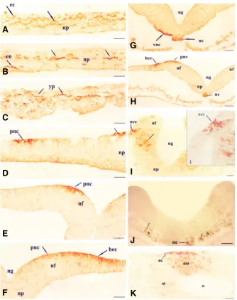
bation (stage-7), positive immunoreaction was detected in the various regions of the embryo. A very strong immunoreaction was seen in the presumptive neural crest cells (pnc) near the dorsal margin of the neural fold and the reaction was membrane bound (Fig. 1 E,F,H). Some of the cells in the ventrolateral part of the neural plate close to the developing notochord showed strong immunoreaction (Fig. 1G). The cells were spherical in shape and reported (Gnidehou et al., 2006). All three
isoforms show similarity in their N-terminal domain, but differ in their cytoplasmic C- termi-nal tail which plays a critical role in the activity of DEHAL1 isoforms (Gnidehou et al., 2006). Till date there are no reports on the occur-rence and role of DEHAL1 in a vertebrate embryo. Here we report the occurrence of DEHAL1 in the discrete regions of the chick embryo during early embryogenesis, before the onset of thyroidal secretion. Our report on the occurrence of DEHAL1 in chick embryo may provide clues to our understanding of the iodine metabolism during early embryogen-esis.
Results
Immunohistochemistry
Primitive streak and head fold stage
Following 18 hrs of incubation, during gas-trulation (stage-4), very strong DEHAL1 posi-tive reaction was observed in the area pellu-cida and also in the area opaca (Fig. 1 A-C). In the embryonic ectoderm (ec) the immunoreaction was localized on the cell mem-brane of the cells (Fig. 1A). A large number of cells in the neural plate (np) showed strong DEHAL1 positive reaction (Fig. 1 A-B). In the center of the neural plate, few cells showed DEHAL1 positive reaction (Fig. 1B). The reac-tion was distinctly localized on the membrane of cells. The yolk platelets (yp) showed strong immunoreactivity (Fig. 1C). Few cells of the embryonic endoderm (en) were also DEHAL1 immunoreactive (Fig. 1B). After 24 hrs of incu-bation, during the head fold stage (stage-6), weak immunoreactive cells were seen in the middle region of embryonic axis. DEHAL1 posi-tive reaction was seen in the presumpposi-tive neural crest cells, dorsal margin of the neural fold, and also in the embryonic ectoderm (not shown). In the posterior part of the embryo, where the primitive streak is regressing few primitive neural crest cells (pnc) and some of the cells on dorsal side of the neural plate (np) were strongly DEHAL1 reactive (Fig. 1D). DEHAL1 immunoreaction in the yolk platelets was similar as seen in stage 4 embryo.
Neurulation
During early neurulation, at 26 hrs of
incu-Fig. 1. Transverse sections of chick embryo from stage 4-10.(A-C) Transverse sections of Primitive streak stage 4. Strong DEHAL1 positive reaction was seen in the neural plate (np), embryonic ectoderm (ec), embryonic endoderm (en), and in the yolk platelets (yp). (D)
Transverse section through the posterior region of stage 6 embryo showing DEHAL1 positive reaction on the cell membrane of the presumptive neural crest cells (pnc) and on the dorsal surface of the neural plate (np). (E-H) Transverse sections of stage 7 embryo. Strong membrane bound DEHAL1 positive reaction was seen in the presumptive head ectoderm (hec) and presumptive neural crest (pnc) cells. DEHAL1 reaction was also seen in the notochord (nc) and along the ventrolateral margin of the neural plate (vnc) close to the developing notochord.
(I-J) Transverse sections of stage 8 embryo showing immunopositive DEHAL1 reaction in the neural crest cells (ncc), lateral parts of the neural plate (thick arrow), notochord (nc) and somite (s). i: Magnified view of neural crest cells (ncc) shown in Fig: I showing membrane bound DEHAL 1 immunoreaction. (K) Transverse section of stage 10 embryo showing DEHAL1 positive cells in the dorsal part of neural tube (dnt) and in the ectoderm (ec). Scale bar, 50m.
G
B
C
D
E
F
H
I
J
the reaction was localized only on the ventral margin of the cells. Strong DEHAL 1 positive reaction was also seen in the cells of notochord (nc) (Fig. 1 G,H). Some of the cells in the head ectoderm (hec) were DEHAL 1 positive (Fig. 1 F,H). As neurula-tion progressed and the neural plate started folding (stage-8), cells in the lateral part of forebrain and midbrain showed strong DEHAL1 positive reaction (Fig. 1I). Some of the neural crest cells (ncc) near the neural fold were strongly DEHAL1 positive (Fig. 1I). Few marginal cells of the developing notochord (nc) and somite (s) were also DEHAL1 reactive (Fig. 1J).
During later stages of neurulation (stage 9-10), when the neural fold fuses, overall immunoreaction pattern was similar in the forebrain and midbrain as seen in stage 8. In the forebrain, reaction was weak and restricted to the lateral part while in the
midbrain region immunoreaction was detected in the dorsal part of the neural tube (Fig. 1K). Similar reaction was also seen in the hindbrain region. A group of ectoderm cells (ec) near dorsal part of the neural tube were positive for DEHAL1 (Fig. 1K). During late neurulation (Stage 11-12), overall reaction was very weak and localized in various parts of the neural tube.
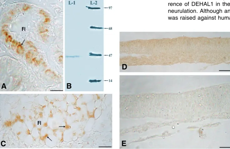
Sections of mouse and chicken thyroid gland, used as a positive control, showed positive DEHAL1 immunoreaction in the thyrocytes of the thyroid follicles (Fig. 2 A,C). Preabsorption of the primary antibody with pure human DEHAL1 antigen resulted in complete loss of immunoreaction (Fig. 2D). Similarly, omission of the primary antibody from the reaction (negative control) resulted in complete loss of immunoreaction (Fig. 2E).
Immunoblot assay
During Immunoblot assay, it was found that anti-DEHAL1 antibody used in this study recognizes a protein with an apparent molecular mass of 33 KD in the chick embryo (Fig. 2B).
Deiodinase assay
During enzyme assay, the DEHAL1 activity could be measured in the various stages (stages 4-12) of the chick embryo (Table 1).
It was found that the enzyme could act on DIT and requires NADPH as cofactor to liberate iodine. At stage 4 maximum enzyme activity (93.493 1.886 S.A. U/mg protein) was measured as compared to stage 12 (22.24 1.131 S.A. U/mg protein).
Discussion
In the present study we demonstrate for the first time occur-rence of DEHAL1 in the chick embryo during gastrulation and neurulation. Although anti-DEHAL1 antibody used in this study was raised against human DEHAL1 protein, it seems to
cross-Fig. 2. Transverse sections of mouse and chick tissue and image of immunoblot analysis. (A)
Transverse section of mouse thyroid, showing DEHAL1 positive immunoreaction in the thyrocytes (arrow) Scale bar, 50 m. (B) Immunoblot analysis. Lane-1 (crude extract of chick embryo 80g protein), lane-2 (molecular weight marker). (C) Transverse sections of chick thyroid showing DEHAL1 immunoreactivity in thyrocytes of thyroid follicles. (D) Transverse section of chick embryo showing no immunoreaction in preabsorption control. (E) Transverse section of chick embryo showing no immunoreaction in negative control (omission of primary antibody).
react with DEHAL1 polypeptide in chicken, perhaps because DEHAL1 has a highly conserved N-terminal domain located between Glu92 and Gly244 (Gnidehou, et.al., 2004). The Chicken (Gallus gallus) and human iodotyrosine deiodinase protein se-quences have shown 84% homology when aligned (GeneID: 421631, GeneID: 389434, NCBI Blast).
The immunoreactivity seems to be specific to DEHAL1, since various control procedures including preabsorption of the primary anti-body with pure DEHAL1 antigen re-sulted in complete loss of immunoreaction. Chicken and Mouse thyroid sections used as a positive control showed immunoreaction on the membrane of thyrocytes in the thyroid follicles. Similar type of DEHAL1 positive reaction was re-ported in the Human thyroid (Gnidehou, et.al., 2004). Presence of DEHAL1 was also confirmed by performing deiodinase assay. Chick Stage S.A (U/mg protein) S.A (U/mg tissue)
Stage 4 93.493 ±1.886 4.674 ±0.094
Stage 6 80.711 ±0.471 1.614 ±0.009
Stage 7 74.804 ±0.965 1.346 ±0.017
Stage 8 32.148 ±0.558 1.285 ±0.022
Stage 10 43.699 ±0.707 1.783 ±0.028
Stage 12 22.24 ±1.131 0.842 ±0.086
TABLE 1
IODOTYROSINE DEHALOGENASE ACTIVITY ON DIT AT DIFFERENT DEVELOPMENTAL STAGES OF CHICK EMBRYO
Enzyme activity was performed thrice from three independent sets and the standard deviation calculated. S.A, specific activity (U/mg protein and U/mg tissue)
B
C
D
homogenate was found to deiodinate DIT in presence of NADPH. Immunoblot analysis revealed that anti-DEHAL1 antibody used in this study recognized a protein with an apparent molecular mass of 33 KD in the chick embryo. Human DEHAL1 was also found to be in the similar molecular weight range (Gnidehou, et.al., 2004). Occurrence of DEHAL1 is documented in the thyroid, liver and kidney of several adult mammals (Friedman et al. 2006; Gnidehou
et al., 2006) and also in the thyroid of reptiles (Chiu and Wong, 1978). In these tissues, DEHAL1 is involved in rapid recycling of iodine by deiodinating diiodotyrosine (L-DIT) and monoiodotyrosine (L-MIT) released during the hydrolysis of thyroglobulin (Dunn and Dunn, 2001; Gnidehou et al., 2004). However, there is no informa-tion on the occurrence and role of DEHAL1 in developing verte-brates. Present study shows the presence of DEHAL1 in the discrete regions of chick embryo during embryogenesis and considers its functional significance.
The distribution pattern and the intensity of DEHAL1 in chick embryo seems to be specific to the developmental stages. During gastrulation, DEHAL1 was located in the presumptive ectoderm, endoderm and neural plate. However, during neurulation occur-rence became restricted and specific to some areas like presump-tive neural crest cells, head mesenchyme cells, and few cells in the neural tube, notochord, ectoderm, endoderm and somites. Yolk platelets in the area opaca were also positive for DEHAL1, from gastrulation till neurulation. Enzyme assay also revealed similar results. Maximum DEHAL1 enzyme activity was mea-sured at gastrulation (stage-4) (93.493 1.886 S.A. U/mg protein) while minimum activity was measured at late neurulation (stage-12) (22.24 1.131 S.A. U/mg protein).
Iodine metabolism seems critical during chick embryo devel-opment. T4 and T3 are deposited in the egg of chicken and quail during oogenesis and are available to the developing embryo before the onset of thyroid function (McNabb and Wilson, 1997; Prati et al., 1992). Reports suggest that the yolk platelets in the area opaca release T3 even before gastrulation (Flamant and Samarut, 1998). The released hormone diffuses from yolk to the area pellucida cells and is available to the developing embryo before the onset of its thyroidal secretion (Prati et al., 1992). T3 hormone is also produced in the Hensen’s node and primitive streak during gastrulation. T3 hormone may be physiologically active since the c-erbA gene, which codes for thyroid hormone receptor, is also expressed during gastrulation and neurulation (Flamant and Samarut, 1998). Recent studies in adult chickens suggest that T3 may be converted to DIT in presences of deiodinase enzymes (D1, D2, D3) (Geyten et al., 2002; Darras et al., 2000, 2006). Thyroid hormone receptor gene is also reported to express during development of frog and influences apoptosis and cell proliferation (Shi et al., 1998). Furthermore, it is also reported that in mammals DIT may be converted to MIT and ultimately MIT into iodine and tyrosine in the presence of DEHAL1 (Dunn and Dunn, 2001; Gnidehou et al., 2004). While the physiological significance of DEHAL1 during development in the chick embryo is not known, we speculate that it might be involved in the active metabolism of DIT and MIT in the developing chick. Furthermore, presence of DEHAL1 in the discrete regions of the chick embryo can be used as a marker for the presence of iodine compounds like DIT and MIT. In addition, presence of DEHAL1 in different components of the chick embryo suggests its involvement in iodine turnover prior to the formation of functional thyroid.
Materials and Methods
All the experiments were performed following ethical guidelines estab-lished for animal usage by University of Pune, India. Fresh fertilized White Leghorn chicken eggs were incubated at 37.5ºC and embryos were
staged according to Hamburger and Hamilton (1951). Embryos at the required developmental stages from gastrulation till neurulation (stage 4-12) were harvested by the filter paper ring technique (Olzanska and Lassota, 1980). For immunohistochemical study, chick embryos (stage 4-12) were washed in saline and fixed in Bouin’s fixative or 4% Paraform-aldehyde (in 0.1 M phosphate buffer, pH 7.4, on ice) for 24 hrs. Embryos were washed to remove the fixative and then dehydrated in alcohol, embedded in Paraffin and serially sectioned (6m) in the transverse plane. Sections were mounted on poly-L-Lysine coated slides and were stored for further use. For immunoblot analysis and enzyme assays, chick embryos (stage 4-12) were harvested and frozen in liquid nitrogen and immediately stored at -80C until further experimentation.
Immunodetection
Rabbit polyclonal antibody was raised against met27-trp180 fragment of
DEHAL1 enzyme. Rabbits were immunized by intradermal injections of 150 g of recombinant DEHAL1 protein. Serum was collected one month after the third injection (in Prof. Corinne Dupuy’s Laboratory, Gnidehou, et.al., 2004). This DEHAL1 antibody was used at a dilution of 1:200 in the immunohistochemistry experiments and 1:5000 in the Western blot analysis.
Immunohistochemistry
Chick embryo sections were deparaffinized, hydrated and then treated with 0.3% H2O2 in methanol for 40 min to deactivate endogenous peroxidase activity. Sections were then washed in 0.01 M phosphate buffered saline (PBS, pH 7.5, 20 min), incubated for 30 min in PBS containing BSA (0.5%) and gelatin (0.5%) and again washed in PBS (10 min). Sections were treated with goat serum (1:40 dilution, 30 min, room temperature, Vectastain, Burlingame, CA, USA). After blotting off excess serum, sections were incubated (16 hrs, 4ºC) in a humid chamber with
anti-DEHAL1 antibody (1:200 dilution), generated in the rabbit against N-terminal domain (met27-trp180 fragment) of human DEHAL1 protein. After
the incubation, slides were washed in PBS (10 min) and treated with biotinylated goat anti-rabbit antibody (30 min), followed first by a 10 min wash in PBS and then incubation (16ºC, 40 min) with ABC reagent (1:100
dilution, Vectastain, Burlingame, CA, USA). After three washes in PBS, sections were reacted in dark for 5–6 min with 0.05% 3,3'-diaminobenzi-dine tetra hydrochloride (DAB) and 0.02% H2O2 dissolved in 0.05 M Tris (pH 7.2). Slides were rinsed in distilled water (5 min), dehydrated in alcohol, cleared in xylene and were mounted in DPX.
To verify the specificity of antibody, several control procedures were performed. These included omission of the primary antibody from the reaction and replacing the antiserum against DEHAL1 with PBS. For preadsorption controls, 1 ml of diluted antibody was incubated with pure DEHAL1 protein at 10-5 M, for 24 hrs prior to the incubation. To test the
antibody, a tissue known to contain DEHAL1, the thyroid gland of mouse and 17 days post hatch chicken/ adult chicken thyroid were processed as a positive control.
Mouse and chicken were anesthetized and the thyroid gland was dissected out. Thyroid tissues were processed and the paraffin sections were cut at 6 m thickness. Sections were processed for immunocy-tochemical staining with DEHAL1 antibody according to the above proto-col.
Immunoblot analysis
centrifuged at 10,000 rpm for 10 min to eliminate the cell debris. Protein estimation was done using Bradford’s method (Bradford, 1976).
A 10% Sodium Dodecyl Sulfate Polyacrylamide Gel Electrophoresis (SDS-PAGE) was performed and 80 g of protein sample was loaded and electrophoresed. The separated proteins were transferred to nitrocellu-lose membrane (MDI Cellunitrocellu-lose Nitrate Blotting membrane, Advanced Microdevices, Ambala, India) by electro-blotting in transfer buffer (192 mM glycine, 25 mM Tris, 0.1% SDS, 20% methanol) for 1.5 hrs at 20V using a minigel electro transfers system (BioRad). After transfer, the membrane was treated with blocking solution in PBS containing 0.1% Triton X-100 and 1% BSA for 30 min at room temperature. Blot was incubated overnight at 4ºC in DEHAL1 antisera (1:5000 dilution). After incubation, membrane was washed in PBS thrice (15 min each) and then incubated with biotinylated goat anti-rabbit antibody (1:1000 dilution) for 2 hrs, followed by incubation with ABC reagent (1:100 dilution) for 2 hrs at room temperature. 3,3'-diaminobenzidine tetra hydrochloride (DAB) was used as chromogenic substrate to visualize the reaction product.
Deiodination assay
Chick embryos of different stages (stage 4-12) were weighed and separately homogenized (1:4, w/v ratio) in ice-cold 0.1M Tris buffer, pH 7.4 containing a PMSF and cocktail of protease inhibitors. The homog-enized samples were centrifuged at 10,000 rpm for 10 min at 4ºC to eliminate cells debris. The supernatants were collected and stored at -20ºC until further use. Protein estimation was carried out according to Bradford (1976). Enzyme activities for DIT were measured in triplicates by iodine liberation method (Bergmann and Sanik, 1957).
The standard assay mixture (total volume of 2.00ml) contained 0.1M Tris buffer (pH 7.4), 0.1mM Dithiothreitol (DTT), substrate solution 5mM and 0.05moles nicotinamide adenine dinucleotide phosphate (NADPH) as final concentration. The reaction was initiated by addition of enzyme and incubated at 30oC for 30 min with constant shaking. The reaction was
terminated by the addition of 10l of 3 M sulfuric acid and the assay mixture was centrifuged at 3000 rpm for 2 min at room temperature. One ml of supernatant was taken and mixed with 0.1ml of 0.25 M Ferric ammonium sulfate and 0.1ml of absolute alcohol saturated with mercuric thiocyanate. Control incubations were carried out in the absence of the enzyme and the activity was corrected for non-enzymatic deiodination. The deiodinating enzyme activity was read spectrophotometrically at 460 nm. The enzyme activity was performed thrice from three independent sets and the standard deviation calculated (enzyme assay performed three times and each set consisted of pool of 20 embryos).
One unit of enzyme activity is expressed as the amount of enzyme that catalyzed the liberation of 1mole of iodine per minute under the given assay conditions (Bergmann and Sanik, 1957).
Acknowledgement
Authors acknowledge financial support from the BCUD-UoP, Univer-sity of Pune, India. We also wish to thank Dr. Ameeta Ravikumar from Institute of Bioinformatics and Biotechnology, University of Pune for her kind help in enzyme assays. We thank Mr. Mandar Paingankar of the Department of Zoology, University of Pune for his help in photography. We also thank Dr. Bhuktar from IVBP Pune, for his help in identifying the thyroid gland in chick
References
ASTERIA, C. (1998). Crucial role for type II iodothyronine deiodinase in the metabolic coupling between glial cells and neurons during brain development.
Euro. J. Endocrinol. 138: 370–371.
BECKER, K., STEPHENS, K., DAVEY, J., SCHNEIDER, M. and GALTON, V. (1997). The type 2 and type 3 iodothyronine deiodinases play important roles in coordinating development in Rana catesbeiana tadpoles. Endocrinol. 138: 2989-2997.
BERGMANN, J. and SANIK, J. (1957). Determination of trace amounts of chlorine
in naphtha. Anal. Chem. 29: 241-243.
BORGAVE, S. and GHASKADBI, S. (2009). Fibroblast growth factor regulates early mesoderm and neural development in chick embryo through its action on brachyury, goosecoid, ERNI and noggin. Curr Sci. 96: 1217-1223.
BRADFORD, M. (1976). A rapid and sensitive method for the quantitation of microgram quantities of protein utilizing the principle of protein-dye binding.
Anal. Biochem. 72: 248-254.
CHIU, K. and WONG, C. (1978). The snake thyroid gland: III. Mono-iodotyrosine deiodinase. Gen. Comp. Endocrinol. 35: 93–95.
COURTIN, F., ZROURI, H., LAMIRAND, A., LI, W., MERCIER, G., SCHUMACHER, M., GOASCOGNE, C. and PIERRE, M. (2005). Thyroid hormone deiodinases in the central and peripheral nervous system. Thyroid 15: 931- 942. DARRAS, V., GEYTEN, S., EDUARD, R. and KUHN, E. (2000) Thyroid hormone
deiodination in poultry. Biotech. Agron. Soc. Environ. 4 13:20.
DARRAS, V., VERHOELST, C., REYNS, G., KUHN, E. and GEYTEN, S. (2006). Thyroid hormone deiodination in birds. Thyroid 16: 25-35.
DUNN, J. and DUNN, A. (2001). Update on intrathyroidal iodine metabolism.
Thyroid 11: 407–414.
ELLIS, R. E., YUAN, J. Y. and HORVITZ, H. R. (1991). Mechanisms and functions of cell death. Annu. Rev. Cell. Biol. 7: 663-698.
ERNFORS, P., KUCERA, J., LEE, K-F., LORING, J. and JAENISCH, R. (1995). Studies on the physiological role of brain-derived neurotrophic factor and neurotrophin-3 in knockout mice Int. J. Dev. BioI. 39: 799-807.
FLAMANT, F. and SAMARUT, J. (1998). Involvement of thyroid hormone and it’s
receptor in avian neurulation. Dev. Biol. 197: 1–11.
FLEMING, A., GERRELLI, D., GREENE NICHOLAS, D. E. and COPP, A. J. (1997). Mechanisms of normal and abnormal neurulation: evidence from embryo culture studies. Int. J. Dev. Biol. 41: 199-212.
FRIEDMAN, J., WATSON, J., LAM, D. and ROKITA, S. (2006). Iodotyrosine deiodinase is the first mammalian member of the NADH Oxidase/Flavin reduc-tase superfamily. J. Biol. Chem. 281: 2812–2819.
GEREBEN, B., PACHUCKI, J., KOLLAR, A., LIPOSITS, Z. and FEKETE C. (2004). Ontogenic Redistribution of type 2 deiodinase messenger ribonucleic acid in the brain of Chicken. Endocrinol. 145: 3619–3625.
GEYTEN, S., EYNDE, I., SEGERS, I., KUHN, E. and DARRAS, V. (2002). Differential expression of iodothyronine deiodinases in chicken tissues during the last week of embryonic development. Gen. and Comp. Endocrinol. 128: 65:73.
GNIDEHOU, S., LACROIX, L., SEZAN, A., OHAYON, R., NOEL-HUDSON, M., MORAND, S., FRANCON, J., COURTIN, F., VIRION, A. and DUPUY, C. (2006) Cloning and characterization of a novel isoform of iodotyrosine dehalogenase 1 (DEHAL1) DEHAL1C from human Thyroid: comparisons with DEHAL1 and DEHAL1B. Thyroid 16: 715-724.
GNIDEHOU, S., CAILLOU, B., TALBOT, M., OHAYON, R., KANIEWSKI, J., NOEL-HUDSON, M., MORAND, S., AGNANGJI, D., SEZAN, A., COURTIN, F., VIRION, A. and DUPUY, C. (2004). Iodotyrosine dehalogenase 1 (DEHAL1) is a transmembrane protein involved in the recycling of iodide close to the thyroglobulin iodination site. FASEB J. 18: 1574-1576.
GOSWAMI, A. and ROSENBERG, I. (1979). Characterization of a flavoprotein iodotyrosine deiodinase from Bovine thyroid. J. Biol. Chem. 254: 12326-12330. HAllBOOK, F., BACKSTROM, A., KUllANDER, K., KYlBERG, A., WilLIAMS, R. and EBENDAl T. (1995). Neurotrophins and their receptors in chicken neuronal development. Int. J. Dev. Biol. 39: 855-868.
HAMBURGER, V. and HAMILTON, H. (1951). A series of normal stages in development of the chick embryo. J. Morphol. 88: 49–92.
HYUGA, M., KODAMA, R. and EGUCHI, G. (1993). Basic fibroblast growth factor as one of the essential factors regulating lens transdifferentiation of pigmented epithelial cells. Int. J. Dev. Biol. 37: 319-326.
LE DOUARIN, N. M. (2001). Early neurogenesis in Amniote vertebrates. Int. J. Dev. Biol. 45: 373-378.
MCNABB, F. and WILSON, C. (1997). Thyroid hormone deposition in avian eggs and effects on embryonic development. AMER. Zool. 37: 553-560.
of early avian development in the quail. Br. Poult. Sci. 21: 395–403. PATWARDHAN, V., GOKHALE, M. and GHASKADBI, S. (2004). Acceleration of
early chick embryo morphogenesis by insulin is associated with altered expres-sion of embryonic genes. Int. J. Dev. Biol. 48: 319-326.
PRATI, M., CALVO, R. and MORREALE DE ESCOBAR, G. (1992). L- Thyroxine and 3, 5, 3'–Triiodothyronine concentration in the chicken egg and in the embryo before and after the onset of thyroid function. Endocrinol. 130: 2651-2659. QUICK, Q. and SERRANO, E. (2007). Cell proliferation during the early
compart-mentalization of the Xenopuslaevis inner ear. Int. J. Dev. Biol. 51: 201-209. ROCHE, J., MICHEL, R., MICHEL, O. and LISSITZKY, S. (1952). Enzymatic
dehalogenation of iodotyrosine by thyroid tissue on its physiological role. Biochem. Biophys. Acta. 9: 161–169.
ROSENKILDE, P. and USSING, A. P. (1996). What mechanisms control neoteny and regulate induced metamorphosis in urodeles? Int. J. Dev. Bioi. 40: 665-673. SANCHEZ-CALDERON, H., MILO, M., LEON, Y. and VARELA-NIETO, I. (2007). A network of growth and transcription factors controls neuronal differentation and survival in the developing ear. Int. J. Dev. Biol. 51: 557-570.
SHI, Y-B., SU, Y., LI, Q. and SDAMJANOVSKI, A. (1998). Auto-regulation of thyroid
hormone receptor genes during metamorphosis: roles in apoptosis and cell proliferation. Int. J. Dev. Biol. 42: 107-116.
SOLIS-S, J., VILLALOBOS, P., OROZCO, A. and VALVERDE-R, C. (2004). Comparative kinetic characterization of rat thyroid iodotyrosine dehalogenase and iodothyronine deiodinase type 1. J. Endocrinol. 181: 385–392.
STANBURY, J. and MORRIS, M. (1958). Deiodination of diiodotyrosine by cell-free systems. J. Biol. Chem. 233: 106–108.
STANBURY, J. (1957). The requirement of monoiodotyrosine deiodinase for triphosphopyridine nucleotide. J. Biol. Chem. 228: 801–811.
VECINO, E., HERNÁNDEZ, M. and GARCÍA, M. (2004). Cell death in the developing vertebrate retina Int. J. Dev. Biol. 48: 965-974.
WASCHEK, J., CASILLAS, R., NGUYEN T., DICICCO-BLOOM, E., CARPENTER. E. AND. RODRIGUEZ, W. (1998). Neural tube expression of pituitary adenylate cyclase-activating peptide (PACAP) and receptor: Potential role in patterning and neurogenesis. Proc. Natl. Acad. Sci. USA 95: 9602–9607.
Further Related Reading, published previously in the
Int. J. Dev. Biol.
See our recent Special Issue Placenta edited by Joan S. Hunt and Kent L. Thornburg at: http://www.ijdb.ehu.es/web/contents.php?vol=54&issue=2-3
Neural crest ontogeny during secondary neurulation: a gene expression pattern study in the chick embryo Liliana Osório, Marie-Aimée Teillet, Isabel Palmeirim and Martin Catala
Int. J. Dev. Biol. (2009) 53: 641-648
EphrinA-EphA receptor interactions in mouse spinal neurulation: implications for neural fold fusion Noraishah M. Abdul-Aziz, Mark Turmaine, Nicholas D.E. Greene and Andrew J. Copp
Int. J. Dev. Biol. (2009) 53: 559-568
Triiodothyronine (T3) action on aquatic locomotor behavior during metamorphosis of the bullfrog Rana catesbeiana Marisabel Fernández-Mongil, Celia J. Venza, Amelia Rivera, José A. Lasalde-Dominicci, Warren Burggren and Legier V. Rojas Int. J. Dev. Biol. (2009) 53: 101-108
TTF-1/NKX2.1 up-regulates the in vivo transcription of nestin
Roberta Pelizzoli, Carlo Tacchetti, Paola Luzzi, Antonella Strangio, Grazia Bellese, Emanuela Zappia and Stefania Guazzi Int. J. Dev. Biol. (2008) 52: 55-62
A newly discovered oxidant defence system and its involvement in the development of Aurelia aurita (Scyphozoa, Cnidaria): reactive oxygen species and elemental iodine control medusa formation
Stefan Berking, Nicole Czech, Melanie Gerharz, Klaus Herrmann, Uwe Hoffmann, Hartmann Raifer, Guy Sekul, Barbara Siefker, Andrea Sommerei and Fritz Vedder
Int. J. Dev. Biol. (2005) 49: 969-976
Neurulation in amniote vertebrates: a novel view deduced from the use of quail-chick chimeras N M Le Douarin, M A Teillet and M Catala
Int. J. Dev. Biol. (1998) 42: 909-916
Mechanisms of normal and abnormal neurulation: evidence from embryo culture studies A Fleming, D Gerrelli, N D Greene and A J Copp
Int. J. Dev. Biol. (1997) 41: 199-212