Copyright2001 by the Genetics Society of America
Fine Structure Analysis of the Yeast Centrin, Cdc31p, Identifies Residues Specific
for Cell Morphology and Spindle Pole Body Duplication
Irena Ivanovska and Mark D. Rose
Department of Molecular Biology, Princeton University, Princeton, New Jersey 08544 Manuscript received August 15, 2000
Accepted for publication October 20, 2000
ABSTRACT
Centrin/Cdc31p is a Ca2⫹-binding protein related to calmodulin found in the MTOC of diverse organ-isms. In yeast, Cdc31p localizes to the SPB where it interacts with Kar1p and is required for SPB duplication. Recent findings suggest that centrin also functions elsewhere in the cell. To dissect the functions of Cdc31p, we generatedcdc31mutations chosen only for temperature sensitivity, but otherwise unbiased as to phenotype. Three phenotypes of thecdc31mutants, temperature sensitivity, G2/M arrest, and cell lysis, were not well correlated, indicating that the mutations may differentially affect Cdc31p’s interactions with other proteins. Alleles near the C-terminal region exhibited high G2/M arrest and genetic interactions withkar1-⌬17, suggesting that this region modulates an SPB-related function. Alleles causing high lysis and reduced Kic1p kinase activity mapped to the middle of the gene, suggesting disruption of aKIC1 -like function and defects in activating Kic1p. A third region conferred temperature sensitivity without affecting cell lysis or G2/M arrest, suggesting that it defines a third function. Mutations in the C-terminal region were also defective for interaction with Kic1p. Mapping the alleles onto a predicted structure of Cdc31p, we have identified surfaces likely to be important for interacting with both Kar1p and Kic1p.
M
ICROTUBULE organizing centers (MTOCs) are ing both the normal cell cycle and the defective cell cycle during cancer.nearly ubiquitous eukaryotic organelles that
nu-cleate microtubules and regulate their dynamics. Al- In general, the MTOCs can be thought of as cyto-plasmic organelles that play a central role in the nuclear though functionally conserved, MTOCs show vast
mor-phological diversity as exemplified by the mammalian division cycle. Because of this duality, MTOCs are ideally positioned to coordinate the nuclear and cytoplasmic centrosome, the Chlamydomonas basal body, and the
divisions, as originally suggested by Boveri in 1903 yeast spindle pole body (SPB). During interphase, the
(Moritz and Sauer 1996). Centrin is a centrosomal MTOC and the microtubules direct intracellular
traf-protein with significant cytoplasmic roles and has been ficking and organelle positioning (for review see
Balc-suggested to provide this coordinating function ( Pao-zon 1996; Reinsch and Gonczy 1998). Just prior to
letti et al.1996). mitosis, the MTOC duplicates and the two MTOCs
estab-Studies in yeast have defined several steps of SPB du-lish the poles of the mitotic spindle. Subsequent
attach-plication as well as key regulatory and structural protein ment of the spindle microtubules to the chromosomes
components. The SPB is a disc-shaped structure embed-ensures equal segregation of the genetic material,
re-ded in the nuclear envelope (Byers and Goetsch sulting in two daughter cells each having a full set of
1974). The half-bridge is a specialized region of the chromosomes and one MTOC. Clearly, proper timing
nuclear envelope found adjacent to the SPB. The nas-and execution of MTOC duplication is essential for
cent SPB forms from a satellite structure that appears mitosis. In the absence of MTOC duplication, a bipolar
on the cytoplasmic side of the half-bridge during G1 spindle cannot be formed and all subsequent steps of
(ByersandGoetsch1974). The satellite grows into a mitosis cannot be executed. Bipolar spindle defects
re-duplication plaque by addition of SPB components and sult in activation of the spindle assembly checkpoint
is eventually inserted into the nuclear envelope, where that causes a cell cycle arrest at G2/M (Wells 1996).
duplication is completed. It has been hypothesized that Hyperamplification of the centrosome, the mammalian
insertion occurs by contraction of the half-bridge medi-MTOC, also occurs during cellular transformation and
ated by Cdc31p (AdamsandKilmartin1999). cancer (Salisburyet al.1999). Therefore, the
elucida-Proteins involved in the earliest steps of SPB duplica-tion of MTOC duplicaduplica-tion is important for
understand-tion include Cdc31p, Kar1p, and the ubiquitin-related proteins Dsk2p and Rad23p. Mutations inKAR1,CDC31, orDSK2andRAD23cause cells to arrest at G2/M, with
Corresponding author:Mark D. Rose, Lewis Thomas Laboratory,
De-large buds and duplicated DNA, but unduplicated SPBs.
partment of Molecular Biology, Princeton University, Washington Rd.,
Princeton, NJ 08544. E-mail: [email protected] The unduplicated SPBs lack a satellite, suggesting that
these proteins are required for satellite formation. Cer- is thought to function during their calcium-mediated contraction. Mutations invfl2, the gene for Chlamydo-tain alleles ofKAR1andCDC31also lack a half-bridge,
suggesting that Kar1p and Cdc31p may also play a role monas centrin, lead to defects in basal body localization and/or segregation (Taillon et al. 1992). In higher in half-bridge formation or maintenance. Kar1p is a
nuclear membrane protein, found on the cytoplasmic plants, a centrin-related protein localizes to microsomes and the plasmodesmata at the cell plate, suggesting that side of the half-bridge (Vallenet al.1992;Spanget al.
1995). Kar1p helps localize Cdc31p to the half-bridge, it may function during cytokinesis or in intercellular transport (Blackmanet al.1999;Stoppin-Melletet al. and a kar1-⌬17 mutant fails in SPB duplication due
to mislocalization of Cdc31p. Cdc31p binds to a small 1999).
In yeast, Cdc31p also physically interacts with a protein region of Kar1p, which is partially deleted inkar1-⌬17
(BigginsandRose1994;Spanget al.1995). Dominant kinase, Kic1p. The kinase activity of Kic1p is defective incdc31mutants, indicating that Cdc31p mediates Kic1p mutations in CDC31 and DSK2 suppress kar1-⌬17 by
relocalizing Cdc31p to the SPB. In addition, genetic function (Sullivan et al. 1998). Strikingly, mutations inKIC1and certaincdc31alleles result in defects in bud interactions implicate the Pkc1p pathway as playing a
positive role in Cdc31p’s function in SPB duplication morphology and cell integrity defects, suggesting that Cdc31p and Kic1p may also play a role in these processes (Khalfanet al.2000). Despite the well-established
re-quirement for Cdc31p at the SPB, the specific role of (Lussieret al.1997;Sullivan et al.1998).
Taken together, the results from diverse systems sug-Cdc31p and its downstream targets are not known.
Centrin/Cdc31p shares homology with calmodulin/ gest multiple cellular roles for centrin. Lack of well-defined alleles ofcdc31that affect a single function has Cmd1p (Baumet al.1986;Huanget al.1988). Cdc31p
and Cmd1p are members of a protein superfamily char- precluded a systematic analysis of these functions. All previouscdc31alleles were obtained in genetic screens acterized by four EF-hand Ca2⫹-binding domains (
Mon-criefet al.1990) contained in two lobes connected by that were biased toward its role in SPB duplication. Three temperature-sensitive alleles,cdc31-1,cdc31-2, and a flexible tether (Head1992). Calmodulin binds to an
amphipathic ␣-helix in ligands through hydrophobic cdc31-5, were isolated on the basis of their uniform cell cycle arrest at G2/M (Byers1981). However, thecdc31-1 domains on each lobe. The flexible tether allows the
two lobes to come together on either side of the ligand mutant required multiple cell cycles at the nonpermis-sive temperature before it arrested at G2/M (Byers (Persechini and Kretsinger 1988). Cdc31p and
Cmd1p are 42% identical (Baumet al.1986), suggesting 1981). Such multiple cycle mutants are thought to be defective for the synthesis or assembly of the protein conservation of structure and ligand-binding properties.
In support of this view, the Cdc31p-binding site in Kar1p (Hartwell 1974). Such alleles may be defective for multiple functions, but may preferentially affect the is closely related to the IQ calmodulin-binding site
(Geieret al.1996). most sensitive function first. After prolonged
incuba-tion, the existing cdc31 alleles were found to exhibit Centrin is found in centrosomal structures across the
eukaryotic phyla. Mammals have three centrin genes allele-specific cell lysis and bud morphology defects (Sullivanet al.1998). The fourth allele,CDC31-16, was that express distinct isoforms. Cen1p is expressed only
in the testis of adult mice, at the time of spermatogene- isolated as a dominant suppressor ofkar1-⌬17and has a recessive loss-of-function defect in SPB duplication sis, suggesting a meiosis-specific role (Hartet al.1999).
HsCen2p is restricted to ciliated cells and is upregulated (Vallen et al. 1994). Because of the requirements of their acquisition, mutations that specifically affect the during ciliogenesis (LeDizetet al.1998). HsCen2p
lo-calizes to the connecting cilium and may play a role in additional functions of CDC31 would not have been identified from these screens. The existence of multiple cellular motility and microtubule severing (Wolfrum
1995;WolfrumandSalisbury1998). However,⬎90% functions for Cdc31p has prevented its further genetic analysis by the acquisition of suppressor mutations. of HsCen2p is found in cytosolic fractions and may play
a role in coordinating the nuclear and cytoplasmic divi- To dissect the multiple functions of Cdc31p, we set out to isolate multiple alleles solely on the basis of the sion cycles (Paolettiet al.1996). HsCen3p is the closest
human homologue of Cdc31p and appears to play a role criterion of temperature sensitivity. This relatively unbi-ased screen has identified mutations in the different in centrosomal duplication (Middendorpet al. 2000)
similar to Cdc31p. Although expression of HsCen3p functions of the protein. We have identified clusters of alleles that appear to be specific for each function. does not suppresscdc31mutants, it does block SPB
du-plication by competing with Cdc31p (Middendorp et Mutations in the central part of the protein are defective in Kic1p kinase activity, whereas mutations in the car-al.2000).
Centrin plays a role in microtubule severing in the boxy-terminal region are defective in Kic1p binding. The carboxy-terminal region also mediates the Kar1p-flagellated green algaChlamydomonas reinhardtii(
Sand-ersandSalisbury1989, 1994). In these cells, centrin related function of Cdc31p, because mutations in this region led to a high G2/M arrest and failed to localize localizes to three different fibrous structures in the
cated, overlapping fragments of theKluyveromyces lactis URA3 did not have a localization defect, suggesting that they
gene (a generous gift of N. Erdeniz) were amplified with can bind Kar1p but fail to execute a downstream step. primers PR299 (5⬘-TCT AAG AAG ATT AAA ATT CGT GAA Mutations in the amino-terminal region led to neither CAC AAA AAG AAA AAG GCA AGA AAG CTG TCC AAA SPB- nor Kic1p-related defects, providing candidates for TTA TTA CGA TAA ATC AAT ATC CGT TTT AAG AGC TTG GTG-3⬘)/PR303 (5⬘-CAT GGT GGT CAG CTG GAA TTC GAT future exploration of novel functions. Our findings
rep-GAT GTA GTT TCT GGT T-3⬘) and PR298 (5⬘-GAG CAA resent significant progress in the dissection of the
func-TGA ACC CAA TAA CGA AAT C-3⬘)/PR304 (5⬘-CAT GGC tions of Cdc31p and may provide insight into the
func-AAT TCC CGG GGA TCG TGA TTC TGG GTA GAA GAT tion of centrins in other organisms. CG-3⬘), resulting in molecules tagged with the adaptomersa orb, respectively. In the second round of PCR, the matching tags on the molecules (AwithaandBwithb) allowed fusion MATERIALS AND METHODS of each allele to either of the taggedURA3 fragments. To integrate each allele into the genome, the fusion molecules Microbial techniques and yeast strain construction: Yeast
were cotransformed into yeast (Gietz and Schiestl 1995) media and microbial techniques were essentially as described
and transformants were selected on synthetic complete (SC) (Roseet al.1990). Bacterial media were as described
(Sam-medium lacking uracil. Homologous recombination between brooket al.1989), and bacterial strain XL1-Blue was used for
the two fusion molecules and the genome resulted in integra-all bacterial manipulations. All restriction enzymes were from
tion of the URA3 marker flanked by the mutant alleles on New England Biolabs (Beverly, MA) and were used according
both sides. A single altered copy of the mutant alleles was to the supplier’s specifications. Primers were from the
obtained by selecting for loss of the URA3 gene on 5-FOA Princeton University synthesis and sequencing facility or from
(Boeke et al. 1987). Integration of the mutant alleles was GIBCO BRL (Gaithersburg, MD). DNA sequencing was
per-scored by temperature sensitivity and by sequencing the geno-formed at the Princeton University synthesis and sequencing
micCDC31locus that had been amplified by PCR. facility. Yeast strains were constructed using standard genetic
All kar1-⌬17 cdc31 double mutants were constructed by techniques and are congenic to S288c.
transforming strain MS6286 (kar1-⌬17 cdc31⌬::LEU2 MATa Site-directed mutagenesis of the phenylalanine residues of
ura3-52 leu2-3, 112 ade2-101 his3-⌬300MR2018 [CDC31,URA3, Cdc31p was performed using thedut-ung method (Bio-Rad
CEN/ARS]) with aHIS3CEN/ARS plasmid containing each Laboratories, Hercules, CA). For random hydroxylamine
mu-allele. Loss of the wild-type CDC31plasmid was selected on tagenesis, plasmid MR3523 [CDC31,HIS3, CEN/ARS] was mu- 5-FOA at 23⬚. Strains that did not grow on 5-FOA after repeated tagenized in vitro (Rose et al. 1990) and transformed into
attempts and extended time were deemed synthetically lethal. strain MS2584 (cdc31⌬::LEU2 MAT␣ura3-52 leu2-3, 112
his3-Strains that grew on 5-FOA at 23⬚ were further analyzed at ⌬200MR2225 [CDC31 URA3CEN/ARS]). We screened 8000
30⬚, 35⬚, and 37⬚ using a 10-fold dilution plate assay on SC colonies and identified temperature-sensitive transformants
medium. after selecting against the wild-type plasmid on 5-fluoroorotic
Microscopic analysis:To examine the G2/M arrest pheno-acid (5-FOA). We identified nine temperature-sensitive
trans-type of the variouscdc31alleles, strains were grown in synthetic formants. One of the mutant plasmids contained two different
medium at 23⬚to early logarithmic phase and one-half of the base pair changes and was not studied further. Two plasmids
cultures were shifted to 37⬚ for 2, 4, 6, or 8 hr. To examine had the same mutation, and only one of them was studied
the nuclear morphology, cells were harvested by centrifuga-further. The remaining six alleles (cdc31-6, -21, -30, -49, -54,
tion and fixed with methanol/acetic acid (3:1 ratio) for 0.5 hr and -65) were further analyzed along with the PCR-generated
on ice and stained with 4⬘,6-diamino-2-phenylindole (DAPI) mutants.
for 0.5 hr on ice. DAPI was obtained from Accurate Biochemi-For PCR-mediated mutagenesis (Leunget al.1989;
Muhl-cals and Scientific Corp. (Westbury, NY). rad et al.1992), plasmid MR3523 was gapped using SnaBI
Previously described indirect immunofluorescence meth-and ClaI and the resulting linear plasmid was transformed
ods were used to visualize tubulin (Roseet al.1990). Thecdc31 into MY2584 along with a mutagenized PCR fragment
overlap-mutant strains were grown to early logarithmic phase at 23⬚ ping the gap on the plasmid. The mutagenized PCR fragment
and were shifted to 37⬚ for 4 hr. Cells were harvested and was amplified using primers PR287 (5⬘-CAC GAC GTT GTA
fixed with 4% formaldehyde for 1.5 hr. Rat anti-␣-tubulin AAA CG-3⬘) and PR288 (5⬘-ATT TAA GCT CGA AAT GGC-3⬘),
antibody (YOL 1/34; Accurate Biochemicals and Scientific plasmid MR3523 as a template, and mutagenic PCR conditions
Corp.) was used at a 1:2 dilution, and fluorescein isothiocya-with 0.9 mmMnCl2. Homologous recombination in yeast
re-nate (FITC)-conjugated goat anti-rat IgG secondary antibody covered circular plasmids. We screened 6345 colonies and
(Boehringer Mannheim Biochemicals, Indianapolis) was used identified 40 temperature-sensitive transformants. Of these,
at a 1:1000 dilution. 14 had single base pair changes, 19 had double base pair
Cell viability was assayed with the cell-permeable two-color changes, and 7 had triple base pair changes. To ensure that
fluorescent probe, FUN-1 (Millardet al.1997). Metabolically only a single function of Cdc31p is defective in each mutant,
active cells convert FUN-1 from a diffuse pool of green fluo-only the single mutants were studied further. All single
mu-rescent stain to orange-red intensely fluomu-rescent intravacuolar tants and their amino acid substitutions are listed in Table 1.
structures. Conversion of FUN-1 to the vacuolar structures The mutations were integrated at the endogenousCDC31
requires both plasma membrane integrity and metabolic capa-locus in strains MS1554 (MATa ura3-52 leu2-3, 112 ade2-101
bility. Metabolically inactive cells with intact plasma mem-his3-⌬300) and MS2290 (MAT␣ura3-52 leu2-3, 112 ade2-101
branes do not form the intravacuolar structures and retain his3-⌬300) by a PCR-based method that uses two sets of
diffuse green cytoplasmic fluorescence. In contrast, dead cells adaptomers, or chimeric oligomers complementary to two
lacking an intact plasma membrane exhibit intense yellow different DNA sequences (Erdeniz et al.1997). In the first
cytoplasmic fluorescence. FUN-1 was added to 1 ml of cell round of PCR, each allele was amplified with primers PR301
culture to a final concentration of 10m(Molecular Probes, (5⬘-GAT CCC CGG GAA TTG CCA TGT TAA CTA TCG GTG
Eugene, OR). The cultures were incubated at room tempera-CAA ATA G-3⬘) and PR302 (AAT TCC AGC TGA CCA CCA
ture in the dark for 0.5 hr. Cells were examined by differential TGA TGA GTA AGA ACA GGT CAT C-3⬘), resulting in
set (Axiophot; Carl Zeiss, Thornwood, NY). Greater than 100 SWISS-MODEL protein modeling server to predict the cells were counted for each sample. three-dimensional structure of Cdc31p. Cmd1p and
Cdc31p was localized in strains containing each allele
inte-Cdc31p are 42% identical (Baumet al.1986) and there-grated at the endogenouscdc31 locus. Cultures were grown
fore are likely to have similar three-dimensional struc-in synthetic complete medium until logarithmic phase and
were shifted to 37⬚for 4 hr. Cells were harvested and prepared ture. The resulting prediction for the structure of for immunostaining as previously described (RoutandKil- Cdc31p was based upon the average of two different martin1990) using rabbit anti-Cdc31p polyclonal antibody NMR structures of calmodulin. Like calmodulin, at a dilution of 1:300 (BigginsandRose1994) and a
FITC-Cdc31p was predicted to fold into a dumbbell-shaped conjugated goat anti-rabbit secondary antibody at a dilution
structure with amino- and carboxy-terminal lobes con-of 1:1000 (Boehringer Mannheim). We counted⬎100
DAPI-stained nuclei for each strain. Nuclei that contained one or nected by an␣-helical loop. Each lobe contains two EF-two dots of FITC signal were included in the “Cdc31p localiza- hand Ca2⫹-binding domains. The dumbbell is predicted tion” data and nuclei with no detectable FITC dots were to curve around to form a doughnut; in calmodulin the counted as “Cdc31p mislocalization.”
central channel is the site for interaction with a peptide Two-hybrid interactions:For two-hybrid interaction analysis,
ligand containing the IQ site (Ikuraet al.1992;Meador thecdc31alleles were cloned into plasmid pGBT9 [PADH1-GAL4
BD,TRP1, 2] and were assayed against a library isolate of et al.1992). In calmodulin, the central␣-helix is flexible KIC1(Sullivanet al.1998) in plasmid pGAD424 [PADH1-GAL4 and adopts a more extended structure in the absence
AD,LEU2, 2] (FieldsandSong1989). The two-hybrid re- of protein ligand. Figure 1C shows three different views porter strain PJ69-4A was used in all instances (James et al.
of the predicted structure. For Cdc31p, the amino-ter-1996). Assays of-galactosidase activity were performed using
minal region is drawn as extended. Colored residues a crude yeast extract and activity was measured as previously
described (Roseet al.1990). reflect different functional groups on the basis of our Protein techniques and kinase assays:Protein extracts were phenotypic analysis described below.
prepared from strains containing eachcdc31allele integrated Generation of multiple cdc31 alleles: To generate into the genome. Cultures were grown in SC medium at 23⬚
multiple alleles ofcdc31, we performedin vitro mutagen-until logarithmic phase and one-half of each culture was
esis and isolated temperature-sensitive alleles, without shifted to 37⬚for 4 hr. To assay Cdc31p mutant protein levels,
85 g of total yeast protein extracts were loaded on 15% bias for cell cycle phenotype. We used three different SDS-polyacrylamide gels and transferred to nitrocellulose mutagenesis protocols to maximize saturation of the (Schleicher and Schuell, Keene, NH). Affinity-purified rabbit screen and broaden the spectrum of possible base pair anti-Cdc31p antibody was used at 1:300 dilution (Bigginsand
substitutions. First, we used a site-directed mutagenesis Rose1994) and horseradish peroxidase-conjugated secondary
protocol similar to that used to define the multiple donkey anti-rabbit IgG was used at 1:5000 dilution (Amersham
Life Science, Arlington Heights, IL). Signal was detected using functions of calmodulin (OhyaandBotstein1994b). ECL Western blotting reagents (Amersham Life Science). In that study, Ohya and Botstein mutated single or multi-For Kic1p kinase assays, plasmids pEGKT [PGAL-GSTURA3 ple phenylalanine (Phe) residues ofCMD1to alanine. 2] (Mitchellet al.1993) and MR3041 [PGAL-GST-KIC1 URA3
The Phe residues in calmodulin are evolutionarily con-2] (Sullivanet al.1998) were transformed into strains
con-served and interact with a peptide ligand in the crystal taining each allele integrated into the genome. Cultures were
grown at 23⬚until logarithmic phase in SC medium lacking structure (Ikuraet al.1992). In calmodulin, single and uracil and containing raffinose as the sole carbon source. The multiple Phe→ Ala mutations generated 14 tempera-cultures were induced by the addition of galactose to a final ture-sensitive alleles that could be grouped into four concentration of 2% for 4 hr at 23⬚and protein extracts were
complementation groups with distinct phenotypes prepared and kinase assays were performed as previously
de-(Ohya and Botstein 1994a). Cdc31p has nine Phe scribed (Lauzeet al.1995) with slight modifications (Sullivan
et al.1998). residues, six of which are conserved between Cmd1p
Protein modeling:The Cdc31p three-dimensional structure and Cdc31p. Accordingly, we mutated the Phe residues was predicted by the SWISS-MODEL protein modeling server in Cdc31p and assayed their phenotypic consequences. (Peitsch1995, 1996;GuexandPeitsch1997) and
manipu-UnlikeCMD1, all but one of these mutations resulted in lated using the RasMol 2.0 program (Sayle and
Milner-a lethMilner-al phenotype. The bMilner-asis of this difference between White 1995). The SWISS-PROT protein database was
searched by a fast alignment program (BLASTP) using the Cdc31p and Cmd1p is unclear. The single temperature-protein sequence of Cdc31p. Amino acids 11–161 of Cdc31p sensitive allele,cdc31-F54A, was in a Phe residue unique were used in the model structure. The templates used to to Cdc31p and arrested with a large-budded G2/M ar-generate the model were the NMR structures forXenopus laevis
rest phenotype similar to the previously identified calmodulin in the absence of calcium (1CFC;Kuboniwa et
cdc31-1allele. al.1995) andDrosophila melanogastercalmodulin complexed
with the calmodulin-binding domain of rabbit skeletal myosin Next, to isolate random temperature-sensitive muta-light chain kinase in the presence of calcium (2BBMA and tions, we used both hydroxylamine and PCR mutagene-2BBNA;Ikuraet al.1992). sis of the genein vitroand identified 21 mutations with
single base pair changes. Twenty-six additional mutants had two or more base pair changes and were not studied RESULTS
further. Because the mutants were isolated on a centro-mere-based plasmid, we next integrated them into the
Prediction of the structure of Cdc31p:To aid in the
un-able to integrate three alleles (cdc31-6, -57, and -89), al.1994; see also Figure 4). Our results corroborate these observations.
suggesting that these alleles might be lethal when
pres-ent in a single copy. We therefore characterized these The previously identified cdc31 mutants arrested at G2/M with monopolar spindles due to the failure in SPB alleles on a plasmid. All random mutations and their
amino acid substitutions are listed in Table 1. duplication. To confirm that the G2/M arrest of the new mutants was also due to a failure in SPB duplication, we The distribution of the alleles in the protein showed
a number of interesting trends. First, 85% of the muta- examined their spindles by indirect immunofluorescence of␣-tubulin. In all of the new cdc31mutants, ⬎95% of tions clustered in the carboxy-terminal half of the
pro-tein (Figure 1, A and B), suggesting that either the the large-budded cells had monopolar spindles, consistent with a failure in SPB duplication (Figure 3, A and B). amino-terminal region does not perform any essential
functions or mutations in the amino-terminal region Interestingly, for a subset of mutants (cdc31-134, -57, -6, -89, -97, and -65) the number of large-budded cells often lead to lethality. It is interesting that, among the
centrins, the amino-terminal region is the most variable observed by immunofluorescent staining of tubulin was consistently lower than when identical cultures were ob-portion of the protein (Bhattacharyaet al.1993). It
is two to three times longer than the amino-terminal served by DAPI staining. The total number of large-bud-ded cells was reduced by 23–56% (x⫽40%). Immuno-region of Cmd1p and it has been proposed to confer
specificity and/or functional diversity (Bhattacharya fluorescence of these mutants also revealed corresponding increases in the numbers of anucleate cells and unbudded et al.1993). Therefore, the few new alleles in the
amino-terminal half may be particularly valuable in elucidating cells with monopolar spindles. The two techniques differ in that the immunofluorescence protocol uses Zymolyase the function of this part of the protein.
Second, the majority of random mutations (65%) to remove the cell walls. Thus, for these mutants, digestion of the cell walls caused a subset of large-budded cells to were on the surface of the predicted protein structure
(cdc31-30,-159,-152,-134,-57,-122,-6,-98,-89,-97,-54, separate into two unbudded cells, one anucleate and one with a monopolar spindle. The separation of the buds may and -65), suggesting that they do not interfere with
protein folding and/or stability, but may affect binding reflect an additional bud neck defect or the completion of cytokinesis in these mutants.
of interacting proteins (Figure 1C). Strikingly, all except
cdc31-30 were on the same side of the protein. The Cdc31p was observed to play a role in cell integrity and cell wall morphogenesis via interaction with Kic1p remainder of the alleles (cdc31-115,-138,-145,-49,-158,
-168, and-113) were on the inside of the protein (Figure (Sullivanet al.1998). We examined cell viability and lysis in the newcdc31mutants using FUN-1, a fluorescent stain 1D). Of these,cdc31-113was buried in the hydrophobic
core of the carboxy-terminal lobe, whereas the rest line that requires cells to have intact plasma membranes and be metabolically active to convert diffuse cytoplasmic green an internal cavity analogous to the hydrophobic domains
of the calmodulin ligand-binding site. In contrast, the fluorescence into intense orange-red vacuolar structures (seematerials and methods). Cells that have lost plasma majority of the site-directed Phe→Ala mutations lined the
internal cavity of Cdc31p (Figure 1E). The only exception, membrane integrity exhibit bright yellow cytoplasmic flu-orescence. Wild-type cultures showed⬎95% cells with the Phe54, mapped to the surface and resulted in a
tempera-ture-sensitive phenotype. orange-red vacuolar structures indicative of metabolically active cells with intact plasma membranes (Figure 3E).
Microscopic analysis of phenotypes:To ascertain the
basis for the temperature sensitivity of the newcdc31alleles For alleles toward the amino- and carboxy-terminal re-gions, most cells were like the wild type, indicating a low and identify the function(s) affected by each allele, we
first examined their cellular morphology. In particular, cell lysis defect (Figure 3F). In contrast, several alleles in the middle of the protein caused a high number of cells we investigated whether the new mutants had phenotypes
similar to the existingcdc31alleles, including G2/M cell to exhibit the intense yellow fluorescence indicative of a loss of cell integrity (cdc31-5, -134, and -145; Figures 2B cycle arrest, cell lysis, and actin cytoskeleton defects.
Cdc31p’s role in SPB duplication is most easily examined and 3G). These results indicate that the central region of the protein is particularly important for association with by determining the percentage of cells arrested in G2/M
with a large bud and a single nucleus. Wild-type cultures proteins required for cell integrity.
Previous analysis showed that the G2/M-arrested cells showed ⬍5% of cells in this category. The new alleles
showed a wide range of G2/M arrest (Figure 2A). On ofcdc31-1have a disrupted actin cytoskeleton (Sullivan et al.1998). Instead of actin rings around the bud neck, average, alleles in the carboxy-terminal half (amino acids
95–161) showed a high G2/M arrest (x⫽60%), whereas characteristic of large-budded cells, these cells had actin patches scattered throughout the mother and daughter alleles in the amino-terminal region showed a lower G2/M
cell cycle arrest (x⫽35%). The amino- and carboxy- cells. We examined actin localization in the new cdc31 alleles and found that whereas unbudded and small-bud-terminal regions of Cdc31p fold into separate lobes and
the carboxy-terminal region had been previously sug- ded cells had a wild-type actin cytoskeleton, the large-budded cells in all alleles had the same pattern of scattered gested to play a role in SPB duplication because of the
dis-Figure1.—(A) Linear representa-tion of Cdc31p. The entire protein (amino acids 1–161) is shown. The numbers below the horizontal line depict the approximate amino acid positions. The vertical bars represent mutation of an amino acid that re-sulted in a temperature-sensitive phe-notype and the height of each bar represents the number of different alleles recovered at the same posi-tion. The number by each bar repre-sents the allele number. The black arrows represent the positions of the four EF hands. (B) The position of the mutations on the amino acid se-quence of Cdc31p. The letters above the sequence represent the amino acid substitutions at each position. The numbers in parentheses represent the allele numbers. (C) Distribution of residues on the predicted three-di-mensional structure of Cdc31p muta-tions, which resulted in temperature-sensitive phenotype. Each part repre-sents a 90⬚ rotation to its neighbor. The color coding corresponds to the predicted functions that are dis-rupted by each allele. Green depicts alleles that may be disrupted for novel functions, yellow depicts alleles that may be disrupted for functions downstream in the SPB duplication pathway, blue depicts alleles that may be disrupted for Kic1p-related func-tions, and red depicts alleles that may be disrupted for multiple functions (see discussion). (D) Distribution of residues in the inner cavity of Cdc31p. The two images are rotated by 180⬚and the front half of the pro-tein has been excluded from each im-age. (E) Distribution of Phe residues on the predicted three-dimensional structure of Cdc31p.
ruption of the actin cytoskeleton may be a secondary con- class ofCDC31mutations. Certain mutants showed signifi-cant SPB localization (e.g., 50% for cdc31-113) although sequence of the G2/M arrest, and not a specific defect
of a subset ofcdc31alleles. the Cdc31-113p was not detectable by Western blot analysis (see below and Figure 5B). This result may reflect the
Localization of mutant Cdc31 proteins:Depending on
the fixation conditions for immunofluorescence, Cdc31p relative difference in sensitivity of the two assays (indirect immunofluorescence vs. Western blot analysis). The shows either diffuse, uniform cytoplasmic staining (
Big-gins andRose 1994; Levy et al. 1998) or localizes as a amount of Cdc31-113p undetectable by Western blot anal-ysis may be easily observed by indirect immunofluores-single dot at the edge of the nucleus, coincident with the
SPB (Biggins and Rose 1994; Spang et al. 1995). To cence when concentrated at the SPB.
Genetic interactions betweenkar1-⌬17andcdc31alleles:
investigate whether the new alleles affect Cdc31p
localiza-tion, we quantified the extent to which each mutant pro- Genetic interactions, such as synthetic lethality or suppres-sion, are important probes of function and physical con-tein localizes to the SPB (Figure 2C). There was a wide
variation in the extent to which the mutations disrupted tacts. We sought to explore the relationship betweenKAR1 and the newcdc31alleles by assessing genetic interactions Cdc31p localization to the SPB. Although most alleles
caused a defect in localization, a subset of alleles (cdc31- in double mutants with kar1-⌬17. The cdc31 alleles fell into four groups with respect to genetic interactions with 21, -30, -49, -89, and -115) was essentially like wild type
Figure1.—Continued.
-115, and -152, the temperature spectrum was not altered sensitivecdc31alleles isolated askar1-⌬17suppressors also mapped to this region (Vallenet al.1994; see also Figure by the presence ofkar1-⌬17(Figure 4A); these alleles did
not show genetic interactions with kar-⌬17.The second 4). A fourth group of alleles, cdc31-30, -113, -134, and -98, showed a suppression phenotype where the double group of alleles, cdc31-21, -49, -57, -65, -89, -125, -145,
-158, -159, and -168were synthetically lethal withkar1-⌬17; mutant grew at temperatures restrictive for either single mutant (Figure 4C). Remarkably,cdc31-30and kar1-⌬17 these double mutants failed to grow at any temperature
(data not shown). A third group of alleles, cdc31-138, behaved as dominant cosuppressors of each other because the double mutant grew in the presence of either wild--122, and -54, showed a marked enhancement of growth
defect in combination withkar1-⌬17 and failed to grow type plasmid. This behavior is similar to the interaction betweenkar1-⌬17andCDC31-16(Vallenet al.1994). In at temperatures permissive for either single mutant
(Fig-ure 4B). Most alleles in the carboxy-terminal region of contrast, cosuppression between cdc31-113and kar1-⌬17 was dominant to KAR1, but recessive to CDC31(Figure Cdc31p were synthetically lethal or showed enhanced
cdc31-TABLE 1 with Kic1p in cell wall morphogenesis and cell integrity (Sullivanet al.1998). Cdc31p and Kic1p interact directly Amino acid substitutions in the temperature-sensitive
and Kic1p kinase activity was reduced in the two cdc31
CDC31alleles
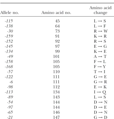
alleles that were previously examined. Therefore, we tested the newcdc31alleles for their effect on Kic1p kinase activ-Amino acid
Allele no. Amino acid no. change ity. To ensure similar levels of Cdc31p, the kinase assays were performed on cultures grown at the permissive
tem--115 45 L→S
perature. Figure 5A shows that the amino-terminal
mu--138 64 L→F
tants (cdc31-115, -138, and -30) contained wild-type levels
-30 73 R→W
-159 91 K→R of Kic1p kinase activity. These results correlate with the -152 92 R→S lack of cell lysis in these alleles, suggesting that the amino--145 97 E→G terminal region of the protein does not have a specific -134 99 K→E cell wall-related function. Alternatively, the temperature
-49 101 A→T
sensitivity of these mutants may be due to reduced protein
-158 105 F→L
Kic1p-complex stability at the nonpermissive temperature,
-168 105 F→Y
rather than to loss of a specific function. To test this we
-57 110 T→I
-122 111 G→E measured Kic1p kinase activity in cultures grown at 37⬚ -6 111 G→R and found that it was comparable to wild-type levels (data -98 112 E→K not shown), indicating that the temperature sensitivity in -113 134 I→Q cdc31-115, -138, and -30 is not due to a defect in Kic1p
-89 143 L→S
kinase activity.
-54 144 D→N
Alleles in the central part of the protein showed
drasti--97 144 D→E
cally reduced levels of Kic1p kinase activity. Five alleles
-65 146 D→N
-21 147 G→D (cdc31-152, -159, -49, -134, and -145) had a defect at both permissive and nonpermissive temperatures, whereas two alleles (cdc31-158 and -168) had a temperature-sensitive defect. A mutation that maps to the region between the 134 kar1-⌬17double mutant was recessive to bothKAR1 third and the fourth EF hands had wild-type levels of Kic1p andCDC31.Finally,kar1-⌬17is a recessive suppressor of kinase activity, suggesting that this part of the protein does cdc31-98.The recessivity of some of the cosuppressors sug- not affect Kic1p function. Certain alleles in the carboxy-gests that they act by a mechanism other than by increasing terminal region also showed a temperatusensitive re-the affinity of binding to Kar1p or a downstream effector. duction in Kic1p kinase activity, most notably cdc31-54
Microscopic analysis ofcdc31 kar1-⌬17double mutants: and-21.In the case ofcdc31-54, the temperature-sensitive
To further analyze the basis of the genetic interactions kinase defect correlates with a temperature-sensitive loss betweenkar1-⌬17and the newcdc31alleles, we examined of Cdc31p. However, this is not the case incdc31-21, sug-the cell morphology of sug-the double mutants. The synsug-theti- gesting that this region may also contribute to Kic1p func-cally lethal combinations could not be analyzed because tion.
they did not grow under any conditions. We therefore Temperature-sensitive alleles may arise from mutations analyzed the alleles that showed enhanced growth defects that disrupt the protein structure, leading to protein mis-(Table 2A). We found that the double mutants containing folding and degradation at the nonpermissive tempera-cdc31-54andcdc31-122had a higher level of large-budded ture. Therefore, one trivial explanation for lack of Kic1p cells at both the permissive (23⬚) and the semipermissive kinase activity is absence of Cdc31 protein. Although we (35⬚) temperatures. The cdc31-138 kar1-⌬17mutant had performed the kinase assays on cultures grown at the per-a higher level of lper-arge-budded cells (60%) compper-ared to missive temperature, we also directly examined the lev-either single mutant [kar1-⌬17 (22%) and cdc31-138 els of Cdc31p at both the permissive and nonpermissive (18%)] at 35⬚. temperatures. The mutants behaved in three different We also analyzed the alleles that did not show any patterns with respect tocdc31protein levels (Figure 5B). growth phenotypes in combination withkar1-⌬17.Double First, some mutants had wild-type protein levels at both mutants between allelescdc31-6, -115, and -152andkar1- temperatures. Other mutants showed temperature-depen-⌬17arrested at G2/M to the same degree as the single dent reduction in protein levels. Finally, some mutants mutants alone (Table 2B). Finally, the cosuppressing al- had low protein levels at both temperatures. Of particu-leles,cdc31-30, -113,-134, and -98, showed a reduction in lar concern, the mutants that showed severe reduction G2/M arrest phenotype as compared tokar1-⌬17(Table in Kic1p kinase activity did express wild-type levels of 2C), suggesting that they suppress the growth defect of Cdc31p at the permissive temperature. Therefore, be-kar1-⌬17by suppressing its SPB duplication defect. cause the protein extracts were prepared from cells at
The central domain of Cdc31p is required for Kic1p the permissive temperature, the lack of Kic1p kinase activity was not due to absence of Cdc31p. In summary,
Figure2.—G2/M arrest and cell lysis phenotypes are not correlated. Each part is a separate linear representation of Cdc31p. Amino acids 45–147 are depicted, because mutations in the first 44 and the last 14 amino acids were not recovered. The vertical bars depict the position of each mutation and the numbers underneath each bar represent the allele number. The height of the bars represents the percentage of each phenotype for each allele. The numbers above A depict the approximate amino acid positions. Greater than 100 cells were counted for each allele for each phenotype. (A) Percentage of large-budded cells with a single nucleus after 4 hr at 37⬚. Alleles in the carboxy-terminal region (gray shading) had a higher G2/M arrest phenotype as compared to alleles in the amino-terminus. (B) Percentage of cell lysis after 4 hr at 37⬚. Alleles in the center of the protein (gray shading) had higher cell lysis then alleles in the amino- and carboxy-termini. (C) Percentage of cells showing Cdc31p mislocalization.
we identified the central part of the protein, encom- higher levels of-galactosidase activity, suggesting that they were not impaired for binding to Kic1p. Alleles in passing the region between the second and the third
EF hands, as being important for activating the Kic1p the central part of the protein that caused severe defects in Kic1p kinase activity exhibited variable phenotypes kinase, with a minor contribution from the
carboxy-terminal region of Cdc31p. with respect to the Kic1p two-hybrid interaction. Alleles cdc31-152, -159, and -158showed wild-type Kic1p
bind-Binding of mutant Cdc31 proteins and Kic1p: We
next tested whether lack of Kic1p kinase activity was ing. Thecdc31-49allele showed a temperature-sensitive defect in-galactosidase activity, whereascdc31-134and due to a defect in Cdc31p-Kic1p binding. For this
pur-pose, we used the yeast two-hybrid system with the -145 showed moderate defects at both temperatures (ⵑ25%-galactosidase activity as compared to the wild Cdc31p mutants fused to the Gal4p DNA-binding
do-main and Kic1p fused to the Gal4p activation dodo-main type). Most alleles in the carboxy-terminal region of Cdc31p (cdc31-57, -122, -6, -98, -113, -54, -97, -65, and (Fields and Song 1989; James et al. 1996). Figure 6
shows the-galactosidase activity levels for the different -21) showed severely reduced-galactosidase levels com-parable to the vector control. Some of these alleles combinations of Kic1p and Cdc31 mutant fusion
pro-teins for cultures grown at 37⬚. Results obtained from showed temperature-sensitive defects in Kic1p kinase activity (cdc31-21and cdc31-54). We were surprised to cultures grown at 30⬚were comparable, except for one
Figure 3.—Characteristic cdc31 phenotypes. (A and B) Mutations in the carboxy-ter-minal region of Cdc31p caused cells to arrest in G2/M as large-budded cells with one nucleus and a monopolar spindle. Rep-resentative cdc31-6 cells are shown (A) DAPI; (B) tubulin. (C and D) Localization of Cdc31p to the SPB was detected by immunofluorescence. Shown is a cdc31-49 mutant, which does not disrupt localization. (C) DAPI; (D) Cdc31p. (E–G) The FUN-1 stain was used to measure membrane integrity and cell viability. In metaboli-cally active cells with intact membranes, FUN-1 accumu-lates in the vacuole and forms characteristic red structures. In cells with disrupted plasma membranes, FUN-1 stains the cytoplasm bright yellow. (E) wild-type cells; (F) cdc31-113 mutant with few inviable cells; (G) cdc31-134 mutant with many inviable cells.
activity was not due to lack of two-hybrid fusion proteins with additional components downstream in the SPB because immunoblot analysis showed the same level of duplication pathway. The cdc31-49andcdc31-21alleles hybrid proteins for all alleles from cultures grown at had additional Kic1p-related defects, suggesting that both 30⬚ and 37⬚ (data not shown). In conclusion, we they affect multiple functions, similarly to previously found that mutations in the carboxy-terminal region of identified cdc31-1, -2, -5, and -16 (Biggins and Rose Cdc31p strongly disrupted binding to Kic1p, whereas 1994). The cdc31-89allele (shown in yellow in Figure mutations toward the central part of the protein had a 1) did not have obvious additional defects, suggesting minor effect. that it may be specifically defective for SPB duplication.
Distinct regions of Cdc31p bind to and activate Kic1p:
Mutations in the middle region of Cdc31p caused severe DISCUSSION defects in Kic1p kinase activity. Most notably, mutations cdc31-152, -159, -168, and -158(line 4 in Table 3, shown Phenotypic studies on temperature-sensitive mutants
in blue in Figure 1) caused specific defects in activating of cdc31revealed that different mutants exhibited
dis-Kic1p because the proteins did bind to dis-Kic1p and the tinct phenotypes. Table 3 summarizes the findings and
mutants did not have a pronounced G2/M arrest defect. groups the mutants on the basis of their phenotypes.
Although Cdc31p was not present at the SPB in these Analysis of the results suggested that different regions of
alleles, the lack of G2/M arrest suggests that adequate Cdc31p mediate distinct functions, described in detail
levels of Cdc31p have localized to the SPB but were below.
not detected by immunofluorescence. These alleles may
Uncoupling of Cdc31p localization and G2/M arrest:
provide a means for elucidating the mechanism by Cdc31p localizes to the SPB where it activates SPB
dupli-which Cdc31p activates Kic1p kinase activity. cation (Spang et al. 1993; Biggins and Rose 1994;
Three additional mutations in the middle of Cdc31p, Vallen et al. 1994). Interestingly, some of the cdc31
cdc31-49, -134, and -145(lines 6 and 7 in Table 3, shown mutations dissociate these two functions. Three alleles
in red in Figure 1), exhibited strong Kic1p activation (cdc31-21, -49, and -89) showed normal localization to
defects and Kic1p-binding defects. However, they also the SPB and a relatively high degree of G2/M arrest
exhibited moderate to high G2/M arrest defects, sug-(49–59%; lines 2, 5, and 6 in Table 3), indicating that
gesting that they are defective for multiple functions. they uncouple SPB duplication from Cdc31p
Figure4.—Genetic interactions betweenkar1-⌬17andcdc31alleles. The genotypes of each strain arecdc31⌬::LEU2 kar1-⌬17 [cdc31*HIS3 CEN/ARS], where * represents the differentCDC31alleles. The column on the right shows the effective genotypes, where strains that arekar1-⌬17contain a URA3vector plasmid, while strains that areKAR1 contain aKAR1 URA3 CEN/ARS plasmid. Three 10-fold serial dilutions are shown for each strain at each temperature. (A) Allelescdc31-115, -6, and -152had the same temperature spectrum alone or in combination withkar1-⌬17, suggesting that they do not interact genetically. (B) Alleles cdc31-138, -54, and -122showed an enhanced growth defect when combined withkar1-⌬17, with the double mutant having a lower nonpermissive temperature than either single mutant. (C) Alleles cdc31-30, -113, -98, and -134 showed cosuppression phenotype withkar1-⌬17, where the double mutant grew at temperatures permissive for either single mutant. (D) Schematic representation of the genetic interaction on the linear sequence of Cdc31p. Amino acids 45–147 are depicted, because the first 44 and the last 14 amino acids were not mutated in any of the alleles. Vertical bars represent mutations of amino acids, and the color of the bar represents the nature of the genetic interaction as depicted on the left. The numbers under each bar represent the allele number and the numbers at the top depict the approximate amino acid positions.
(cdc31-57,-122,-6,-98,-54,-97,-65, -113, and -21; lines interact with and be activated by other proteins. By this model, the alleles in the middle of Cdc31p interact with 5 and 8 in Table 3; shown in red in Figure 1) caused a
strong defect for Kic1p-binding in the two-hybrid assay. Kic1p, resulting in a dead-end complex, and therefore Kic1p is no longer free to interact with other proteins. On the basis of the previous analysis, we proposed that
these alleles also affect Kar1p binding, suggesting that One candidate for such a surrogate activator is calmodu-lin, because it shares sequence homology with Cdc31p the Kic1p- and Kar1p-binding domains overlap on the
carboxy-terminal surface of Cdc31p. This conclusion is and is known to interact with and activate kinases. Al-though a two-hybrid interaction between Kic1p and not unprecedented for two reasons. First, calmodulin
binds most of its substrates on the same surface, in the Cmd1p was not detected (Sullivan et al. 1998), this was done in the presence of Cdc31p. Second, the two central region of the protein. Second, Kic1p and Kar1p
share a 15-residue motif that is similar to calmodulin- assays may have different levels of sensitivity. For exam-ple, the kinase assay may be sensitive enough to detect binding domains that may mediate binding to the same
region of Cdc31p for both proteins (Geieret al.1996; activation of Kic1p kinase activity even when binding to Cdc31p is greatly impaired. The alleles that caused Sullivanet al.1998).
Strikingly, some alleles that showed a strong defect moderate defects in Kic1p binding and strong defects in kinase activity suggest that Kic1p requires Cdc31p for in Kic1p binding were not defective in Kic1p kinase
activity (cdc31-54, -113, and -6). We offer four possible activity. Presumably, alleles exhibiting wild-type kinase activity but low levels of-galactosidase activity still bind interpretations of these data. One possibility is that
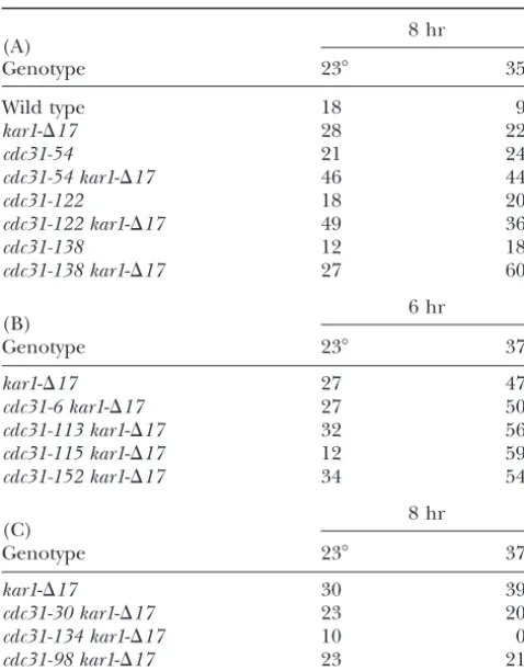
TABLE 2 negatively. By this model, Kic1p has constitutive activity when not bound to Cdc31p, regulated activity when G2/M cell cycle arrest inkar1-⌬17 cdc31: Double mutant
bound to Cdc31p, and is not active when bound to combinations (A) that showed enhanced growth defects,
mutant Cdc31p defective for activation. Mutations (B) that did not show any genetic interactions,
and (C) that showed suppression cdc31-115 and cdc31-6 may be defective in negatively regulating Kic1p because they have higher than wild-8 hr type kinase activities. Interestingly, whereas cdc31-115 (A)
exhibited a higher than wild-type-galactosidase activity
Genotype 23⬚ 35⬚
in the two-hybrid assay,cdc31-6 was defective in Kic1p
Wild type 18 9 binding. Because these two mutations map to distinct
kar1-⌬17 28 22 regions of Cdc31p, they may affect Kic1p kinase activity
cdc31-54 21 24 by distinct mechanisms.
cdc31-54 kar1-⌬17 46 44
A surface on the carboxy-terminal region of Cdc31p
cdc31-122 18 20
may be important for binding Kic1p and Kar1p:Eleven
cdc31-122 kar1-⌬17 49 36
alleles (cdc31-138, -134, -145, -57,-122, -6, -98,-54, -97,
cdc31-138 12 18
cdc31-138 kar1-⌬17 27 60 -65, and -113) showed both Cdc31p mislocalization and high G2/M arrest defects (lines 3, 7, and 8 in Table 3, 6 hr
(B) shown in red in Figure 1). Strikingly, when projected
Genotype 23⬚ 37⬚ onto the predicted three-dimensional structure of
Cdc31p, 8 of these 11 alleles mapped in close proximity,
kar1-⌬17 27 47
on the same surface on the carboxy-terminal lobe. In
cdc31-6 kar1-⌬17 27 50
cdc31-113 kar1-⌬17 32 56 addition,cdc31-113andcdc31-21mapped to the core of cdc31-115 kar1-⌬17 12 59 the carboxy-terminal lobe, suggesting that they may lead cdc31-152 kar1-⌬17 34 54 to destabilization of the domain. The carboxy-terminal
region of Cdc31p had been previously predicted to play 8 hr
(C) a role in SPB duplication and Kar1p function (Biggins
Genotype 23⬚ 37⬚
andRose1994;Vallenet al.1994). The surface on the kar1-⌬17 30 39 carboxy-terminal lobe may represent the Kar1p-binding cdc31-30 kar1-⌬17 23 20 site on Cdc31p. Alternatively, this surface may be part cdc31-134 kar1-⌬17 10 0 of an oligomerization region, because centrin is the cdc31-98 kar1-⌬17 23 21 major component of contractile filamentous structures
associated with MTOCs in Chlamydomonas and mam-The percentage of large-budded cells for each culture is
shown, representing the G2/M arrest phenotype. (A) All malian cells (Wrightet al.1985;BaronandSalisbury strains are cdc31⌬::LEU2 kar1-⌬17 [cdc31* HIS3 CEN/ARS], 1988). The remaining 2 alleles in this class, cdc31-138 where * represents the differentCDC31alleles. Strains
desig-andcdc31-145, line the surface of an internal cavity of nated as kar1-⌬17 contain a URA3 vector, while the other
Cdc31p, analogous to the calmodulin ligand-binding strains contain a [KAR1 URA3 CEN/ARS] plasmid. The strain
site. Interestingly, these 2 alleles have only a moderate designated wild type contains a [CDC31 HIS3] and a [KAR1
URA3] plasmid. The strain designated kar1-⌬17 contains a degree of mislocalization as compared to the alleles on [CDC31 HIS3] plasmid and a URA3 plasmid. Strains were the external surface (x⫽47%vs. x⫽71%). One possi-grown in synthetic medium lacking uracil and histidine to
bility is that they affect binding to additional SPB com-midlogarithmic phase at 23⬚and one-half of each culture was
ponent(s) that provide a secondary means of Cdc31p shifted to 35⬚ for 8 hr. The relatively low G2/M arrest of
localization to the SPB. Alternatively, these alleles may kar1-⌬17 may be because the experiment was performed at
the intermediate temperature of 35⬚. (B) All strains are also affect Kar1p binding, if Kar1p and Cdc31p have cdc31⌬::LEU2 kar1-⌬17[cdc31*HIS3 CEN/ARS], where * repre- extensive interactions on different surfaces of Cdc31p. sents the differentCDC31alleles. The strain designatedkar1
-The amino-terminal region of Cdc31p mediates novel
⌬17contains a [CDC31 HIS3] plasmid. Strains were grown in
functions:In two mutants,cdc31-115and -30(green in synthetic medium lacking histidine to midlogarithmic phase
Figure 1, line 1 in Table 3), Cdc31p localized to the at 23⬚ and one-half of each culture was shifted to 37⬚ for 6
hr. (C) All strains are cdc31⌬::LEU2 kar1-⌬17 [cdc31* HIS3 SPB, and the cells exhibited a low G2/M cell cycle arrest CEN/ARS], where * represents the differentCDC31alleles. The (31 and 35%, respectively), suggesting that these alleles strain designatedkar1-⌬17contains a [CDC31 HIS3] plasmid.
most likely do not affect SPB duplication-related func-Strains were grown in synthetic medium lacking histidine to
tions. Consistent with this, these alleles map far from midlogarithmic phase at 23⬚and one-half of each culture was
the putative Kic1p/Kar1p-binding surface and the al-shifted to 37⬚for 8 hr.
leles with high G2/M arrest. In addition,cdc31-115and -30did not show defects in cell integrity, Kic1p binding, or Kic1p kinase activity, suggesting that these alleles may Third, the fusion proteins in the two-hybrid assay may
Figure5.—(A) The cen-tral part of Cdc31p activates Kic1p kinase activity. The top represents kinase assays performed for each allele at 23⬚and 37⬚. The placement of each part corresponds to the position of the allele on the linear representation of the protein. The bottom is a summary of the kinase assay results depicted on the lin-ear sequence of Cdc31p. Each bar corresponds to a mutation. ( ) Wild-type Kic1p kinase activity; ( ) temperature-sensitive defect; (䊏) temperature-indepen-dent defect. (B) Steady-state Cdc31p mutant protein lev-els after growth at 23⬚or 37⬚ for 4 hr.
to the genetic interactions with the Pkc1p pathway plain the synthetic lethality and growth defects between kar1-⌬17 and the different cdc31 alleles. One type of (Khalfanet al.2000).
Insights from the phenotypes ofcdc31 kar1-⌬17dou- allele may cause a partial defect in binding to Kar1p. Deleting the Cdc31p-interaction domain on Kar1p with
ble mutants:The newcdc31alleles showed a variety of
genetic interactions when combined withkar1-⌬17.In the kar1-⌬17 mutation may further compromise the Kar1p-Cdc31p interaction, resulting in enhanced growth principle, we imagine three nonexclusive ways to
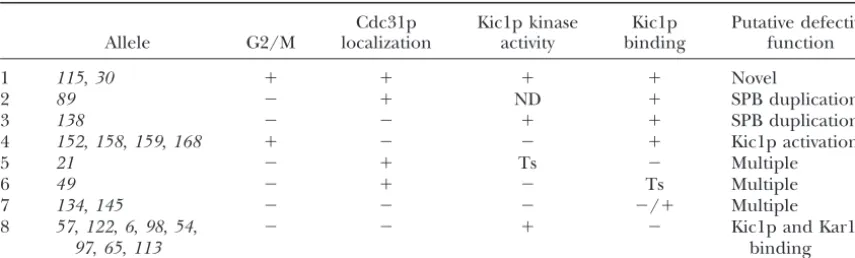
TABLE 3
Summary of defects of temperature-sensitivecdc31alleles and the predicted function that they disrupt
Cdc31p Kic1p kinase Kic1p Putative defective
Allele G2/M localization activity binding function
1 115,30 ⫹ ⫹ ⫹ ⫹ Novel
2 89 ⫺ ⫹ ND ⫹ SPB duplication
3 138 ⫺ ⫺ ⫹ ⫹ SPB duplication
4 152,158,159,168 ⫹ ⫺ ⫺ ⫹ Kic1p activation
5 21 ⫺ ⫹ Ts ⫺ Multiple
6 49 ⫺ ⫹ ⫺ Ts Multiple
7 134,145 ⫺ ⫺ ⫺ ⫺/⫹ Multiple
8 57,122,6,98,54, ⫺ ⫺ ⫹ ⫺ Kic1p and Kar1p
97,65,113 binding
⫹ indicates that the mutants behaved like the wild type with respect to that phenotype.⫺ in the G2/M column indicates that the mutants exhibited⬎50% large-budded arrest.⫺in the Cdc31p localization column indicates that the mutant exhibited⬎50% mislocalization. Ts, a temperature-sensitive defect; ND, not deter-mined.
and G2/M defects. Becausecdc31-54andcdc31-122map a defect in binding to other ligands and are therefore free to bind Kar1p.
to the carboxy-terminal region, we propose that they
may be partially defective in binding Kar1p. The second Are cell lysis and Kic1-kinase activity correlated?The cluster of alleles causing the highest cell lysis defect type of allele may cause a defect in binding to an
addi-tional SPB component but may allow localization to the coincided with the cluster of alleles exhibiting defects in Kic1p kinase activity in the middle of the protein. SPB by interacting with Kar1p. Thekar1-⌬17mutation
may sever this residual localization, resulting in the en- However, this correlation was not absolute. Whereas cdc31-134showed the highest lysis defect (55%) and a hanced defect. On the basis of the distant position of
cdc31-138, we propose that it may be defective in binding strong kinase activity defect, cdc31-159 had a similar kinase defect but negligible lysis. One possibility is that additional SPB components. Finally, other alleles may
severely reduce the level or stability of Cdc31p to the these phenotypes are not related and that cdc31-134 is defective for two different interactions. This would extent that there is insufficient protein to interact with
Kar1p. suggest that Cdc31p has Kic1p-unrelated roles in cell
wall morphogenesis. In support of this model, the preex-The suppressing alleles represent a particularly
inter-esting class because they mapped to an unexpected re- istingCDC31alleles have Kic1p-independent cell mor-phogenesis defects (Sullivanet al.1998). Another pre-gion of the protein. At first glance, suppressor mutations
might be expected to suppress kar1-⌬17 by restoring diction of this model is that the lysis and Kic1p kinase defects should be separable phenotypes. Whereas we binding to Kar1p, and as such they are predicted to be
rare and to cluster to the Kar1p binding region. How- found alleles that are defective in Kic1p kinase activity only, we have not found alleles that lyse but exhibit ever, a relatively high proportion of the
temperature-sensitive mutations (19%) suppressed kar1-⌬17 and a wild-type Kic1p kinase activity. Therefore, a second interpretation of our results is that the two phenotypes most of them did not map to the putative
Kar1p-inter-acting region. Instead, they mapped to the second EF are related, but that the ability to activate Kic1p is com-promised to different degrees in the different cdc31 hand (cdc31-30), the central loop (cdc31-134), the third
EF hand (cdc31-98), and the region between the third alleles, in a way that is not measured by the kinase assay. By this model, cdc31-134 would have the most severe and the fourth EF hands (cdc31-113). This is in contrast
to the more carboxy-terminal location of the earlier set defect in activating Kic1p, resulting in appreciable cell lysis, whereas the other alleles are only partially defec-of suppressors (Figure 4 and Vallenet al.1994). The
modes of suppression were also different, withcdc31-30 tive. A third possibility is that lysis is a secondary conse-quence of multiple defects. Therefore,cdc31-134, which andcdc31-113being dominant andcdc31-98and
cdc31-134being recessive. This is also in contrast to the origi- has high G2/M arrest and a Kic1p kinase defect, may be defective for multiple functions that in combination nal set of non-temperature-sensitive suppressors that
were all dominant (Figure 4 andVallen et al. 1994). lead to lysis, whereas the neighboring alleles are defec-tive only in Kic1p kinase activity.
Three of the four new suppressors showed a Cdc31p
mislocalization defect, suggesting that they do not sup- Cdc31p and calcium:Although Cdc31p contains four EF-hand domains, only the first and the fourth EF hands press by increasing the affinity to Kar1p. One possible
aspartate residues in the first and fourth EF-hand do- sion (Ohya and Botstein 1994a). In contrast, in re-peated trials we failed to detect any intragenic comple-mains that severely reduced the affinity for calcium were
lethal at 23⬚ or 30⬚, whereas the analogous mutations mentation between the differentCDC31alleles. However, Cdc31p may oligomerize, precluding simple intragenic in the second or third EF hand had no growth defects
(Geieret al.1996). Interestingly, we recovered tempera- complementation. Alternatively, all of the mutants may exhibit sufficient overlap in their defects such that no ture-sensitive mutations in the second, third, and fourth
EF hands. Mutations in the aspartate residues critical mutant combination is restored to wild-type function. In conclusion, fine structure analysis of Cdc31p has for binding calcium were not recovered. We observed
extensive clustering of alleles to the third and fourth identified regions of the protein important for binding its known ligands, as well as regions that potentially EF hands, indicating that both of these regions perform
important functions. Most mutations in both the third mediate interactions with novel targets. Future analysis of the new alleles may identify the novel targets of and fourth EF-hand clusters caused severe defects in
Kic1p binding. Thus, calcium binding by an EF hand Cdc31p and elucidate the specific roles of Cdc31p in SPB duplication and cell wall biogenesis.
domain,per se(e.g., third EF hand), is not essential for
function. This is consistent with the finding that Kic1p We thank Sean Clark, Waheeda Khalfan, and Trisha Davis for critical kinase activity is not affected by the presence of calcium reading of the manuscript and Stephen T. Miller for discussions about the predicted protein structure. This research was supported by
Na-(Sullivan et al.1998).
tional Institutes of Health grant GM52526 to M.D.R. I.I. was supported Comparisons between Cdc31p and calmodulin:Cdc31p
by a fellowship from the New Jersey Commission on Cancer Research.
and Cmd1p share 42% sequence identity (Baumet al. 1986), and the structure of Cdc31p was predicted to be similar to that of calmodulin. In addition, the two
LITERATURE CITED proteins may be similar in that they can both bind
multi-ple substrates. The substrate specificity of calmodulin Adams, I. R.,andJ. V. Kilmartin,1999 Localization of core spindle pole body (SPB) components during SPB duplication in
Saccharo-seems to be determined by specific Phe residues in the
myces cerevisiae.J. Cell Biol.145:809–823.
binding site (Okano et al. 1998). By analogy, it may Balczon, R.,1996 The centrosome in animal cells and its functional be possible to identify residues in the carboxy-terminal homologs in plant and yeast cells. Int. Rev. Cytol.169:25–82.
Baron, A. T.,andJ. L. Salisbury,1988 Identification and
localiza-surface or the internal cavity of Cdc31p that do not lead
tion of a novel, cytoskeletal, centrosome-associated protein in
to temperature sensitivity but that are important for PtK2 cells. J. Cell Biol.107:2669–2678.
binding either Kar1p or Kic1p. Baum, P., C. Furlong and B. Byers,1986 Yeast gene required for spindle pole body duplication: homology of its product with
One major difference between Cdc31p and
calmodu-Ca2⫹-binding proteins. Proc. Natl. Acad. Sci. USA83:5512–5516.
lin is the importance of the conserved Phe residues. Bhattacharya, D., J. SteinkotterandM. Melkonian,1993 Mo-Whereas mutating single Phe residues in calmodulin did lecular cloning and evolutionary analysis of the calcium-modu-lated contractile protein, centrin, in green algae and land plants.
not lead to any observable phenotypes, the analogous
Plant Mol. Biol.23:1243–1254.
mutations incdc31led primarily to lethality. In contrast, Biggins, S.,andM. D. Rose,1994 Direct interaction between yeast single mutations in residues adjacent to the Phe in the spindle pole body components: Kar1p is required for Cdc31p localization to the spindle pole body. J. Cell Biol.125:843–852.
internal cavity and on the surface of Cdc31p caused a
Blackman, L. M., J. D. HarperandR. L. Overall,1999
Localiza-temperature-sensitive phenotype. Temperature-sensi- tion of a centrin-like protein to higher plant plasmodesmata. tive point mutations were extremely difficult to isolate Eur. J. Cell Biol.78:297–304.
Boeke, J. D., J. Trueheart, G. NatsoulisandG. R. Fink,1987
5-Flu-inCMD1(Daviset al.1986). The greater severity of the
oroorotic acid as a selective agent in yeast molecular genetics.
Phe→Ala alleles inCDC31may reflect the greater ease Methods Enzymol.154:164–175.
of acquisition of conditional mutants inCDC31relative Byers, B.,1981 Multiple roles of the spindle pole bodies in the life cycle ofSaccharomyces cerevisiae, pp. 119–131 inMolecular Genetics
toCMD1.
in Yeast, edited byD. von Wettstein, J. Friis, M.
Kielland-Interestingly, although Phe105→Ala substitution in BrandtandA. Stenderup.Munksgaard, Copenhagen. Cdc31p resulted in lethality, Phe105→Leu (cdc31-158) Byers, B.,andL. Goetsch,1974 Duplication of spindle plaques
and integration of the yeast cell cycle. Cold Spring Harbor Symp.
or Phe105→ Tyr (cdc31-168) substitutions resulted in
Quant. Biol.38:123–131.
temperature sensitivity. Phe105 in Cdc31p corresponds Davis, T. N., M. S. Urdea, F. R. MasiarzandJ. Thorner,1986 Isola-to Phe92 in Cmd1p that is mutated in cmd1-226 and tion of the yeast calmodulin gene: calmodulin is an essential
protein. Cell47:423–431.
results in abnormal actin organization. Cmd1-226p is
Erdeniz, N., U. H. MortensenandR. Rothstein,1997
Cloning-defective in binding the Ca2⫹/calmodulin-dependent
free PCR-based allele replacement methods. Genome Res. 7:
protein kinase, but not calcineurin (Okanoet al.1998). 1174–1183.
Fields, S.,and O. Song,1989 A novel genetic system to detect
Strikingly, the analogous mutations inCDC31,cdc31-158
protein-protein interactions. Nature340:245–246.
andcdc31-168, are also defective in activating a kinase,
Geier, B. M., H. WiechandE. Schiebel,1996 Binding of centrins
Kic1p. and yeast calmodulin to synthetic peptides corresponding to
bind-ing sites in the spindle pole body components Kar1p and
Intragenic complementation is often used as a means
Spc110p. J. Biol. Chem.271:28366–28374.
to define alleles defective in nonoverlapping functions.
Gietz, R. H.,andR. H. Schiestl,1995 Transforming yeast with
Calmodulin alleles that affect distinct functions were DNA. Methods Mol. Cell. Biol.5:255–269.
Guex, N.,andM. C. Peitsch,1997 SWISS-MODEL and the