2 8 1 02 15 39 2
The Structure and Properties of Soluble Phosphate Based Glasses
Thesis submitted by Katrin Franks For the degree of DOCTOR OF PHILOSOPHY
In the
Faculty o f Dentistry University of London
Department o f Biomaterials, Eastman Dental Institute For Oral Healthcare Sciences,
University College London, 256 Gray’s Inn Road,
London W CIX 8LD
-All rights reserved
INFORMATION TO ALL USERS
The quality of this reproduction is dependent upon the quality of the copy submitted.
In the unlikely event that the author did not send a complete manuscript and there are missing pages, these will be noted. Also, if material had to be removed,
a note will indicate the deletion.
uest.
ProQuest U133779
Published by ProQuest LLC(2016). Copyright of the Dissertation is held by the Author.
All rights reserved.
This work is protected against unauthorized copying under Title 17, United States Code. Microform Edition © ProQuest LLC.
ProQuest LLC
789 East Eisenhower Parkway P.O. Box 1346
Unser Kopf ist rund, damit das Denken die Richtung wechseln kann.
.. .our head is round to allow our thoughts to change directions.
Antoine de Saint-Exupéry
Phantasie ist wichtiger als Wissen, derm Wissen ist begrenzt.
.. .imagination is more important than knowledge because knowledge is limited.
Abstract
The recent discipline of tissue engineering has developed innovative degradable
and non-degradable (dependant upon application) materials in combination with low
toxicity and good biocompatibility. The major focus of these new technologies is to guide
the regenerative process. The aim of this project was to take the regenerative tissue
healing process a step further by developing a material which not only guides the tissue
regeneration process, but also enhances it and has a degradation profile that is tailored to
the tissue and on degradation, leaves no toxic or irritating debris behind to cause any
tissue reaction. The chosen material was a soluble phosphate based glass, modified with
CaO, NazO, MgO, K2O and/or Cap2. Materials have been developed in order to work as
closely as possible to the natural phase of bone, within the limitations imposed by the
glass forming process.
5 different glass systems have been synthesised via conventional glass making
procedures and the solubility process has been investigated via weight loss experiments
and ion and pH measurements. All materials were soluble with different degradation
processes, depending on the composition of the glass. Glasses with low CaO content
showed a linear relationship between weight loss per unit area and time. Glasses with
higher CaO content show an increasing non-linearity in their weight loss behaviour. The
pH showed a significant increase in the first stages of degradation, which was explained
by cation-exchange processes taking place from the material to the solution and vice
versa. The ion concentration in solution was found to increase with time as expected and it
mirrored the weight loss curves.
Preliminary cell culture tests (MTT tests) using the MG63 human osteoblast cell
line were established to test the biocompatibility of the soluble extracts from the different
however similar to the control line or above for glasses with CaO contents higher than 30
mol%, i.e. low solubility. The best test results, with enhanced proliferation was seen for
glass with the highest CaO content. The MTT test results look very promising for
materials with a high calcium oxide content, indicating biocompatibility with enhanced
cell proliferation. There was also evidence that small amounts of K2O and MgO affected
cell proliferation.
Structural analysis was carried out using DTA and MAS-NMR spectroscopy. The
results from the glasses were found to be in line with X-ray analysis of similar glass-
ceramics, which had been analysed in earlier studies. Thermal analysis revealed multi
crystallisation events as evidenced by the presence of one or two crystallisation peaks with
more than two or three corresponding melting points. The use of MAS-NMR spectroscopy
showed that two species, Q* and formed the basis of the glass structure and it was
possible to identify the dominant species as Na4Ca(P0 3)6. The Q’ species is
represented by (Ca3(P04)2 or Na2P2 0?). However, it was up to the present time not
possible to identify the Q' species unambiguously.
The work has shown that it is possible to synthesise biodegradable glasses for hard tissue
surgery, whose composition is close to the inorganic phase of bone. Biocompatibility
studies have helped to define optimal compositions and it is therefore hoped that the
results from this study will contribute towards future implant research utilising soluble
Acknowledgements
I would like to thank Dr. Jonathan Knowles and Dr. Isaac Abrahams for their
supervision and support throughout the my Ph.D.
I very much enjoyed the work on the MAS-NMR Spectroscopy and would like to
thank Dr. Geoff Hawkes at Queen Mary and Westfield College for his support for
the analysis and Dr. Abil Aliev at University College London for his help and
guidance.
I am extremely grateful for the MTT test on my material, carried out by Dr. Vehid
Salih at the Eastman Dental Institute. I am indebted, many thanks.
Furthermore, I would like to thank Colebrand for the scholarship, without this the
Ph.D. would not have been possible.
I also would like to thank Dr. Mike Braden and Dr. Showan Nazhat for the
discussions regarding aspects of diffusion and also Jill Williams for the advice for
the fluoride measurement, of which I am very grateful.
C H A P T E R 1 ...8
IN T R O D U C T IO N ...9
1.1 Materialsfo rh a r dt issues u r g e r y... 9
1.1.1 Host /Tissue Response...10
1.1.2 Bioactive Glasses...12
1.1.3 Controlled Released Glasses...18
C H A P T E R 2 ... 20
A IM S O F T H IS W O R K ... 21
C H A P T E R 3 ... 24
G L A S S ... 25
3.1 Glassing e n e r a l... 26
3.1.1 Single Oxide Glass System...27
3.1.2 Binary glass systems:...29
3.2 Glass Structure Th eo ries... 32
3.3 Solid State N M R Spectroscopy (M A S -N M R )... 35
3.3.1 Beginning o f MAS-NMR Spectroscopy with Commercial Phosphates 36 3.3.2 Structural Investigation by MAS-NMR Spectroscopy fo r Binary Glass Systems...37
3.3.3 Structurallnvestigation by MAS-NMR Spectroscopy o f Ternary and Quaternary Glass Systems...44
3.4 X-RAY Photoelectron Spectroscopy ( X P S )... 45
3.4.1 Structural Investigation o f Glasses using X P S...46
3.5 Gla ss Form ation Pr o c e s s...47
3.5.1 Glass Transition Temperature...49
3.6 Differential Therm al An a l y sis (D T A )...50
C H A P T E R 4 ...54
4.1 So lu ble Gl a s s e s... 55
4.1.2 Solubility o f silicate glasses...56
4.1.2 Solubility o f Phosphate Glasses...60
C H A P T E R 5 ...6 8 5.1 M A T E R IA L S A N D M E T H O D S ... 69
5.1 General Re m a r k s... 69
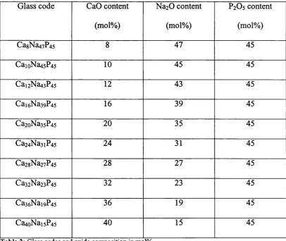
5.1.1 Selection o f Glass Composition...71
5.2 Prelim inary Gla ss Mak ing Studiesfo rtheter n a r y Sy s t e m...72
5.2.1 Establishment o f glass production methodology...72
5.3 Chem ical Eq uationsa n d Com positional Ca l c u l a t io n s...73
Katrin Franks Ph.D. Thesis
5.4.1 Standard Glass Preparation...75
5.4.2 Glass Cutting and Storage...76
5.4.3 p H Measurements...78
5.4.4 Ion Measurements...78
C H A P T E R 6 ... 82
T E R N A R Y S Y S T E M C A O -N A2O -P2O5...83
6.1 S p e c ia l P r o c e d u r e s f o r t h e T e r n a r y S y ste m CAO-NA2O-P2O5...83
6.1.1 Solubility Tests in Distilled Water. Preliminary Experiments...85
6.1.2 Preliminary Studies on the Solubility o f the Na2 0-Ca0-P205 System...86
6.1.3 p H Measurements...91
6.2 Io n Me a s u r e m e n t s...93
6.2.1 Calcium Measurements...93
6.2.2 Sodium Measurements...95
6.2.3 Solubility Test in H B SS...96
6.3 D is c u s s io n o f t h e T e r n a r y S y ste m NA2O-CAO-P2O5... 100
6.4 D e t a i l e d S t u d y o f t h e N o n -L in e a r B e h a v io u r o f t h e CAO-NA2O-P2O5 S y s t e m ...101
6.4.1 Discussion...108
6.5 Co m par iso nwithfindingsofothera u t h o r s...109
6.5.1 Solubility values...109
C H A P T E R 7 ...113
7.1 T E R N A R Y S Y S T E M K2O -C A O -P2O5... 114
7.1.1 Solubility test results fo r ternary K2 0-Ca0-P205 glass system...116
7.1.2 p H measurements...119
7.1.3 Ion measurements...120
C H A P T E R 8 ...123
Q U A T E R N A R Y S Y S T E M N A2O -K2O -C A O -P2O5... 124
8.1 S o l u b i l i t y t e s t f o r Q u a t e r n a r y G l a s s S y ste m NA2O-K2O-CAO-P2O5...127
8.1.1 p H Measurements...139
8.1.2 Ion Measurements...144
8.2 Disc u ssio n...155
C H A P T E R 9 ...159
Q U A T E R N A R Y S Y S T E M N A2O -C A O -M G O -P2O5... 160
9.1 Solubility Test Re su l t s...162
9.2 pH Me a su r e m e n t s...166
9.3 Ion Me a s u r e m e n t s... 167
9.4 Disc u ssio n...170
C H A P T E R 1 0 ... 172
10.1 F L U O R H )E S Y S T E M C A O -N A2O -C A F2-P2OS... 173
10.1.1 Pr epara tio n...173
10.1.2 So lubility Testfor Fluoride Gl a s s e s...176
10.1.4 Disc u ssio n... 181
C H A P T E R 1 1 ... 183
S T R U C T U R A L A N A L Y S I S ... 184
11.1 Differential Therm al An a l y sisDTA... 184
11.1.1 Materials and M ethods...184
11.2 S S-N M R Spe c t r o sc o p y...185
11.3 Differential Therm al An a l y s is...185
11.3.1 DTA Results fo r Ternary System Ca0-Na20-P20s...185
11.3.2 Discussion...187
11.3.3 DTA Results fo r the Ternary Glass System K20-Ca0-P20s...190
11.3.4 DTA results fo r the Quaternary Glass System Ca0-Na20-K20-P20s System ...192
11.3.5 DTA Results fo r Quaternary Glass System Ca0~Na2 0-Mg0-P2 0 5...195
11.4 ^'p-M A S-N M R Sp e c t r o sc o py...198
11.4.1 Chemical Shift Assignment...199
11.5 St r uctura l In v e s t ig a t io n s...200
11.5.1 Ternary Ca0-Na2 0-P2 0 5 glass system...200
11.5.2 Ternary Ca0-K20-P20s glass system...202
11.5.3 Discussion fo r Ternary Glass System Na2 0-Ca0-P2 0 5...203
11.5.4 Discussion fo r ternary glass system 304 11.6 Com positionoftheg l a s s...205
11.6.1 Combined discussion o f NMR and DTA results...205
C H A P T E R 1 2 ... 208
12.1 T H E M T T T E S T - C E L L C U L T U R E T E S T IN G ...209
12.2 IN T R O D U C T IO N ... 209
12.3 Re s u l t s...212
12.4 Ternary glass system Ca0-Na20-P20s...212
12.5 Quaternary system Ca0-K20-Na20-P20s...216
12.6 Quaternary system Ca0-Mg0-Na2 0-P205...220
12.7 Disc u ssio n: ... 223
C H A P T E R 1 3 ... 225
13.1 Co n c l u s io n s...226
13.2 Futurew o r k... 229
R E F E R E N C E L I S T ... 230
A P P E N D IX 1 ... 240
N M R S P E C T R A ... 241
T e r n a r y g l a s s s y s t e m NA2O-CAO-P2O5... 241
T e r n a r y g l a s s s y s t e m K2O-CAO-P2O5... 244
A P P E N D IX 2 ... 247
Katrin Franks Ph.D. Thesis
List of Figures
Figure 1. Shows the cellular progression after implantation...1 1
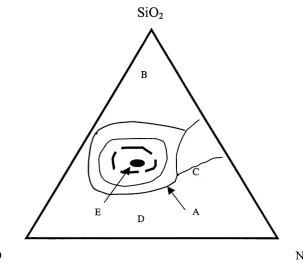
Figure 2. Region D represents technical glasses with limited biological use...14
Figure 3. Surface reaction stages for Bioglass®... 15
Figure 4: (a) Monosilicate structure and (b) two linked (condensed) m onosilicate...27
Figure 5: Phosphate mono unit... 28
Figure 6: Structural changes by adding alkaline oxides to P2O5 with Q-nomenclature for appropriate sp ecies...31
Figure 7; Structure o f glass with modifying ions. Black circles: metal or non-metal, white circles: oxygen atoms, M"^ or modifying oxides (Zachariasen, 1932)...33
Figure 8: Schematically representation o f electron transfer from oxygen to phosphorus within the double bond and single bond caused by modifying cation...41
Figure 9: Proposed structure for inter (a) and intra (b) -molecular hydrogen bonding...43
Figure 10 Figure 11 Figure 12 Figure 13 Figure 14 Figure 15 Figure 16 Figure 17 Figure 18 Figure 19 Figure 20 Figure 21 Figure 22 Figure 23 Figure 24 Figure 25 Figure 26 Figure 27 Figure 28 Figure 29 Figure 30 Figure 31 Figure 32 Figure 33 Figure 34 Figure 35 Figure 36 Figure 37 Figure 38 Figure 39 Figure 40 Figure 41 Figure 42 Figure 43 Figure 44 Figure 45 Figure 46 Figure 47 Figure 48 Glass formation process... 48
Typical DTA trace with two temperature events for a polymer... 50
Calibration curve for calcium standard... 79
Calibration curve for sodium standard...79
Calibration curve for potassium standard... 80
Caibration curve for fluoride standard...80
Weight loss curve for Cai2Na4 3P4s ... 86
Weight loss curve for Ca,6Na3 9P4 5... 87
Weight loss curve for Ca2oNa3sP4 5... 87
Weight loss curve for Ca2 4Na3iP4s ... 88
Weight loss curve for Ca2gNa2 7P4 5... 88
Weight loss curve for Ca3 2Na2 3P4s ... 89
Weight loss curve for Ca3 6Nai9P4s ... 89
Weight loss curve for Ca4oNaisP4 5... 90
pH progress with time for the ternary Ca0-Na20-P20s system ... 91
pH progress with time for the ternary Ca0-Na20-P20s system... 92
pH progress with time for the ternary Ca0-Na20-P20s system ... 92
Ca^^ ion measurement for ternary system Ca0 -Na2 0-P2 0 5...93
Ca^^ ion measurement for ternary system C a0-Na20-P20s...94
Ca^^ ion measurement for ternary system Ca0-Na20-P20s...94
Na"^ ion measurement for ternary system Ca0 -Na2 0-P2 0 5... 95
Na"^ ion measurement for ternary system Ca0-Na20-P20s... 95
Na^ ion measurement for ternary system C a0-Na20-P20s... 96
Weight loss plot for Ca2oNa3 5P4s...97
Weight loss plot for Ca2gNa2 7P4 5...97
Weight loss plot for Ca4oNa,5P4 5...98
Solubility plot for Cag Na4 7P4s ... 103
Solubility plot for CaioNa4 5P4s... 103
Solubility plot for Cai2Na4 3P4s... 104
Solubility plot for Cai6Na3 9P4s ... 106
Solubility plot for Ca2oNa3sP4 5... 105
Solubility plot for Ca2 4Na3iP4s... 105
Solubility plot for Ca2gNa2 7P4s... 106
Solubility plot for Ca3 2N2 3P4s... 106
Solubility plot for Ca3 6N ai9P4s... 107
Solubility plot for Ca4oNai5P4s... 107
Ca^^ concentration p lo t... 114
Na^ concentration p lo t...112
Solubility plot for Ca24K3i? 4 5...117
Solubility plot for Ca32K2 3 ? 4 5...118
pH measurement for o Ca,6K39P45 ° Ca24K3iP45 A Ca32K23P45... 119
Ca^^ ion measurement for o C a i6K39P45 aCa24K3iP45 A Ca32K23P4s... 120
K^ ion measurement for o Cai6K39P4s ° Ca24K3iP4s A Ca32K23P45...121
Solubility plot for Na2oKi5Ca2oP4 5...127
Solubility plot for N ai5K2oCa2oP4s ...128
Solubility plot for NaioK25Ca2oP4 5...128
Solubility plot for Na5K3oCa2oP4 5...129
Solubility plot for Na26KsCa24P4 5... 130
Solubility plot for Na2iKio Ca2 4P4s...130
Solubility plot for NaieKio Ca24P4s...132
Solubility plot for NanK,gCa24P4 5...131
Solubility plot for Na6K2 5Ca24P4s...132
Solubility plot for Na22K;Ca2gP4 5... 133
Solubility plot for NanKioCa2gP4 5...133
Solubility plot for N a,2K;5Ca2gP4 5...134
Solubility plot for Na7K2oCa2gP4s... 134
Solubility plot for Na2oK3Ca32P4s... 135
Solubility plot for Nai5KgCa32P4 5...136
Solubility plot for NaioKi3Ca32P4 5...136
Solubility plot for Na5K,gCa32P4 5...137
pH progress for o Na2oKi5Ca2o P45 and ° NasK3oCa2oP4 5... 139
pH progress for o N ai5K2oCa2o P45 and ° NaioK25Ca2oP4 5... 140
pH progress for o Na2iKioCa24P45 and ° Na6K24Ca24P45...141
pH progress for o Na26KgCa24P45 and □ Na,6K,5Ca24P4s A NanK2oCa24P45... 141
pH progress for o Na22K5Ca2gP4s and ° Na?K2oCa2gP45...142
pH progress for o Nai7KioCa2gP45 and ° N a,2K ,3Ca2gP4 5... 142
pH progress for o N ai5KgCa32P45 and ° NaioKi3Ca32P4s...143
Ca^^ ion measurement for o Na2oKi5Ca2oP45 and □ Na5K3oCa2oP4s...144
Ca^^ ion measurement for o NaigK2oCa2oP45 and ° NaioK25Ca2oP4s...145
Ca^^ ion measurement for o Na2iKio Ca24P45 and ° NaôK24Ca24P45...145
Ca^^ ion measurement for o Na26K5Ca2 4P4s ° Nai6Ki5Ca24P45 and A NanK2oCa24P45 146 C a.^* ion measurement for o Na22Ks a2gP4s and ° Na7K2oCa2gP4s... 147
Ca^^ ion measurement for o N ai7KioCa2gP45 and ° Nai2Ki5Ca2gP45... 147
ion measurement for o Na2oK3Ca32P45 and ° NajKig Ca32P4s... 148
Figure 49 Figure 50 Figure 51 Figure 52 Figure 53 Figure 54 Figure 55 Figure 56 Figure 57 Figure 58 Figure 59 Figure 60 Figure 61 Figure 62 Figure 63 Figure 64 Figure 65 Figure 66 Figure 67 Figure 68 Figure 69 Figure 70 Figure 71 Figure 72 Figure 73 Figure 74 Figure 75 Figure 76 Figure 78 Figure 79 Figure 80 Figure 81 Figure 82 Figure 83 Figure 84 Figure 85 Figure 86 Figure 87 Figure 88 Figure 89 Figure 90 Figure 91 Figure 92 Figure 93 Figure 94 Figure 95 Figure 96 Figure 97 Figure 9Î Figure 99 Figure 100: Ion concentration plotted against t^^^ o Na22K5Ca2gP4 5 and ° Na7K2oCa2gP45... 158
Figure 101: Solubility plot for Ca3oMg2Na2 3P4s ... 162
Ca" Ca^^ ion measurement for o Na,5KgCa32P4s and ° NaioK^ Ca32P4s... 149
Na"^ ion measurement for o Na2oKi5Ca2oP45 and ° Na5K3oCa2oP45...150
Na^ ion measurement for o NaisK2oCa2oP45 and ° NaioK25Ca2oP45...150
Na^ ion measurement for o Na2iKioCa24P45 and ° Na6K24Ca24P45...151
Na"^ ion measurement for o Na26KsCa24P45 ° Nai6Ki5Ca24P45 and A Na, ,K2oCa24P45...152
Na"^ ion measurement for o Na22K5Ca2gP4s and ° Na7K2oCa2gP4s ... 152
Na^ ion measurement for o Nai7KioCa2gP45 and ° Nai2Ki5Ca2gP45...153
Na"^ ion measurement for o Na2oK3Ca32P4s and ° Na^Kig Ca32P4s...154
Na"^ ion measurement for o Na,5KgCa32P45 and ° NaioKi3Ca32P45...154
CaO content 20mol% ... 157
CaO content 24 mol% ... 1557
CaO content 28 mol% ... 158
CaO content 32 mol% ... 155
Ion concentration plotted against t''^^ for o Na22K5Ca2gP4s and ° Na7K2oCa2gP4s... 157
Katrin Franks Ph.D. Thesis
Figure 103: Solubility plot for Ca2oMgi2Na23P4 5... 163
Figure 104: Solubility plot for Cai5M gi7Na23P4 5... 164
Figure 105: Solubility plot for CaioMg22Na23P4s... 165
Figure 106: pH measurement for o Ca3oMg2Na2 3P4s and ° Ca,oMg22Na23P4 5... 166
Figure 107: pH measurement for o Ca2sMg7Na2 3P4 5, ° Ca2oMgi2Na23P4s and A Ca,5Mg]7Na23P4 5...167
Figure 108: Ca^^ ion measurement for o Ca3oMg2Na23P45 and ° CaioMg22Na2 3P4 5...167
Figure 109: Ca^^ ion measurement for o Ca2sMg7Na23P4 5, ° Ca2oMg,2Na23P45 and A Cai5M gi7Na23P45 168 Figure 110: Na"^ ion measurement for o Ca3oMg2Na23P4s and ° CaioMg22Na2 3P4 5...169
Figure 111: Na^ ion measurement for o Ca25Mg7Na2 3P4 5, ° Ca2oMgi2Na2 3P4s and A Cai5M gi7Na23P45 ...169
Figure 112: Solubility test for code 4 ... 176
Figure 113: Solubility plot for code 5 ... 177
Figure 114: F ion measurement for A code 4 ° code 5 ...179
Figure 115: pH measurements for A code 4 a code 5 ... 179
Figure 116: Ca^^ measurements for A code 4 ° code 5 ... 183
Figure 117: Na"^ ion measurement for A code 4 ° code 5 ...180
Figure 118: Shows the glass transition point Tg o plotted against the CaO content... 185
Figure 119: Shows the crystallisation temperatures Tgl o and Tg2 ° plotted against the CaO content. 186 Figure 120: Shows melting points T^l o, 7^2 a and 7^3 A plotted against CaO content... 187
Figure 121: 7 g plotted against CaO content... 190
Figure 122: 2 crystallisation points 7c 1 o and 7c 2 a plotted against CaO content... 191
Figure 123: Shows melting points 7m 1 o, 7^ 2 □ and 7m 3 A plotted against CaO content... 191
Figure 124: : Thermal parameters for Na20-K20-Ca0-P20s with fixed 20mol% CaO and varying K2O ...196
Figure 125: Thermal parameters for Na20-K20-Ca0-P20s with fixed 24mol% CaO and varying K2O ...193
Figure 126: Thermal parameters for Na2 0-K2 0-Ca0 -P2 0 5 with fixed 28mol% CaO and varying K2O ...194
Figure 127: Thermal parameters for Na20-K20-Ca0-P20s with fixed 32mol% CaO and varying K2O ...194
Figure 128: Shows increasing Tg o against increasing MgO content... 195
Figure 129: Crystallisation points against increasing MgO content...196
Figure 130: Melting points plotted against MgO content...197
Figure 131: Show a typical ^*P-MAS-NMR spectrum for ternary glass system Na20-Ca0-P20s with Band A and Band B and their corresponding side b ands...198
Figure 132: o Isotropic chemical shift ôjso plotted against CaO content for the ternary glass system N a20-C a0-P 20s... 203
Figure 133: o isotropic shift ôjso plotted against CaO content for ternary K 20-Ca0-P20s glass system ... 204
Figure 134: Suggested structure for Na4Ca(P0 3)ô...206
Figure 135: MTT test result for glass Ca24Na3iP4s .(test result after 2 days o f incubation)...212
Figure 136: MTT test result for glass Ca2gNa27P4 5 (test result after 2days o f incubation)... 213
Figure 137: MTT test result for glass Ca32Na2 3P4s (test result after 2 days o f incubation)... 214
Figure 138: MTT test result for glass Ca3ôNai9P4 5 (test result after 2 days o f incubation)...215
Figure 139: MTT test result for glass Ca4oNaisP4 5 (test result after 2 days o f incubation)...215
Figure 140: MTT test result for glass Ca28Na22KsP45 (test result after 2 days incubation)... 217
Figure 141: MTT test result for glass Ca2gNai7KioP45 (test result after 2 days o f incubation)... 218
Figure 142: MTT test result for glass Ca2gNai2K i5P45 (test result after 2 days o f incubation)... 218
Figure 143: MTT test result for glass Ca2gNa7K2oP4s (test result after 2 days o f incubation)... 219
Figure 144: MTT test result for glass Ca3oMg2Na2 3P4s (test result after 2 days incubation)...220
Figure 145: MTT test result for glass Ca2sMg7Na2 3P45 (test result after 2 days o f incubation)... 221
Figure 147: MTT test result for glass Cai5M gi7Na2 3P4 5 (test result after 2 days o f incubation)...222
Figure 148: MTT test result for glass CaioMg22Na2 3P45 (test result after 2 days o f incubation)... 222
List of Tables Table 1: Weight gain check... 76
Table 2: Glass codes and oxide composition in m ol% ... 83
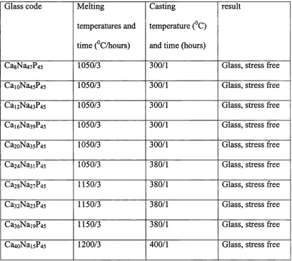
Table 3: Melting and casting temperatures... 84
Table 4: Overview o f measured solubilities for the ternary Na2 0-Ca0 -P2 0 5 system...90
Table 5: Solubility overview for ternary system in H B S S ... 98
Table 6: Overview o f Fickian/case II diffusion portion... 108
Table 7: Solubility values...110
Table 8: Glass codes and oxide composition in mol% ... 114
Table 9: Melting and casting temperature overview ...115
Table 10: Solubility overview for ternary C a0-K 20-P20;...118
Table 11: Glass codes and oxide compositions in mol%...125
Table 12: Melting and casting temperatures... 126
Table 13: Solubility overview for the quaternary Na20-K20-Ca0-P20s system ... 138
Table 14: Glass composition with oxide contents... 160
Table 15: Melting and casting temperatures... 161
Table 16: Overview o f solubilities for quaternary system CaO- MgO-Na20-P2O$...165
Table 17: Preliminary compositions attempted...173
Table 18: Glass codes and chemical compositions... 175
Table 19: Melting and casting temperatures... 175
Table 20: Solubilities for code 4 and code 5 ... 177
Table 21: Listed chemical shifts, % o f amount and anisotropic shift parameters with asymmetry parameter for the ternary Na2 0-Ca0 -P2 0 5 glass system ...201
Katrin Franks Ph.D. Thesis
Introduction
1.1 Materials for hard tissue surgery
The quest for materials, which replace parts of the human skeleton, is not new. There
is a general need for some type of bone replacement or regeneration for patients who
have suffered from trauma or chronic degenerative diseases, which lead to bone loss
or abnormalities.
There are many requirements that these materials must meet if they are to be
implanted in the human body.
The main requirements are:
1.) Chemical and physical stability for the required lifetime o f the device
2.) A wound healing tissue response with no toxic, allergic or chronic
Katrin Franks Ph.D. Thesis
1.1.1 Host /Tissue Response
No implant material is completely inert. Many years of research have made it clear
that any changes in a material, which is implanted into a living tissue, can be linked to
the tissue response.
According to Hench (1 )4 types o f response can occur:
1.) The implant material is toxic and the surrounding tissue dies
2.) The implant material is non-toxic and almost inert, fibrous tissue forms
3.) The implant material is non-toxic and biologically active and a tissue
attachment is formed
4.) The implant material dissolves and the surrounding tissue replace it.
These response types are not discrete and do overlap, but clearly, responses type 1 and
2 are far from ideal and can lead to loss o f the implant. Only responses 3 and 4 with
either a direct stable attachment of tissue to the implant, or a complete replacement of
the implant by natural bone will be useful and successful.
Figure 1 shows the general series of overlapping cellular events, which occurs during
Fibrin
Injury/
Operation Days Post - T raum a
Figure 1. Shows the cellular progression after implantation. PMN Leukocytes
I Macrophages Fibroblasts Collagen
Lymphocytes Bone Mineral
Stiffness
0 5 10 1 5 20 2 5 30
Initial events are concerned with cleaning of debris from the wound side, followed by
soft tissue formation, such as the laying down of collagen.
The bone formation usually starts around the 14th day. An ideal implant for bone
regeneration will show low levels of fibrous capsule formation and support and
enhance the bone matrix repair process. Recently, the discipline of Tissue Engineering
has been established to develop degradable and non-degradable biomaterials, which
support and guide the tissue healing process. One example of a biocompatible but
stable polymer is a PTFE fibrous membrane, which is used for treating periodontal
disease. An example of degradable polymers widely used for internally suturing of
wounds would be a polymer or copolymer such as polyglycolic/polylactic acid and is
Katrin Franks Ph.D. Thesis
in place o f PTFE as they are degradable. The major aim of the technologies above is to
guide the regenerative process in terms o f reproducing new, fully functioning living
tissue. This newly formed tissue will be formed on top of a polymer matrix or in a
porous scaffold. Degradable polymers, as mentioned above, show the advantage of
initiating and supporting new guided tissue formation, however their disadvantage is
the unpredictable degradation process.
As one can see from Figure 1, the bone mineral matrix formation is generated after 14
days. An ideal material would be a scaffold material, which is stable up to 14 days and
then slowly dissolves and is replaced by the newly developing bone matrix. An
additional advantage would be a material, which dissolves in a way that it will release
beneficial ions, which may be used by the cells to produce mineralised tissue.
There have been developments in the use o f glasses for hard and soft tissue
regeneration, which show evidence of at least a partial degradation mechanism.
Developments and features o f these materials will be reviewed in the following
1.1.2 Bioactive Glasses
In 1969, Hench (1) developed the well known and clinically used Bioglass® and it was
the first synthetic material to bond with living bone. Bioglass® is a bioactive SiOz
based glass. With compositional features such as SiO: content <60 mol.%, high
CaO/PzOg ratio and high CaO and NazO content, this distinguishes Bioglass® firom
any other “normal” soda-lime silica glass. Today bioactive glasses are clinically used
in non-load bearing situations, and have been used for alveolar ridge augmentation,
periodontal pocket obliteration and maxillofacial reconstruction.
Over several years many other bioactive silica glasses have been developed, however
they are still based on the original 45S5 (Bioglass®) composition, which is today used
as a baseline for many further developments, for example the glass with the
composition SiOz: 45mol%; P2O5: 6mol%; CaO: 24.5mol% and NazO: 24.5mol%
Bonding to bone in the compositional range of 45S5 glass was first demonstrated by
Katrin Franks Ph.D. Thesis
SiO:
CaO NaiO
Figure 2. Compositional dependence (in weight percent) o f bone bonding. A ll compositions in region A have a constant amount o f 6 wt% P 2 O 5 . All compositions within this region bond to bone and the region is termed the bioactive-bone-bonding boundary. Region E (inside the dashed line) is the soft-tissue bonding region. Silica glasses in region B behave as nearly inert materials and will elicit fibrous capsule formation. Glasses in region C are resorbable, 10-30 days after implantation. Region D represents technical glasses with limited biological use.
Bioactive glasses show a common characteristic feature o f a time dependent surface
modification after placing in tissue. When implanted, the surface of the glass
undergoes a transformation to form a biologically active hydroxy apatite layer (HCA).
It forms the basis o f a strong bond with bone. Hench (1) concluded that bioactivity
only occurs if certain amounts and ratios of glass forming or modifying oxides are
present. If the tissue forms a bond to the implant a layer o f biologically active HCA
must form. “This is perhaps the only common characteristic o f all known bioactive
12 reaction steps are needed to successfully bond to bone and these are shown in the
following figure (3).
B ioactive glass surface
1 + 2 Formation o f SiO H bonds
T
P olycondensation o f SiOH + SiOH Si - O - Si
A dsorption o f amorphous Ca3^ + POz»^' + CO3
2 T
-C rystallisation o f hydroxyl carbonate apatite H -CA
A dsorption o f b iological m oieties in H C A layer
A ction o f m acrophages
A ttachm ent o f stem cells
T
D ifferentiation o f stem cells
J _________________
Generation o f matrix
C rystaîisation o f matrix V_
Proliferation and growth o f bone
11
l 2
Figure 3. Surface reaction stages for Bioglass
The alkali-ion-hydrogen-ion exchange is the first reaction process stage and occurs
quite rapidly. After 20 min. all of the Si-O-Si bonds are broken and transformed to
give Si-OH (silanols). The result is a silica gel-layer of polycondensated silanol
groups. As early as 10 minutes after implantation a calcium phosphate layer appears
due to migration of Ca^^ and P0 4^' ions through the SiOz layer. This formed calcium
Katrin Franks Ph.D. Thesis
from solution. With time, this amorphous calcium phosphate layer will transform into
a hydroxy-carbonate-apatite layer (5), (6), (7).
Bioglass® has the advantage of being able to bond to hard tissue via the HA layer
formed, however the disadvantage o f this material is its brittleness. As a consequence,
difficulties in shaping arise and so its use for load bearing applications is limited.
Improvements have been made by incorporation of Bioglass® particles in a high-
density polyethylene (HOPE) matrix. According to Wang et al. (8) microhardness
increased with increased Bioglass® volume while tensile strength and fracture strain
decreased. These composite materials still develop the same biological apatite layer
like the bulk glass when placed in simulated body fluid.
A modification o f Bioglass® by Kim (9) was developed. This was a fluoride containing
glass, which might be beneficial for dental applications. With the use of fluoride it was
hoped to form a fluoro-hydroxyapatite instead o f a hydroxyapatite layer. A fluoro-
hydroxyapatite can release fluoride, which may help to stabilise the mineral phase in
teeth and prevent decay.
In this study 40mol% of the original CaO content was replaced by Cap2. After soaking
in Tris-Buffered Solution for several hours a crystalline fluoroapatite phase was
formed on the surface of the glass.
Toxicology and biocompatibility studies of Bioglass® took place as early as 1981 by
Wilson et a l (10) on Bioglass® and modified Bioglass®, where CaF2 substitutes CaO.
This work studied the in vitro and in vivo response with different cell types. In
types proved to show only 2-5% toxic effects to cells. Similar test results were
observed for the in vivo testing where after 2.5 years no toxic effect was seen.
In contrast, a paper on the toxic effect o f silica-containing glasses was demonstrated
by Nagase et al. in 1991 (11). This was an extension of a preliminary study published
in 1987 (12) on a fine powder o f silicate containing glass-ceramics, which showed
toxic effects in mice. However the mechanism o f toxicity and the exact composition of
the silica-containing glass-ceramic, which evoked the toxicity remains unclear.
Suspensions of calcium phosphate glasses with various amounts of SiO] were injected
into mice to determine the mortality rate. The mortality of the mice was directly
proportional to the dissolved amount o f released Si^^ ions. The conclusion was drawn
that SiO] when dissolved in the body has a high toxic potential.
A less strong toxic effect for Bioglass was seen on human synoviocyte cultures.
Bendall et ût/ (13) described the effect o f particulate Bioglass® in a concentration range
o f 1 .0 -1 0 pg/ml as having low cytotoxicity. However these concentrations induced
Katrin Franks Ph.D. Thesis
1.1.3 Controlled Released Glasses
Phosphate based glasses - a possible biomaterial?
What can be seen from the above discussion is that it is still not clear what effects the
release o f Si"^^ ions has on human tissue.
Glasses, which consist only of biologically related phosphorus, calcium and sodium
ions, offer great potential as a biomaterial. They show characteristics making them
potentially useful as a temporary device such as predictable solubility, controllable
chemical composition and therefore controllable solubility, biodegradability without
toxic residues and most importantly it might be possible to reproduce the
stoichiometric phase of natural bone within the glass matrix.
Vogel and Holand (14) developed in 1987 silicon free phosphate based glasses and
glass ceramics o f various compositions for biomedical applications. Ca0 -Al2 0 3 -? 2 0 5
formed the basic system and gradually this system has been extended to Na2 0-Ca0 -
AI2O3-P2O5 and Na2 0-Ca0 -Al20 3-P2 0 5"F. Na2 0 was used to break down chains within the structure in order to enhance the possibility of apatite formation when heat-
treated to convert the glasses to glass-ceramics.
Preliminary in vivo tests showed in general very promising results. However, the idea
o f avoiding silicon in bioactive glasses or glass-ceramics for biomedical use is
relatively new and therefore no precise and systematic toxicity studies are available, as
this is the case with silicon based glass materials. Work carried out in this thesis will
Soluble phosphate glasses can be used not only for guided tissue regeneration, they
can also be used for localised release o f specific ions, for example platinum for cancer
treatment or silver in order to treat bacterially infected tissue.
In the sixties phosphate based glasses were originally investigated for their electrical
properties for communication use. From the late seventies onwards many pesticides
commonly used were banned because they were considered to be too hazardous. At
the Standard Telecommunication Laboratories, Essex, UK, Drake and Graham (15-
17) developed slowly releasing biocidal glasses, doped with biocidal ions such as
copper, silver or boron to be considered as a substitute for the banned pesticides or
fungicides. As the glass dissolves, biocidal ions constantly leach out until the glass
completely dissolves leaving no residue behind. A decade later Allen et al (18-22) was
involved in another investigation field to cure trace element deficiencies in cattle and
sheep. Classic metabolic diseases such as milk fever, fatty liver syndrome and ketosis
are caused by nutritional deficiency o f either major or trace elements. To prevent for
example milk fever, it is important to ensure an adequate intake of magnesium for the
lactating animal. With the use of slow release phosphate glasses, which are doped with
essential ions, they hoped to cure such diseases and indeed showed that a significant
Katrin Franks Ph.D. Thesis
Aims of This Work.
This project was mainly aimed at the development of novel soluble glasses for
periodontology treatment (Periodontitis). The before mentioned use of a GoreTex®
PTFE fibrous membrane which act as a scaffold to promote hard and soft tissue re
growth is up to day the best choice for patients who suffer from reduced jaw lines
caused by chronic degenerative diseases which lead to bone loss. The use of this
membrane is proven to rebuild tissue matrix however there is a major drawback. Once
the membrane is set in place by a surgical procedure the tissue starts to regenerate.
This membrane has to be removed, hence this is on the one hand very inconvenient for
the patient and it will on the other hand remove parts of the newly formed tissue. It
seems obvious that improvements are necessary. A possible alternative would be a
system which promotes bone re-growth in the same way the membrane functions but
with the difference that the material itself degrades after the bone healing process
starts.
One aspect o f this project is to develop a soluble scaffold matrix, which acts in a
similar way to the GoreTex® membrane in terms of its biocompatibility but also
degrades slowly in an appropriate time. An additional consideration is the careful
choice o f the material. When the implant is placed and it starts to degrade and release
structural units, one should bear in mind the toxic or allergic effect this might cause to
the surrounding tissue. The question which will be related to this consideration is: Is
there a material, which not only avoids any toxic and/or allergic reaction, furthermore,
Katrin Franks Ph.D. Thesis
A potential alternative to a GoreTex® membrane might be a soluble phosphate based
glass modified by chemical elements, found in the natural inorganic phase of bone. It
is well known in the literature that phosphate salts are soluble, however the solubility
of a phosphate based glass matrix has not been fully investigated. Therefore some
questions surfaced here:
1 Is it possible to develop a soluble glass which lasts for the duration of the bone
healing process and
2 is it possible to incorporate beneficial elements such as calcium, magnesium and
sodium into the phosphate glass matrix in order to enhance biocompatibility? In
addition,
3 how do these elements affect the solubility o f these phosphates glasses and is
one able to calculate and predict their solubility,
4 how will the solubility change by adding different alkaline and alkaline earth
oxides? Most important o f all,
5 how will cells react towards a basic phosphate glass and how will they react
towards modified phosphate glass systems? Therefore preliminary cell tests such
as the MTT test has been set up to determine how cells will react towards a
system in vitro.
MTT tests are of great importance; the results help us to understand how cells respond
and to predict if a material is biocompatible or not. Furthermore they can be of great
help to decide if additions of ions are beneficial or even enhance cellular activity.
There is also a more theoretical approach o f the project. Up to today the structure of
Structural investigation with the use o f Solid State Nuclear Magnetic Resonance
Spectroscopy (SS-NMR), Differential Thermal Analysis (DTA), X-ray Diffraction and
Infrared Spectroscopy (IR) helped at least to understand parts of the structural
arrangements. However, more work needs to be carried out to understand this
completely. With further knowledge o f the structure it might be possible to relate the
structure o f the glasses with their solubility.
Further questions arise, such as (1) how are structural units arranged in a phosphate
based glass matrix, (2) what kind o f analytical tool is best to analyse it (3) how will
additional modifiers intervene and (4) is there a noticeable trend by systematically
incorporating different modifiers to the glass network?
If there is a trend in structural changes, can the same trend be found for the solubility
Katrin Franks Ph.D. Thesis
Glass
This chapter was drawn up in order to provide information regarding glass science,
including a survey of the literature of glass-structure theories investigated using X-ray
Photoelectron Spectroscopy (XPS), MAS-NMR spectroscopy and Differential
Thermal Analysis (DTA).
MAS-NMR is a relatively new technique allowing close observation of the local
environment around a particular element and will therefore provide new insights to the
glass structure theories. Differential Thermal Analysis (DTA) can be utilised for two
things:
(1) It may be used for measurement o f classical polymer characteristics such as glass
transition temperature (Tg), melting temperature (Tm) and crystallisation
temperature (Tc) which are vital for the process of glass making.
(2) On the other hand, phenomena such as phase separation in the glass are still not
fully understood and to a certain extent DTA can help to investigate this effect.
The following pages will start with a simple glass forming oxide, explaining important
terminology and parameters in glass science. The complexity o f the system will be
increased and binary systems will be considered. It will also be at this stage that the
glass structure theory with a literature survey will be found. As was mentioned above,
no clear structural theory is today available. However, with the use o f MAS-NMR in
Katrin Franks Ph.D. Thesis
Finally, DTA and the significance of parameters such as Tg, Tm and Tc will be
explained as well as the glass forming process. The DTA was extensively used for the
observation of these parameters but also the phenomena of phase separation will be
considered.
3.1 Glass In general
Man-made glasses appeared around 4000 BC in Egypt but were merely decorative.
Around 1500 BC there were significant developments in both the art and the
technology of glasses and this was followed by further technological developments in
glass science by Faraday, Zeiss, Abbe and Scott to name but a few. Their primary
interest was in glasses for optical use. Nowadays, glasses are widely used both for
everyday applications such as containers, automobiles and windows, but also for
special applications, such as fibre optics. However, the idea o f using a soluble glass as
a biomaterial is a relatively new one.
A glass is in general an amorphous supercooled molten mass, which has not
undergone crystallisation. This supercooled liquid softens very slowly on heating and
its structure shows only short-range order. The “non-crystalline elastic solid” typically
has a maximum ordering range of 2.0 nm with a viscosity >10 poise (10^^^ Ncm'^)
(23). Glasses based on oxides such as M2O3 (e.g. B2O3, AI2O3), MO2 (e.g. Si0 2, 0 e0 2)
and M2O5 (e.g. P2O5, AS2O5), where M is an element o f the third, fourth or fifth chemical group o f the periodic table have extremely low crystal formation velocities.
The rates o f crystallisation are lowest for substances easily cooled to glasses, such as
silica and germania, whereas they are higher for glasses incorporating alkali and
alkaline earth compounds which must be cooled more rapidly to avoid crystallisation
(24).
3.1.1 Single Oxide Glass System
Vitreous oxides:
Due to their relative abundance in nature (silicon and oxygen are very common on
earth (24) ), silicates have been extensively studied. Historically, silica (SiO]) has been
treated as a classic network-forming oxide in both crystals and glasses. This is due to
the fact that silicon has a high affinity to oxygen and secondly The structural unit of
silica is tetrahedral in which silicon is four-fold co-ordinated by oxygen. This
tetrahedron is the basic unit and one mono silicate is cross linked with its neighbours
over comers to form a whole network.
O 0 0
o
/
o
o
/
o
o
o
o
(a) (b)
Figure 4; (a) Monosilicate structure [ S iO /‘] and (b) two linked (condensed) monosilicate [Si2 0 7^ ]
The ability of Si0 4 to cross-link is a well-known principle, which is confirmed by the
existence of many known crystals. An example o f a crystal is the mineral Beryl
Be3Al2Si6 0i8- This cross-link principle forms not only the basis for crystals, it can also
Katrin Franks Ph.D. Thesis
Vitreous silica can be made, but from an industrial point is not routinely used due to
the very high temperatures involved to form such a glass. Modifying oxides are able to
break down the structure and significantly reduce the melting temperature. This is a
major benefit for the production process.
But silicon in conjunction with oxygen atoms is not the only element, which forms
crystals or glasses. Phosphorus is another example o f a chemical element which can be
found in both crystals and glasses with a similar structural relationship.
Phosphorus is an element o f the fifth group o f the Periodic Table and has an electron
configuration o f 3s^p^, showing that it has one extra electron in comparison to Si
(3s^p^). In consequence, phosphorus tends to form a P -0 double bond but will still be
co-ordinated tetrahedrally as found in silica (24).
o
o
Figure 5: Phosphate mono unit
Biscore et al (25) have shown that the unit formation in all P2O5 based glasses is the
[PO4]- tetrahedron which is linked to form a three-dimensional network similar to mica glasses. The incorporation of alkali ions into a three-dimensional network leads
In comparison to S1O2 in its vitreous state, P2O5 is very difficult to obtain in a glassy
form. This is due to the fact that P2O5 is very hygroscopic and volatile and hence the
number o f papers investigating the structure of this state is rather limited. DeDecker
(26) observed in 1942 that for a crystalline P2 0 5(c-P2 0 5) system 2 crystalline
structures exist.
A rhombohedral polymorph will change at high temperature at around 260^C into a
orthorhombic system. He claimed that the volatile, low temperature modification is
built o f P4O10 molecules, whereas the high temperature modification consists of two-
dimensional planes, which have a more network-like nature. This latter form is easily
supercooled to form a glass. At this stage no infrared or no detailed MAS-NMR
spectroscopy observations on vitreous P2O5 have been reported.
3.1.2 Binary glass systems:
Oxides, which readily form glasses are Si0 2, Ge0 2, B2O3, P2O5 and AS2O3 However,
most glasses are made from a mixture of oxides including network modifiers and
network intermediates.
N etw ork m odifying oxides
Technical glasses consist of additional compounds. These additions are called
modifying oxides, as they modify the glass structure, and are in general alkali- and
alkali earth metal oxides, which break down parts of the network.
Katrin Franks Ph.D. Thesis
Using a Nu20 modifier:
RsSi - O - SiRs + NazO -> 2 R3S1 - ONa
Using a CaO modifier:
R3S1 - O - S1R3 + CaO R3S1 - O - Ca - O - S1R3
R = oxygen linkage
NazO as well as CaO are able to break down the strong Si - O - Si bonds and therefore
change the structure and characteristics o f the glass. If Na2 0 is used as a modifier the
corresponding Na^ ion has only one positive charge and will terminate the network at
a certain point. Ca^^ however can cross-link the network as it is a “species with two
arms” (2 positive charges) hence it can re-stabilise the glass network. This may
manifest itself in a significant increase in Tg with CaO addition.
Intermediate oxides
Other groups are oxides such as AI2O3, Fc203, MgO and BeO, to name but a few,
which cannot form glasses on their own but they can to a certain extent substitute for
the Si0 2. Colour changes or conductivity changes are examples of the properties
modified by these oxides.
The addition of modifiers will, with respect to silicate based glasses, reduce
production costs. Lime for instance is extremely cheap to manufacture and thus is a
modifiers will make this glass less hygroscopic, hence structural information may be
more easily obtained. The hygroscopic nature o f phosphate glasses remains a problem
when the modifying oxide is below 30 mol% (27).
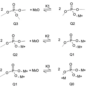
When adding alkali oxides to P2O5, 3 main reactions can occur which are shown in
figure 6 (28).
2 p _ o — + M2O V. ^ 2 O
O.
/
U-IVI+
o03
02
O K2 9
2 —O — + M2O ^ ^
0-M+ / 0-M+
02
01
M K3
2 ^ R —O - M + + M2O 2 Q ^ P —0 - M + O ^ ' \
(
Q1 QO
0 - M +
Figure 6: Structural changes by adding alkaline oxides to P2O5 with Q-nomenclature for appropriate species
According to these equations, stmctural changes continue to occur with increasing
Katrin Franks Ph.D. Thesis
oxides the structure changes to linear, pyro- and ortho phosphates and finally to ortho
phosphate as single ionised PO4 groups (Q°).
3.2 Glass Structure Theories
Glasses may be regarded as polymeric oxides with varying degrees o f three-
dimensional cross-linked network structures. It is therefore possible to regard oxide
glasses as polymers of oxygen. In contrast to organic polymers which are formed from
chains o f adjacent carbon atoms, oxygen polymers contain networks o f alternating
oxygen atoms and intermediate, multivalent atoms, such as silicon, boron and/or
phosphorus. Since the early decades o f this century many workers have been studying
the structure o f glasses, unfortunately no definitive structure is yet apparent. Two
major structure theories exist and over the years they have been reviewed and refined.
In 1926 Goldschmidt (29) formed a hypothesis that any oxide AmOn with a radius ratio
o f Ra/Ro is in the range 0.2-0.4 will form a glass ( R= radius, A= metal or non-metal
and 0 = oxygen). This work showed that oxides were tetrahedrally co-ordinated and
that this special co-ordination was necessary to form a glass. However, a vitreous BeO
has not been made, although the radius ratio is 0.3, this should according to
Goldschmidt’s theory form a glass. In 1932 Zachariasen (30) published work on the
glass structure concerning simple glass systems such as Si0 2. This work considered
glasses as continuous, irregular networks where oxygen polyhedra were linked
together over their comers. Addition of modifiers such as alkaline or earth alkaline
ions were placed in the hollow space o f the network without forming any special
Q
\Q
/
o ' ' ^ v
o
M+
O
Q
O
\
\
O
o
/
o
\ .
o
o
o
M++
\
o
o
o
/
o
\ o
Figure 7: Structure o f glass with modifying ions. Black circles: metal or non-metal, white circles: oxygen atoms, M"^ or modifying oxides (30)
According to this theory, any metal or non-metal oxide should form a glass. The non
existence of a glass form of TiO] or AI2O3 could not have been explained with this
Katrin Franks Ph.D. Thesis
was presented one year later in 1933 by Warren (31) who regarded glasses as micro
heterogens with the existence o f defined chemical phases which are linked together
and it was these that therefore built up the glass structure. However, again this theory
could not explain the non-existence of TiO] in a glassy form.
A refinement o f Warren's glass structure interpretation took place one year later in
1935 when Hagg (32) focused on the glass forming process from the point of the
cooling process. He believed that glasses consist o f chains or two-dimensional nets
and it was this that was the main difference to the interpretation o f Zachariasen. With
this theory he was able to explain why species like SiO] form a glass and TiOz do not.
He believed that the symmetry and the bulkiness of the glass forming units above the
melting temperature are responsible for the glass forming capacity. He supported his
theory with the example o f KNO3. Above the melting point the ions and NO3 are
small and mobile enough that they can easily come together and form KNO3 and if
that molten mass cools down rapidly enough it will ffeeze-in as a glass. Only materials
which will have one-dimensional or two-dimensional unlimited cross-linked units in
the corresponding crystals will be able to form a glass. For example, SiO] exists in
crystalline form as chain and ring silicates (Beryl Be3Al2Si6 0ig), which has unlimited
cross-linking. TiO] however exists “only” in the form of 3 defined crystal
modifications, where oxygen atoms co-ordinate Ti in an octahedral form. There are no
long chains or ring structures known.
Still, there is no precise structural model, which covers all glass types. The
development of modem analytical techniques such as solid state NMR-spectroscopy
Differential Thermal analysis (DTA) has helped to produce a more coherent model for
glass structures.
3.3 Solid State NMR Spectroscopy (MAS-NMR)
Nuclear magnetic Resonance (NMR) spectroscopy is one of the most powerful
methods for structural analysis of inorganic and organic compounds and especially of
disordered materials such as glasses. This technique gives very detailed information
about the very nearest environment around the nucleus under investigation. Even
different surrounding atoms with different charges such as Na^ or Ca^^ can be
detected. Only magnetically active nuclei such as isotopes o f H, C, N, F, and P, which
have a nuclear spin different from zero, can be observed using this technique. NMR
spectroscopy has recently been revolutionised by improved electronics and design,
which have dramatically improved sensitivity and chemical shift separations. With the
development o f magic-angle spinning NMR technique (MAS-NMR) chemists no
longer have to rely on liquid NMR spectroscopy techniques to investigate structures.
If normal NMR methods were applied to solids broad peaks would be the result,
hiding the desired information. This is due to chemical shift anisotropy, which leads to
a simultaneous absorption for all possible random orientations for a powder sample.
With magic angle spinning NMR however, the sample is spun at high velocity around
an axis inclined at an angle of 54^44 to the magnetic field. This effect leads to a
Katrin Franks Ph.D. Thesis
3.3.1 Beginning o f MAS-NMR Spectroscopy with Commercial Phosphates
In the early eighties Grimmer and Haubenreisser (33) reported their work on
polycrystalline P4O10 and binary phosphates in the compositional range of P2O5.XK2O
(0 < X < 3). Samples included phosphates of P4O10, K3PO4, K4P2O7, K5P3O10, K3P3O9
and (KP0 3)x and were measured at 109.3 MHz. ^^P shielding values gave isotropic
chemical shifts for K3PO4 isolated groups of PO4 at -11.7 ppm and K4P2O7 show end
groups at 0.3, 2.2, 2.4 and 4.9 ppm. K5P3O10 consisted o f end and middle groups at
1.2, 4.2 and 19.4 ppm and K3P3O9 showed a cyclic structure with middle groups at
18.2, 19.9 and 21.2 ppm. The polymer (KP0 3)% consisted o f middle groups in a chain
at 18.5 and 20.7 ppm.
In the same year as Grimmer and Haubenreisser published their results, another
publication by Duncan and Douglas (34) dealt with the structural investigation of
condensed potassium phosphates such as K4P2O7, Na4P407, K5P3O10, (NaP03)% and
pure P4O10, using MAS-NMR spectroscopy. The samples were commercial chemical
products and were measured on a CXP-200 Bruker spectrometer using around 4-4.6
kHz rotation fi*equency. Experimental results for K4P2O7 and Na4P207 showed a centre
band at 2.7 and -1.6 ppm, respectively. K5P3O10 showed a different pattern with three
different species, having isotropic shifts o f 1.2, 4.0 (end units) and 19.5 ppm (middle
group). The chain polyphosphate (NaP03)x gave a centre band at 19 ppm (middle
group) with minor species at -2 ppm (end units). The cage phosphate P2O10 was
centred at 33 ppm. Unfortunately, the authors did not mention exactly the nature of
In 1986Mudrakovskii et al (35) used M A S-N M R to analyse a series o f com m ercial phosphates such as AIPO4, A1(P03)3, Zr(H P04)2x2H 20, Bc3(P04)2 x 6H2O and Mg2P2Û7 etc. T hey recorded on a B ruker C X P-300 spectrom eter at 121.46 MHz
(m agnetic field 7.05 T) and the spinning fi*equency w as 3.5-4 kHz. The anisotropy Act
is negative o r positive depending on the individual sample, and the isotropic chem ical shifts ranged fi*om -13 to 52 ppm. They found that both anisotropy Act and the asym m etry param eter r| equal 0, except for the Mg3(P04)2 sample. This indicated the equivalence o f four P-0 bonds for each PO4 group. W ith the help o f crystallographic data, they interpreted that the P- 0 bonds are close to a regular tetrahedron. A dditionally, they also m entioned that the lack o f anisotropy in spectra o f AIPO4 x 2H2O and Bc3(P04)2 x 6H2O is an indication o f high sym m etry. The nearest oxygen atom s, w hich are surrounding the phosphorus atom, are arranged sym m etrically. D istortion in the phosphorus environm ent w ith the appearance o f anisotropy appeared in the spectra o f Zr(HP04)2 x 2H2O, Mg3(P04)2 and KH2PO4. A ccording to the chem ical shift values observed for M g(H P04) x 3H2O, C aH P04 and Ca(H2P04) x H2O there w as evidence for tw o crystallographically non-equivalent phosphorus atoms present.
3.3.2 Structural Investigation by MAS-NMR Spectroscopy for Binary Glass Systems
Villa et al (36) were the first to use MAS-NMR for structural investigations o f alkali
boron phosphate and silver boron phosphate glasses. They examined glasses prepared
Katrin Franks Ph.D. Thesis
increasing the alkaline content the polymerisation of the phosphate units was reduced.
In this same work, it was mentioned that binary phosphate glasses o f the type M2O-
P2O5, consist of
(1) branching units 0 =P-0 3 which have 3 bridging oxygen’s and are neutral in their
charge,
(2) middle units with 2 bridging oxides and 1 negative charge and
(3) end units with 1 bridging unit and 2 negative charges.
Furthermore binary phosphate glasses consist o f monomeric units, which can be found
in orthophosphates M3PO4.
On the basis of a binary silver phosphate glass, MAS-NMR revealed that these glasses
consisted o f middle units at -10- (-2 0) ppm and end phosphate units at + lOppm which
gave curves fitted by nearly two Gaussian lines. More complicated glass systems were
prepared by doping this binary silver phosphate glass with B2O3 Here, monomeric
units appear at +30ppm and units at +10ppm, middle units connected to one boron at
around 0 ppm and another middle unit at -1 0 to -2 0 ppm connected to one boron atom
and one phosphorus atom.
In 1987 ^^P-MAS-NMR investigations o f glassy inorganic phosphates were carried out
by Parbhakar et a l (37). Glasses were prepared using (NH4)2HP0 4 as a starting
reagent, heating to 920 K for several hours and the melt was quenched by pressing it
onto a stainless steel plate. The ^^P -NMR spectra, recorded at 300 MHz show ortho-