O R I G I N A L R E S E A R C H
Open Access
A convenient and efficient total solid-phase
synthesis of DOTA-functionalized
tumor-targeting peptides for PET imaging of
cancer
Subhani M. Okarvi
*and Ibrahim AlJammaz
Abstract
Introduction:An efficient and cost-effective synthesis of the metal chelating agents that couple to tumor-targeting peptides is required to enhance the process of preclinical research toward the clinical translation of molecular imaging agents. DOTA is one of the most widely used macrocyclic ligands for the development of new metal-based imaging and therapeutic agents owing to its ability to form stable and inert complexes under physiological conditions. Although solid-phase synthesis compatible DOTA-tris-(t-Bu ester) is a commercial product, it is expensive and contain chemical impurities. There is a need to explore new and cost-effective methods for the preparation of metal chelating agents, i.e., DOTA, directly on solid support to facilitate rapid, cost-effective, and high purity preparation of DOTA-linked peptides for imaging and therapy. In the present study, we describe a facile synthetic strategy of DOTA preparation and its linkage to peptides directly on solid-phase support.
Methods:Bombesin (BN) peptides were functionalized with DOTA chelator prepared from cyclen precursor on solid-phase and from commercial DOTA-tris and radiolabeled with68Ga. In vitro BN/GRP receptor binding affinities of the corresponding radiolabeled peptides were determined by saturation binding assays on human breast MDA-MB-231, MCF7, T47D, and PC3 prostate cancer cells. Pharmacokinetics were studied in Balb/c mice and in vivo tumor targeting in MDA-MB-231 tumor-bearing nude mice.
Results:DOTA was prepared successfully from cyclen on solid-phase support, linked specifically to BN peptides and resultant DOTA-coupled peptides were radiolabeled efficiently with68Ga. The binding affinities of all the six BN peptides were comparable and in the low nanomolar range. All68Ga-labeled peptides showed high metabolic stability in plasma. These radiopeptides exhibited rapid pharmacokinetics in Balb/c mice with excretion mainly through the urinary system. In nude mice, MDA-MB-231 tumor uptake profiles were slightly different; the BN peptide with Ahx spacer and linked to DOTA through cyclen exhibited higher tumor uptake (2.32% ID/g at 1 h post-injection) than other radiolabeled BN peptides investigated in this study. The same leading BN peptide also displayed favorable pharmacokinetic profile in Balb/c mice. The PET images clearly visualized the MDA-MB-231 tumor.
Conclusions:DOTA prepared from cyclen on solid-phase support showed comparable potency and efficiency to DOTA-tris in both in vitro and in vivo evaluation. The synthetic methodology described here allows versatile, site-specific introduction of DOTA into peptides to facilitate the development of DOTA-linked molecular imaging and therapy agents for clinical translation.
Keywords: DOTA, cyclen, Bombesin, Solid-phase peptide synthesis, Biodistribution, Tumor imaging
© The Author(s). 2019Open AccessThis article is distributed under the terms of the Creative Commons Attribution 4.0 International License (http://creativecommons.org/licenses/by/4.0/), which permits unrestricted use, distribution, and reproduction in any medium, provided you give appropriate credit to the original author(s) and the source, provide a link to the Creative Commons license, and indicate if changes were made.
* Correspondence:[email protected]
Introduction
Timely and accurate detection of tumor lesions is a challen-ging task in nuclear oncology. Molecular imachallen-ging tech-niques, such as radionuclide imaging, have shown great clinical potential for noninvasive precise detection of tumor lesions in early stages with the use of modern and sophisti-cated hybrid imaging modalities, for instance PET/CT/MRI and SPECT/CT/MRI. The key requirement for any success-ful molecular imaging procedure is a chemical agent, called imaging probe (e.g., peptide, antibody, etc.). The macrocyc-lic agents capable of complexing both diagnostic and thera-peutic radionuclides have shown important applications in diagnostic and therapeutic medicine in the form of metal complexes [1–4]. One of the most widely used macrocyclic ligands for the development of new metal-based imaging and therapeutic agents is DOTA (1,4,7,10-tetraazacyclodo-decane-N,N′,N′′,N′′′-tetraacetic acid). DOTA (Fig.1) is an octadentate ligand with four carboxylate and four amino groups, which holds the ability to form thermodynamically stable and kinetically inert complexes with transition metals and lanthanides, under physiological conditions [5,6].
Nonetheless, the commercially available macrocyclic metal chelating agents, triprotected DOTA-tris-(t-Bu ester) with a free carboxyl group forms DOTA-monoamide derivatives upon reaction with free amines [7], is one of the most fre-quently used bifunctional chelating agents and is fully com-patible with Fmoc-based solid-phase peptide synthesis (SPPS). However, the commercial DOTA chelator is expen-sive and suffers from chemical impurities of both the dialky-lated and tetra-alkydialky-lated cyclen [8]. An example of mass spectrometric analysis of the commercially available DOTA is presented in the Fig.2. It can be seen that besides the main product peak at 573, a DOTA (tetraxetan) peak at 405, monoalkylating product peak at 461, and dialkylating prod-uct peaks at 517 are clearly visible in the mass spectrum. In addition, the peak found at 635 may be correlated with tetraalkylating product 629 (M + t-But)+. Thus, there is a need to explore alternatives route of DOTA synthesis that can provide cost-effective, high purity, and be able to at-tach directly to the peptide sequence during SPPS, which is the ultimate method of choice for preparing tumor tar-geting peptides for clinical and preclinical applications [9,
10]. Solid-phase synthesis of a wide variety of tumor-targeting peptides radiolabeled with medically useful
radionuclides for diagnosis and therapy of various human diseases has opened a new scope in the field of molecular imaging [2,5].
An effective synthetic procedure where the DOTA-linked peptide amide is synthesized on a single solid-phase sup-port offers several advantages: the ease of coupling and time to synthesize one DOTA-peptide amide is dramatically re-duced because DOTA is introre-duced to the peptide back-bone [3, 5, 9, 10] while the peptide still attached to resin support, resulting in the formation of relatively pure DOTA-peptide product. Also, a large molar excess of DOTA will not be required as in solution-phase manual conjugation step. Owing to the well-defined peptide chemistry and excellent synthetic purity and yields obtained by the SPPS, our primary efforts in this study have been directed toward the development of DOTA-coupled peptides totally on solid-phase syn-thesis approach.
It is worth pointing out that direct solid-phase synthesis of DOTA-peptides is described previously [9]. Here in this study, we used bromoacetic acid instead of bromoacetyl bromide for N-terminus acylation, which is relatively easy to handle as well as provided efficient acylation and better yield, hence making this synthetic approach more suitable for preparing DOTA-coupled peptides. DOTA was effi-ciently prepared in the following three main synthetic steps: (i) acylation of the amino terminus of the peptide-resin with bromoacetic acid activated ester (activated in situ with HOBt/DIC). (ii) The bromoacetylated peptide-resin was then reacted with cyclen (1,4,7,10-tetraazacyclododecane) (Fig.1) resulted in the nucleophilic displacement of brom-ide function with the amine of cyclen. (iii) Trialkylation of cyclen amines with tert-butyl bromoacetate followed by TFA-mediated cleavage and removal of protecting groups [11–13] (see details in“Experimental procedures” section) to afford target DOTA-linked peptides.
The main aim of this study is the comparison between the commercially available DOTA-tris-(t-Bu ester) and the one synthesized in this study on solid-phase support in order to validate and confirm that the DOTA-peptides prepared via cyclen are as potent as peptides prepared from commercially available DOTA-tris. Three new bom-besin (BN) peptides analogs were selected as our model tumor targeting peptides for coupling with DOTA directly
on solid phase as well as to commercially available DOTA-tris-(t-Bu ester). These BN peptides differ in terms of the presence of a specific spacer group in each peptide. It has been shown that the type and the na-ture of spacer group influence the receptor binding and the tumor-targeting characteristics of the result-ant peptides [14, 15]. The two sets of BN peptides were then compared for their synthesis and radiola-beling characteristics, binding affinity to various BN/GRP receptor-positive cancer cell lines and in vivo biodistribu-tion and tumor-targeting properties. The results of these comprehensive comparisons between the DOTA-peptides prepared in-house on solid-phase support and the com-mercially available DOTA-coupled peptides are presented in this article.
Experimental Procedures General
All standard reagents, solvents, and Fmoc-amino acids for the peptide synthesis were purchased from commercial sources and used as received. DOTA-tris-(t-Bu ester) was obtained from a reputable manufacturer (Macrocy-clics, Dallas, USA). 68Ge/68Ga generator was bought from Eckert & Ziegler (Berlin, Germany). The struc-ture of each synthesized peptide was confirmed by positive-ion electrospray ionization–mass spectrometry (ESI-MS) and acquired on Agilent 6125 single quad-rupole LC/MS system (Agilent Technologies, Santa Clara, CA, USA). Reversed-phase high-performance liquid chromatography (RP-HPLC) analyses were performed on a Shimadzu HPLC system (Shimadzu Corporation, Kyoto, Japan) equipped with a UV-VIS detector (Shimadzu
Corporation, Kyoto, Japan), set at 220 nm, a γ
radioactivity detection system, and the Lauralite chro-matogram analysis program (LabLogic Systems Ltd., Sheffield, UK). Radioactive samples from in vitro and
in vivo studies were measured using a γ-counter
(Mucha, raytest Isotopenmessgeräte GmbH, Strauben-hardt, Germany).
Synthesis of DOTA-peptides
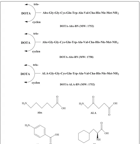
All six BN peptides (Fig. 3) were synthesized manu-ally using a standard peptide synthesis glass reaction vessel (Peptides International, Louisville, USA) by SPPS following standard Fmoc (9-fluorenylmethoxy-carbonyl) chemistry, using Rink-amide MBHA
(4-methylbenzhydrylamine) resin (100–200 mesh) on a
0.2 mmol scale according to a general method of peptide synthesis described previously [15]. Briefly, the rink amide resin (0.2 mmol, 455 mg, loading:
0.44 mmol/g) was swollen in anhydrous DMF
protecting group was removed to facilitate condensation of the BN peptides with DOTA ligand. The DOTA chelate was assembled in peptides during SPPS in a three-step process (Scheme1) as described below.
Step 1: acylation with bromoacetic acid
The peptide resin was divided into two equal por-tions, each with 0.1 mmol scale. To the first portion of the peptide (0.1 mmol), the free amino terminus of
DCM. This coupling procedure was repeated once more to ensure quantitative coupling. The coupling efficiency was determined by the Kaiser ninhydrin test and a negative Kaiser ninhydrin test indicated the coupling reaction was successful. The product was used directly in the cyclen reaction step.
Step 2: reaction of bromoacetyl bombesin peptides with cyclen
h; resulted in the displacement of bromide function with the amine function of the cyclen [4, 9, 12]. One convenient and useful method for the synthesis of DO3A (1,4,7,10-tetraazacyclododecane-1,4,7-triacetic acid) (Fig. 1) functionalized biomolecules is the direct cyclen monoalkylation, followed by the substitution of the remaining three amine positions with tert-butyl bromoacetate as we have utilized this approach in the present study [4, 5, 8].
Step 3: trialkylation of cyclen amines with tert-butyl bromoacetate
The cyclen BN peptides were then alkylated with tert -butyl 2-bromoacetate (3.3 equiv. to cyclen) in the presence of DIEA (3.5 equiv. to cyclen) for 3 h, resulting in the dir-ect alkylation of the three secondary amine positions in the cyclen ring and the formation of tert-butyl-protected DOTA-coupled peptides [3, 9, 10]. Following alkylation, the BN peptides were cleaved from the polymer support together with the deprotection of the side-chain protect-ing groups by the treatment with a cleavage mixture con-taining TFA-TIS-H2O (95: 2.5: 2.5) to afford the desired
unprotected form of DOTA-coupled tumor-targeting pep-tides (Scheme1).
The second portion of the BN peptide resin (0.1 mmol) was reacted manually with commercially avail-able DOTA-tris-(t-Bu ester) (named here ′′tris′′). For coupling of DOTA-tris-(t-Bu ester) to free amino terminus of the peptide-resin, DOTA-tris-(t-Bu ester) (0.3 mmol, 3 equiv.) was first preactivated with HATU (0.3 mmol, 3 equiv.) in the presence of DIPEA (0.6 mmol, 6 equiv.) in DMF for 15 min prior to manual conjugation with free N-terminus of BN pep-tides bound on the polymer resin. After 5 h of coup-ling reaction, the coupcoup-ling efficiency of DOTA to BN peptides was assessed by the Kaiser test. A negative Kaiser test indicated that the DOTA-peptide coupling was successful. Finally, the BN peptides were cleaved from the solid support with TFA-cleavage mixture as described above to provide the desired DOTA-linked peptides. The purity of each BN peptide was con-firmed by HPLC analysis and their structural identity by mass spectrometry.
Radiolabeling with68Ga
For radiolabeling with 68Ga, a 20 μL of each DOTA-coupled BN peptide solution (1 mg/mL CH3CN/H2O (1:1))
was mixed with 400 μL of 2.5 M sodium acetate buffer, followed by the addition of68GaCl3(~ 5 mCi) (eluted from 68
Ge/68Ga generator). The pH of the reaction mixture was adjusted between 4.5 and 5.0. The mixture was then heated for 20 min at 90 °C and cool to room temperature. The la-beling mixture was filtered through a 0.2-μm pore syringe filter to remove any particulate prior to radio-HPLC
analysis. ITLC-SG with 1 M NH4OAc:MeOH (1:1) was
per-formed to determine colloids formation.
A nonradioactive gallium complex, natGa-DOTA-BN
was prepared by reacting for instance, DOTA-BN peptide (0.5 μM, 1 equiv.) with Ga(III)Cl3 (1.0 μM, 2
equiv.) dissolved in 400 μL of 0.05 M HCl. To this
400 μL of 0.2 M ammonium acetate buffer (pH 5.0)
was added. Heating for 60 min at 90 °C was needed to complete the complex formation reaction.
HPLC purification and analysis
The HPLC analysis and purification of the peptides were performed on a Shimadzu HPLC system using
econosphere C18 reversed-phase column (10 μm,
250 × 4.6 mm). For all HPLC experiments, a gradi-ent system of 0.1% (v/v) TFA in water (solvent A) and 0.1% (v/v) TFA in acetonitrile (solvent B) at a flow rate of 1.1 mL/min was used. The HPLC gradi-ent began with a solvgradi-ent composition of 95% A and 5% B from 0 to 2.5 min followed by a linear gradient of 95% A and 5% B to 5% A and 95% B over 30 min. The gradient remained at this position for 3 min before switching back to 95% A and 5% B for another 7 min. The major peak isolated and the acetonitrile was then slowly evaporated under a stream of nitrogen gas. Radiochemical purity was es-timated by evaluating radioactivity peaks eluted for
each 68Ga-labeled peptide from the HPLC column
and calculating the area under the peak (ROI). The main peak of each radiopeptide was isolated and reconstituted in sterile saline for in vitro and in vivo experiments.
Measurement of partition coefficient
For lipophilicity determination, each HPLC-purified radiopeptide (~ 20 μL, ∼25μCi) was added into a glass tube containing an equal volume mixture of n-octanol and water (1 mL each). The samples were vigorously vortexed for 5 min and subsequently centrifuged (5000 rpm, 5 min) to yield two immiscible layers. Duplicate samples (100 μL) from each layer were carefully taken (to avoid cross-contamination between the layers) for radioactivity measurement using a γ-counter. Partition coefficient was determined by the function: partition co-efficient = Log10 (radioactivity in octanol layer/radio-activity in aqueous layer) [15].
In vitro metabolic stability in human plasma
mixture of CH3CN/EtOH (1:1v/v, 400μL) and the
sam-ple was centrifuged (7000 rpm, 7 min). The supernatant layer was removed, filtered through Millex GP filter (0.22 μm), and analyzed by radio-HPLC under the con-ditions described above to determine the proteolytic sta-bility of the68Ga-radiolabeled peptides.
In vitro tumor cell binding
The cell binding of 68Ga-radiolabeled peptides into
BN/GRP receptor-positive MDA-MB-231, MCF7,
T47D human breast cancer, and PC3 human prostate cancer lines (American Type Culture Collection, Rockville, MD, USA) was performed according to the method described previously [15]. In brief, ∼300,000 cells (in 300 μL in low-serum media) were incubated with various amounts of radiopeptides, ranging be-tween 0.1 and 25 nM of peptide (prepared from the serial dilutions of HPLC-purified radiopeptides), in duplicate for 60 min at room temperature. The initial concentrations of 68Ga-labeled peptides were deter-mined according to a well-established HPLC tech-nique with simultaneous detection by UV absorbance [18, 19]. As the amount of BN peptide in the reaction mixture was too small to be detected by UV absorption at 220 nm, the radioactive peptide chro-matograms from the HPLC analysis were compared (with UV chromatograms) with those of known concentrations of the corresponding nonradioactive peptides for the mass determination. Incubation was terminated by dilution with cold saline (300 μL) and the cells were pelleted by centrifugation. The cell pel-lets was then rapidly washed with cold saline to re-move any unbound peptide and centrifuged to collect supernatants. Radioactivity in the cell pellet and the washings was measured in a γ-counter. Non-specific binding was determined in the presence of ~ 200-fold molar excess of unlabeled BN peptide. Specific binding is calculated by subtracting the non-specifically bound radioactivity from that of the total binding. The dis-sociation constant (Kd) was calculated using a plot of
cell bound activity versus the concentration of the radioligand using GraphPad Prism software (GraphPad Software Inc., San Diego, CA, USA).
In vivo animal biodistribution
Approval for the animal protocol used in this study was obtained from the Institutional Animal Care and Use Committee. Animal studies were conducted according to the international regulations governing the safe and proper use of laboratory animals [20]. In vivo biodistri-bution were performed on healthy Balb/c mice (n= 3–5 in each group, body weight 19–22 g) at 1 and 2 h after intravenous injection of the HPLC-purified radiopeptide (100 μL, 15–20 μCi, total peptide dose ~ 20 ng) via the
lateral tail vein. The mice were sacrificed by cervical dis-location at specified time points and blood samples were withdrawn with a syringe from the heart. Major organs such as lungs, stomach, spleen, pancreas, intestines, liver, kidneys, etc. were isolated, weighed, and radioactivity in
each organs and tissues were measured using a γ
-scintillation counter. Uptake of radioactivity in the tis-sues and organs was expressed as the percent injected dose per gram (% ID/g) of tissue/organ, which was cal-culated by comparison with standard solutions repre-senting 10% of the injected dose per animal. For the clearance studies, radioactivity in the urine with bladder and feces is expressed as the percent of the injected dose per tissue (% ID). Values are expressed as mean ± SD for a group of 3-5 animals at each time point.
Cell lines and tumor models
BN/GRP receptor-positive MDA-MB-231 and PC3 cell lines (American Type Culture Collection, Rockville, USA) were grown as monolayers at 37 °C in a hu-midified atmosphere containing RPMI-1640 culture media with 10% fetal bovine serum (FBS) in tissue culture flasks. Twenty-four hours prior to conducting the tumor implantation, the media was replaced with RPMI-1640/10% FBS. The cells were grown to 80-90% confluency and harvested by trypsinization. After centrifugation, about 50 million cells were suspended in 1 mL media for implantation into mice. To pre-pare cells for inoculation, MDA-MB-231 or PC3 can-cer cells with media were centrifuged, the media
decanted, and the cell pellet was mixed with
phosphate-buffered saline (PBS) to reach a concentra-tion of approximately 5 million cells per 100 μL PBS.
Human MDA-MB-231 breast cancer xenografts female mouse models were used for in vivo tumor-targeting experiments. For the implantation of tumor xenografts, approximately five million MDA-MB-231 cells in suspen-sion of 100 μL sterile PBS were injected subcutaneously into the left thigh of each mouse. Tumors were allowed to grow for 4-6 weeks by which tumors had reached weights of 200-300 mg. After sufficient growth of tumors, animals were injected with 20-30μCi of the radiotracers and the animals were sacrificed at specified time points. The up-take of68Ga-radiolabeled peptides in the tumors and other major body organs was calculated by γ-counting as de-scribed above.
For the receptor blocking studies, each animal was injected with excess cold of BN peptide (~ 200 μg) 20 min prior to the radiotracers injection. The
ani-mals (n = 2 per group) were sacrificed at 60 min
Nano PET/CT imaging
PET/CT scans were performed using a preclinical NanoPET/CT scanner (Mediso, Hungary) on MDA-MB-231 tumor-bearing ~ 8-week-ld female nude mice. Mice (19–23 g) were anesthetized prior to the imaging with a mixture of (100 μL, subcutaneous injection) ketamine and xylazine. One hour after the intravenous injection of radi-olabeled BN peptide (100 μL peptide, 100–150 μCi, total peptide dose ~ 100 ng) via the tail vein into the anesthe-tized mouse, each mouse was placed in the camera in prone position and static image was acquired for 30 min. The ac-quired raw data in these imaging studies were recon-structed to visible image form using Interveiw Fusion software (Mediso, Hungary).
After completion of imaging, mice were sacrificed by cervical dislocation and in vivo quantitative tissue bio-distribution were performed (as described above) in order to confirm and compare the findings of the im-aging with quantitative tissue biodistribution.
Statistical analysis
Results are expressed as mean ± S.D. where appropriate. For data comparisons, a Student’s ttest was performed for comparing mean values using GraphPad Prism soft-ware (GraphPad Softsoft-ware, San Diego, CA, USA). A probability value (P) of less than 0.05 was considered statistically significant.
Results and discussion
There is an ongoing interest in the development of new methods for facile and cost-effective synthesis of metal chelating agents, their conjugation to peptides, and ul-timate use of these radiometal complexes in imaging and targeted therapy to accelerate the research and clinical translation of emerging molecular imaging agents [4,5]. As stated earlier, DOTA is one of the most widely used macrocyclic bifunctional ligands for the de-velopment of new metal-based imaging and therapeutic agents [2, 6, 21]. DO3A-tri-t-butyl ester (1,4,7,10-tetraa-zacyclododecane-1,4,7-tris (t-butyl acetate)) is an
im-portant starting material for the preparation of
macrocyclic chelating agents and magnetic resonance imaging contrast agents [5, 8]. Commercially available DOTA-tris-(t-Bu ester) is not an optimal ligand as it contains impurities and is costly [8]. In order to make DOTA-coupled compounds more readily available, we describe herein a simple and cost-efficient three-step solid-phase synthetic route of DOTA-linked peptide preparation starting from the condensation reaction of bromoacetylated peptides with cyclen followed by the al-kylation of cyclen-peptide with ter-butylbromoacetate. We chose BN peptides as our model tumor-targeting peptides because of the overexpression of BN/gastrin-re-leasing peptide (BN/GRP) receptors on various human
cancer cell lines including breast, prostate, and ovarian carcinomas. The preclinical evaluation and comparison of the newly synthesized DOTA-BN peptide analogs pre-pared from cyclen precursor and commercial DOTA-tris-(t-Bu ester) are presented in this paper.
Synthesis of peptides
Our synthetic findings suggest that DOTA can be con-veniently and successfully prepared by solid-phase syn-thetic method and attached to the amine terminus of the peptide-resin in satisfactory yields (up to 40%). It should be noted that for acylation reaction, chloroacetic acid can also be used efficiently and it is as potent acylating agent as bromoacetic acid [11]. Cyclen is the key precur-sor and provides a useful bifunctional ligand framework for synthesis of DOTA-related macrocyclic ligands. The nitrogens of the cyclen ring can be selectively alkylated with various ligating pendent groups, allowing an effect-ive strategy for ligand design. It is worth highlighting that monoalkylation of cyclen is the crucial step during solid-phase DOTA synthesis and the selective monoalky-lation of cyclen was achieved when an excess molar amount of cyclen (10-15 equiv. to resin) was used. For the cyclen alkylation step, it is imperative to use proper molar ratios of tert-butyl 2-bromoacetate to the resin (3.0-3.3 equiv. to resin) and optimized reaction time (~ 3 h) in order to obtain a fully alkylated target compound [9,10,21].
It is should be noted that when DOTA-coupled pep-tides are prepared from the commercially available DOTA-tris, beside the main peptide product peak, a peak at [M + 56]+was usually found, indicating tetraalk-ylating product formation. Whereas in the case of DOTA-coupled peptides synthesized with the cyclen procedure, we occasionally see this overalkylating/tetra-alkylating product. An example of the mass spectromet-ric analysis of DOTA-coupled peptides prepared from DOTA-tris and DOTA-cyclen is presented in the Fig.4. The identity and purity of each peptide was confirmed by mass spectrometry and HPLC analysis. It was found that the experimentally determined molecular weights correlated well with the theoretically calculated values (Table1).
Radiolabeling with68Ga
Radiolabeling of DOTA-coupled BN peptides with 68Ga was accomplished simply by mixing ~ 20μg BN peptide with sodium acetate buffer and 68GaCl3 followed by
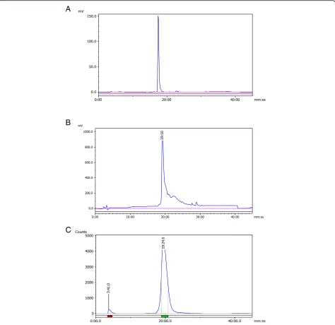
along with some minor peaks (Fig. 5). A radiolabeling efficiency of greater than 90% was obtained in all cases, and it ranged between 90 and 95%. All the complexes were found to be stable at least 4 h after the radiolabeling as determined by radio-HPLC. RP-HPLC retention times of the major peaks, ranging from 18.3 to 20.3 min, reflected slightly variable lipo-philicity of these 68Ga-complexes (Table 1). The col-loids formation was below 2% as revealed by ITLC-SG analysis.
In addition, identification of 68Ga-DOTA-BN complex was achieved by comparison of the retention time with the coldnatGa-DOTA-BN compound eluted with identical re-tention time under the same HPLC condition. Representa-tive HPLC analysis chromatograms are given in Fig.5.
Partition coefficient determination
The partition coefficient values of the six radiopeptides investigated are listed in Table 1. The lipophilicity was determined by measuring the radioactivity distribution Fig. 4An example of the mass spectrum profile of the DOTA-Ahx-BN prepared from cyclen procedure (above) and with DOTA-tris (t-Bu ester) (below). It can be seen that no overalkylating product was observed when the DOTA-Ahx-BN synthesized by the cyclen approach. While tetraalkylating product 1813 (M + t-Bu + H)+is clearly visible in the spectrum when the DOTA-peptide was prepared using DOTA-tris (t-Bu ester)
Table 1Characteristics of the68Ga-labeled BN peptides
Parameter 68Ga-DOTA-Ahx-BN 68Ga-DOTA-Abz-BN 68Ga-DOTA-ALA-BN
Cyclen Tris- Cyclen Tris- Cyclen
Tris-Labeling efficiency (%) > 92.0 > 92.0 > 90.0 > 91.0 > 94.0 > 95.0
Radio-HPLCtR (min) 18.3 18.3 19.2 19.3 20.3 20.3
ESI-MS [M + H]+ 1753.0 1753.0 1759.0 1759.0 1753.1 1753.0
ratios of 68Ga-labeled BN peptides between n-octanol and water layers. A small variation in the partition coef-ficient values was obtained for the BN peptides under in-vestigation possibly due to the similarity of the amino acids sequence and differ mainly in terms of the specific spacer group (i.e., Ahx, Abz, and ALA) in the sequence (Fig. 3), which might contribute to slight different de-grees of lipophilicity of these peptides.
The BN peptide, with Ahx spacer group was found to be the most hydrophilic (−1.21 ± 0.11), whereas
the peptide containing ALA group was less
hydrophilic (−0.80 ± 0.05) among the tested BN
peptides. The obtained log P values varied between
−0.80 ± 0.05 and −1.21 ± 0.11 (see Table 1), demon-strating variable lipophilicity of the 68Ga-complexes. In most cases, a good correlation was observed be-tween the obtained log P values and the RP-HPLC re-tention times of the radiopeptides; as the most hydrophilic BN peptide (with Ahx group) showed the shortest retention time (18.3 min), whereas a less
A
B
C
hydrophilic BN peptide (with ALA group) displayed a somewhat higher retention time (20.3 min) on RP-HPLC (Table 1).
The partition coefficient measurement is important criteria as it usually predicts the possible route of excre-tion of a radiopeptide. Peptides with high hydrophilicity are usually excreted by the urinary system, the preferred route of excretion of a tumor-targeting agent. Indeed, our biodistribution findings also demonstrated that
68
Ga-DOTA-Ahx-BN exhibited a high degree of renal excretion, possibly due to the hydrophilic nature of the complex (Table3).
In vitro metabolic stability in human plasma
In order to produce maximum tumor-targeting effect, it is important that tumor-targeting peptides exhibit high metabolic stability in human plasma and reach intended target intact to deliver maximum tumor-targeting effect. The proteolytic degradation of all six radiolabeled BN peptides were investigated in vitro in human plasma. The radio-HPLC analysis revealed that more than 80% of radioactivity remained bound to peptides after 2 h of incubation, with a low reformation to free 68Ga and some unknown hydrophilic metabolites (up to 20%), demonstrating the high metabolic stability of 68 Ga-la-beled BN peptides. The estimated percent of radiopep-tides remaining intact in plasma was found to be between 89 and 94% at 1 h and between 81 and 87% at 2 h, indicating a low enzymatic degradation of 68 Ga-complexes by plasma proteases.
In vitro tumor cell binding
In vitro cell binding of 68Ga-labeled BN peptides were tested for their ability to bind with BN/GRP receptor-expressing human breast and prostate cancer cell lines. These include estrogen receptor-positive MCF7, estrogen receptor-independent MDA-MB-231, human
ductal breast carcinoma T47D, and androgen
receptor-independent PC3 prostate cancer cell lines.
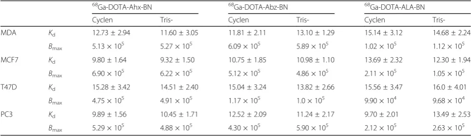
These human cancer cell lines presented variable receptor expression for BN peptides [22, 23]. The binding of radiolabeled BN peptides to the respective receptor-positive cell lines was determined by satur-ation binding assays, and the results of the binding affinity (Kd) and the number of binding sites/cell
(Bmax) was determined by GraphPad software and are
summarized in Table 2.
It is worth mentioning here that all the six BN peptides share nearly the same receptor binding sequence (Fig.3), but differ in terms of methods of DOTA synthesis (cyclen vs. tris) and the particular spacer group introduced be-tween the peptide sequence and DOTA chelating moiety. The results suggest that the68Ga-labeled BN peptide de-rivatives exhibited high affinity binding (Kd values up to
16 nM) to both human breast and prostate cancer cell lines under investigation. The data demonstrate that the BN peptides based on either DOTA-cyclen and their counterparts DOTA-tris displayed quite similar binding affinities to both estrogen receptor-positive and estrogen-independent breast cancer cells, with the binding values ranging between 9.32 and 15.14 nM. A general trend of slightly lower binding affinity was observed (range, 13.82 to 16.0 nM) for all the radioconjugates toward human ductal T47D breast cancer than MDA-MB-231 and MCF7 cancer cells. In addition to good binding to breast cancer cells, the radiolabeled BN peptides under investigation showed equally good binding affinities toward androgen-independent PC3 prostate cancer cell line, with binding affinities varied from 9.70 to 13.49 nM.
The receptor density (Bmax) is generally found to be
high to moderate for MCF7, MDA-MB-231, and PC3 and slightly low for T47D cell line (Table2). The max-imum Bmax value of 6.90 × 10⁵ sites/cell was obtained
for MCF7, and the lowest value of (9.68 × 10⁴sites/cell) was obtained for T47D among the tested cell lines.
From these studies, it seems that the cell binding char-acteristics are not compromised by the method used for preparation of DOTA (cyclen or tris). Furthermore,
Table 2In vitro tumor cell binding characteristics: determination of dissociation constants (Kd) in nM and apparent number of binding sites per cell (Bmax) of the68Ga-labeled BN peptides (n= 3-5 experiments; mean values ± SD;BmaxSD < 15%)
68Ga-DOTA-Ahx-BN 68Ga-DOTA-Abz-BN 68Ga-DOTA-ALA-BN
Cyclen Tris- Cyclen Tris- Cyclen
Tris-MDA Kd 12.73 ± 2.94 11.60 ± 3.05 11.81 ± 2.11 13.10 ± 1.29 15.14 ± 3.12 14.68 ± 2.24 Bmax 5.13 × 105 5.27 × 10⁵ 6.09 × 10⁵ 5.89 × 10⁵ 1.02 × 10⁵ 1.12 × 10⁵ MCF7 Kd 9.80 ± 1.64 9.32 ± 1.50 10.75 ± 1.85 10.98 ± 1.10 13.69 ± 2.32 12.30 ± 1.94
Bmax 6.90 × 105 6.22 × 10⁵ 5.12 × 10⁵ 4.86 × 10⁵ 2.11 × 10⁵ 1.05 × 10⁵ T47D Kd 15.28 ± 3.42 14.51 ± 2.40 15.04 ± 3.24 13.82 ± 2.66 15.56 ± 3.47 16.0 ± 4.01
Bmax 4.75 × 105 4.91 × 10⁵ 1.17 × 10⁵ 1.0 × 10⁵ 9.90 × 10⁴ 9.68 × 10⁴
PC3 Kd 9.89 ± 1.56 10.45 ± 1.71 12.52 ± 2.09 11.24 ± 2.17 9.70 ± 2.01 13.49 ± 2.53
despite the modifications in the peptide sequence, such as the introduction of a chelating moiety for radiolabel-ing and the addition of specific amino acids and spacer groups (Fig. 3), the BN peptides maintained their po-tency and held high affinity and specificity toward breast and prostate cancer cells. This underlines the potential of 68Ga-labeled BN peptides for targeting various GRP-receptor-positive cancers.
Biological evaluation
Pharmacokinetic studies in normal Balb/c mice
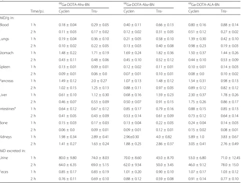
In vivo biodistribution findings of all six 68 Ga-la-beled BN peptides in normal Balb/c mice at 1 and 2 h post intravenous injection are summarized in Table 3. In vivo biodistribution kinetics of 68 Ga-la-beled BN peptides were performed first in normal Balb/c mice (without tumors) to determine normal tissue uptake patterns and clearance kinetics. All the
six radiolabeled BN peptides displayed a somewhat identical biological behavior, characterized by rapid and efficient clearance from the blood both at 1 h and 2 h post-injection (p.i.), with less than 1% ID/g remaining in the blood after 2 h p.i. The
DOTA-coupled BN peptides with different degree of
lipophilicity (Table 3) displayed variable uptake in the liver. The radioactivity retained by the liver ranged from 0.61 ± 0.10% to 2.30 ± 0.37% ID/g at 1 h and from 0.46 ± 0.07% to 1.75 ± 0.26% ID/g after 2 h of radiopeptide injection. Kidney was the only
organ that showed the highest radioactivity
accumulation, which ranged from 1.98 ± 0.34% to 5.89 ± 1.0% ID/g at 1 h and from 1.41 ± 0.27% to 3.05 ± 0.41% ID/g at 2 h p.i, demonstrating varying
degree of accumulation and retention of these
complexes by the kidneys (Table 3). A low to
mod-erate uptake of radioactivity was observed in the
Table 3In vivo biodistribution of68Ga-DOTA-labeled BN peptides in Balb/c mice at 1 h and 2 h post-injection (n= 3-5). Data are expressed as % injected dose per gram of tissue (mean values ± SD). Urinary excretion values (% injected dose) were calculated by measuring the radioactivity associated with the excreted urine and bladder contents at the time of sacrifice at 1 and 2 h post-injection
68Ga-DOTA-Ahx-BN 68Ga-DOTA-Abz-BN 68Ga-DOTA-ALA-BN
Time/p.i. Cyclen Tris- Cyclen Tris- Cyclen
Tris-%ID/g in:
Blood 1 h 0.18 ± 0.04 0.29 ± 0.05 0.40 ± 0.11 0.66 ± 0.13 0.80 ± 0.16 0.88 ± 0.14
2 h 0.11 ± 0.03 0.17 ± 0.02 0.12 ± 0.02 0.31 ± 0.05 0.51 ± 0.12 0.27 ± 0.02
Lungs 1 h 0.19 ± 0.04 0.36 ± 0.10 0.21 ± 0.05 0.58 ± 0.10 1.39 ± 0.30 0.42 ± 0.10
2 h 0.10 ± 0.02 0.22 ± 0.05 0.13 ± 0.03 0.40 ± 0.08 0.98 ± 0.23 0.19 ± 0.03
Stomach 1 h 1.48 ± 0.22 1.71 ± 0.19 1.69 ± 0.24 1.82 ± 0.36 1.50 ± 0.37 1.44 ± 0.26
2 h 0.43 ± 0.11 0.48 ± 0.06 0.45 ± 0.10 0.52 ± 0.12 0.44 ± 0.10 0.53 ± 0.09
Spleen 1 h 0.13 ± 0.01 0.09 ± 0.01 0.12 ± 0.02 0.11 ± 0.01 0.10 ± 0.01 0.14 ± 0.03
2 h 0.09 ± 0.01 0.06 ± 0.0 0.07 ± 0.01 0.10 ± 0.01 0.08 ± 0.0 0.10 ± 0.02
Pancreas 1 h 1.49 ± 0.12 2.0 ± 0.27 1.07 ± 0.13 1.48 ± 0.12 1.54 ± 0.31 0.98 ± 0.13
2 h 1.02 ± 0.15 1.25 ± 0.13 0.88 ± 0.11 0.97 ± 0.05 0.89 ± 0.12 0.82 ± 0.12
Liver 1 h 0.61 ± 0.10 1.12 ± 0.30 0.68 ± 0.16 1.59 ± 0.23 2.30 ± 0.37 1.78 ± 0.26
2 h 0.46 ± 0.07 0.53 ± 0.09 0.50 ± 0.07 0.91 ± 0.15 1.75 ± 0.26 0.86 ± 0.17
Intestinesa 1 h 0.64 ± 0.12 0.67 ± 0.12 0.85 ± 0.17 0.79 ± 0.16 0.88 ± 0.15 0.85 ± 0.13
2 h 0.41 ± 0.05 0.43 ± 0.09 0.53 ± 0.14 0.61 ± 0.09 0.73 ± 0.12 0.64 ± 0.14
Bone 1 h 0.15 ± 0.03 0.17 ± 0.03 0.13 ± 0.04 0.22 ± 0.05 0.24 ± 0.04 0.14 ± 0.03
2 h 0.06 ± 0.0 0.09 ± 0.01 0.09 ± 0.01 0.12 ± 0.01 0.15 ± 0.02 0.08 ± 0.01
Kidneys 1 h 1.98 ± 0.34 2.89 ± 0.41 2.96±0.30 4.0 ± 0.82 5.89 ± 1.0 3.83 ± 0.67
2 h 1.41 ± 0.27 1.63 ± 0.24 1.88 ± 0.25 2.86 ± 0.37 3.05 ± 0.41 2.76 ± 0.49
%ID excreted in:
Urine 1 h 80.0 ± 9.80 74.0 ± 8.03 70.0 ± 8.60 43.0 ± 8.70 53.0 ± 6.80 71.0 ± 12.45
2 h 64.0 ± 6.35 69.0 ± 5.15 62.0 ± 9.54 50.0 ± 3.45 46.0 ± 9.12 78.0 ± 15.0
Feces 1 h 0.85 ± 0.17 0.83 ± 0.19 1.01 ± 0.20 0.90 ± 0.10 1.07 ± 0.17 1.03 ± 0.12
2 h 0.76 ± 0.11 0.69 ± 0.10 0.88 ± 0.12 0.59 ± 0.08 0.91 ± 0.14 0.77 ± 0.10 a
BN/GRP receptor-expressing organ pancreas at both the time points (range, 0.82% to 2.0% ID/g). Another
BN/GRP receptor-expressing organ stomach also
showed relatively low accumulation of radioactivity at both the time points (up to 1.82 ± 0.36% ID/g). The accumulation in the lungs was also low (below 1.5% ID/g) at both 1 h and 2 h p.i., indicating low colloidal particles formation by these complexes. Radioactivity found in the intestines (measured with-out their contents), varied between 0.64 ± 0.12% and 0.88 ± 0.15% ID/g at 1 h and between 0.41 ± 0.05% and 0.73 ± 0.12% ID/g at 2 h p.i. All the radioconju-gates displayed low bone activity both at 1 h and 2 h p.i. of 68Ga-DOTA BN peptides indicating the high in vivo stability of these complexes, which correlates well with the high metabolic stability attained in hu-man plasma in vitro.
It is important to mention here that precautions were taken for the maximal collection of urine, but due to the practical difficulties, complete collection of urine from mice after sacrifice is not always possible, which may result in underestimation of the urinary excretion. Urinary excretion values were calculated using the radioactivity associated with the excreted urine and bladder contents at the time of sacrifice. Any urine expelled at the time of sacrifice was also collected and counted together with the excreted urine. Rapid urinary excretion of radioactivity was seen for all the radiocomplexes, with more than 40% ID already in the urine after 1 h p.i. The most favor-able clearance pattern was observed for the hydro-philic (log P = −1.21) BN peptide analog, with Ahx (aminohexanoic acid) spacer group, as 80% ID already in the urine at 1 h p.i. Another hydrophilic BN pep-tide (log P = −1.10), containing Abz (aminobenzoic acid) spacer and with glycine residue adjacent to it; formulate a hippurate-like structural spacer group, ex-hibited equally good renal excretion properties since 70% ID was found in the urine after 1 h of radio-tracer injection. A hippurate-type biomolecule is known to facilitate clearance through kidneys as is generally preferred for radiopeptides. The clearance kinetics studies highlight the significance of the de-gree of lipophilicity as one of the deciding factors in the clearance route of a radiopharmaceutical.
In general, a rapid and efficient clearance of radio-activity was achieved from most of the
tissues/or-gans, highlighting the favorable biodistribution
profiles of these radiolabeled BN peptides. More importantly, no major difference between the bio-logical behavior of the BN peptides linked with DOTA from cyclen precursor and the commercial DOTA-tris-(t-Bu ester) was observed (Table 3), indi-cating the equal efficiency and potency of these
peptides and the low impact of the method of DOTA preparation on the biological properties of the BN peptides under investigation.
In vivo tumor targeting 68
Ga-labeled BN peptides were further investigated in nude mice carrying estrogen-independent and highly ag-gressive MDA-MB-231 xenografts in order to determine their potential for diagnosis of BN/GRP receptor-positive breast cancer in vivo (Table4). In nude mice, all six radi-olabeled BN peptides exhibited rapid and efficient clear-ance from the blood (< 1.0% ID/g) at both 1 h and 2 h p.i., as it was the case in the normal Balb/c mice. The highest uptake in the tumors was achieved for 68 Ga-DOTA(cy-clen)-Ahx-BN (2.32 ± 0.25% ID/g) as early as 1 h p.i, which reduced to 1.65 ± 0.19% ID/g at 2 h p.i. (with 28% washout from the tumors over 2 h). The same leading BN peptide also exhibited favorable pharmacokinetics in nor-mal mice (Table 3). When a blocking dose of 300 μg of nonradioactive BN peptide was injected intramuscularly for in vivo saturation of receptors 20 min prior to the ad-ministration of radiopeptide, it was observed that the up-take in the tumors was reduced by approximately 80% (0.47 ± 0.06% ID/g blocked vs. 2.32 ± 0.25% ID/g unblocked, P = 0.002), highlighting the specificity of the
68
Ga-labeled BN peptide for respective BN/GRP receptor-positive breast cancer cells. The corresponding 68 Ga-DOTA(tris)-Ahx-BN also showed nearly identical tumor uptake value 2.09 ± 0.33% ID/g at 1 h and 1.62 ± 0.18% ID/g at 2 h p.i. The tumor uptake values of other BN pep-tides varied from 1.78 ± 0.40% to 2.15 ± 0.33% ID/g at 1 h p.i and from 0.84 ± 0.13% to 1.37 ± 0.25% ID/g at 2 h p.i. (Table4). With the blocking dose, good reduction in the tumors (from 51 to 80%) was attained validating the in vivo tumor receptor specificity of these radiolabeled BN peptides for BN/GRP receptor-positive breast cancer cells. The accumulation in the kidneys was moderate (up to 6.52% ID/g) but still somewhat higher than the values ob-served in the Balb/c mice. A low to moderate uptake of radioactivity was found in the BN/GRP receptor-expressing organ pancreas (up to 2.94% ID/g) (range, 0.71 to 2.94% ID/g). It was found that the same blocking dose significantly reduced the uptake in the receptor-positive organ, pancreas, with the maximum blockade of 80% oc-curred for68Ga-DOTA-Ahx-BN analog. Moreover, a vari-able influence of the blocking dose was seen in the stomach, intestines, and kidneys probably related to the low receptor expression by some of these organs [24].
Although the uptake in tumors was not very high for any of the BN peptides evaluated (up to 2.32 ± 0.25% ID/g), a somewhat decent tumor retention with slow washout from the tumors was observed for some of these peptides and especially for 68
Nonetheless, the uptake values in the tumors was al-ways higher than the radioactivity found in the blood and muscle for all the radiopeptides. Tumor-to-nontumor uptake ratios were also calculated and graphically present in Fig. 6. A mixed trend of in-creased tumor-to-blood and tumor-to-muscle uptake ratios over time was observed for all the 68 Ga-la-beled BN peptide analogs. The tumor-to-blood up-take ratios ranged between 1.9 to 3.0 at 1 h, 2.0 to 4.2 at 2 h p.i., whereas tumor-to-muscle uptake ra-tios ranged between 4.6 to 10.3 at 1 h and 4.0 to 8.8 at 2 h p.i. A low bone uptake was detected for all the radioconjugates indicating high in vivo stability of these 68Ga-complexes. Similar to normal mice, the main route of excretion in tumor-bearing mice was through the kidneys into the urine, with the radio-activity excreted into the urine was up to 78% ID, whereas the hepatobiliary excretion (liver + intes-tines) of these radiopeptides were below 12% ID. A fairly good uptake and retention by the tumors
combined with good tumor to background uptake ratios advocating the possible potential of these BN peptides in general, and BN peptide with Ahx spacer in particular, for targeting breast cancer in vivo. Again, the data from this set of experiments further confirm that the method of DOTA synthesis (cyclen or tris) has minimal effect on the biological and tumor-targeting properties of the BN peptides under investigation as both exhibited equally good tumor-targeting potential.
It is worth emphasizing that the scope of this article was not to compare the newly synthesized BN peptides with the published BN peptides but to compare the two different synthetic approaches (DOTA-tris and cyclen) for the preparation of DOTA-BN peptides. Since the se-quences of the BN analogs used in this study are new in terms of the presence of some specific amino acids, therefore, a direct comparison with the previously pub-lished BN may be misleading. On the other hand, most
of the DOTA-coupled BN (7–14) normal sequence
Table 4In vivo biodistribution of68Ga-labeled DOTA-BN peptides in nude mice with MDA-MB-231 tumors at 1 h and 2 h post-injection (n= 3-5). Data are expressed as % injected dose per gram of tissue (mean values ± SD). The radioactivity in the urine + bladder is expressed as % injected dose per tissue
68Ga-DOTA-Ahx-BN 68Ga-DOTA-Abz-BN 68Ga-DOTA-ALA-BN
Time/p.i. Cyclen Tris- Cyclen Tris- Cyclen
Tris-Blood 1 h 0.92 ± 0.15 0.70 ± 0.12 1.0 ± 0.16 0.93 ± 0.22 0.79 ± 0.13 0.85 ± 0.15
2 h 0.51 ± 0.07 0.60 ± 0.09 0.55 ± 0.10 0.64 ± 0.16 0.37 ± 0.10 0.30 ± 0.04
Lungs 1 h 0.55 ± 0.10 1.86 ± 0.14 2.80 ± 0.55 1.82 ± 0.30 1.05 ± 0.17 2.35 ± 0.33
2 h 0.36 ± 0.03 0.61 ± 0.07 1.15 ± 0.23 1.26 ± 0.25 0.67 ± 0.13 0.70 ± 0.16
Stomach 1 h 0.73 ± 0.13 0.87 ± 0.11 1.34 ± 0.29 1.0 ± 0.20 0.87 ± 0.19 0.83 ± 0.21
2 h 0.34 ± 0.05 0.60 ± 0.07 0.49 ± 0.12 0.52 ± 0.13 0.46 ± 0.05 0.37 ± 0.07
Spleen 1 h 0.25 ± 0.02 0.33 ± 0.02 0.22 ± 0.07 0.18 ± 0.03 0.26 ± 0.03 0.44 ± 0.06
2 h 0.14 ± 0.01 0.16 ± 0.01 0.12 ± 0.02 0.09 ± 0.02 0.17 ± 0.02 0.35 ± 0.03
Pancreas 1 h 1.65 ± 0.14 1.30 ± 0.18 2.94 ± 0.43 1.81 ± 0.22 1.05 ± 0.12 1.19 ± 0.15
2 h 0.82 ± 0.10 0.75 ± 0.11 1.18 ± 0.31 0.93 ± 0.15 0.88 ± 0.11 0.71 ± 0.10
Liver 1 h 1.02 ± 0.16 2.20 ± 0.33 2.54 ± 0.46 2.28 ± 0.31 1.40 ± 0.26 2.31 ± 0.38
2 h 0.91 ± 0.10 1.83 ± 0.24 2.0 ± 0.37 1.71 ± 0.20 1.12 ± 0.19 1.50 ± 0.27
Intestinesa 1 h 0.86 ± 0.12 0.89 ± 0.12 0.95 ± 0.27 1.01 ± 0.17 0.90 ± 0.12 1.05 ± 0.18
2 h 0.61 ± 0.06 0.50 ± 0.10 0.54 ± 0.16 0.76 ± 0.12 0.60 ± 0.10 0.73 ± 0.13
Muscle 1 h 0.23 ± 0.03 0.28 ± 0.04 0.32 ± 0.06 0.36 ± 0.07 0.30 ± 0.06 0.39 ± 0.03
2 h 0.19 ± 0.02 0.20 ± 0.02 0.26 ± 0.05 0.28 ± 0.04 0.21 ± 0.03 0.23 ± 0.02
Kidneys 1 h 4.93 ± 0.38 4.85 ± 0.43 6.03 ± 1.0 6.52 ± 0.60 5.94 ± 0.63 5.12 ± 0.69
2 h 4.01 ± 0.41 3.74 ± 0.24 5.0 ± 0.77 3.96 ± 0.48 3.20 ± 0.32 3.56 ± 0.50
Urine + bladder 1 h 64.0 ± 9.15 66.0 ± 6.60 61.0 ± 9.93 58.0 ± 6.25 62.0 ± 6.28 54.0 ± 5.78
2 h 70.0 ± 8.70 68.0 ± 7.55 78.0±5.89 65.0 ± 9.13 71.0 ± 9.68 61.0 ± 7.10
Tumor 1 h 2.32 ± 0.25 2.09 ± 0.33 1.84 ± 0.40 2.15 ± 0.33 1.82 ± 0.26 1.78 ± 0.40
2 h 1.65 ± 0.19 1.62 ± 0.18 1.30 ± 0.37 1.37 ± 0.25 0.84 ± 0.13 1.25 ± 0.21 a
(Gln7-Trp8-Ala9-Val10-Gly11-His12-Leu13-Met14-CONH2)
exhibited the tumor uptake value in PC-3 tumor xeno-grafts in the range of 2.5 to 4% ID/g [14, 15], which is somewhat similar to the values obtained with the BN peptides prepared and evaluated in this study. This indi-cates that that the different synthetic approaches used here has minimal effect on the tumor-binding character-istics of the resultant BN peptide analogs.
PET imaging of68Ga-DOTA-Ahx-BN in MDA-MB-231 tumor-bearing nude mice models
PET images of BN/GRP receptor-positive MDA-MB-231
tumor-bearing nude mice injected with 68
Ga-DOTA-Ahx-BN peptide alone or along with a large excess of unlabeled BN peptide to block BN/GRP receptor sites obtained at 1 h p.i. are shown in Fig.7.
Accumulation of radioactivity in the tumor lesion is clearly visible in PET image of the unblocked MDA-MB-231 tumor-bearing mouse at 1 h p.i., underlining the tumor-targeting ability of this radiotracer. Nonetheless, the radiopeptide showed considerable accumulation in the abdominal region including stomach and intestines. High amount of radioactivity was also seen in the urinary blad-der, suggesting that the radiopeptide excreted mainly by the renal system.
Blocking studies in the presence of 200 μg of un-labeled BN peptide was also performed to demonstrate BN/GRP receptor-specific tumor uptake of68 Ga-DOTA-BN peptide. Unlike in the unblocked mouse PET im-aging, where the tumor was clearly seen in the image, tumor was hardly detectable in the PET image under blocking conditions as shown in the Fig.7. It was found that the presence of a pharmacological dose of BN pep-tide greatly reduced the tumor uptake, demonstrating the tumor specificity of 68Ga-DOTA-BN peptide. A low uptake was observed in the abdominal region but the radioactivity accumulation in the kidneys was higher than the unblocked mouse (Fig. 7). Intense radioactivity was also noted in the urinary bladder as the radiopeptide excreted primarily through the kidneys into the urinary bladder.
Immediately after imaging, the mice were sacrificed and tissue biodistribution were carried out in order to Fig. 6Tumor-to-blood and tumor-to-muscle uptake ratios for the68Ga-DOTA-BN peptides in MDA-MB-231 xenografted nude mice at 1 and 2 h post-injection
confirm the findings of PET-imaging. The data from the in vivo imaging was in agreement with the in vivo quan-titative biodistribution studies (Table4).
Based on encouraging biological and
tumor-targeting properties, 68Ga-DOTA(cyclen)-Ahx-BN was further tested in nude mice bearing PC3 prostate car-cinoma xenografts. Again in PC3 animal models,
68
Ga-DOTA(cyclen)-Ahx-BN displayed fast and effi-cient clearance from the blood and other major
or-gans. Kidney was the only major organ which
retained most of the radioactivity (up to 6% ID/g) after 2 h p.i. The uptake of this radiotracer in the PC3 tumors was found to be 2.05 ± 0.54% ID/g at 1 h and 1.34 ± 0.22% ID/g at 2 h p.i., respectively. The tumor-to-blood and tumor-to-muscle ratios were 3.27 and 12.62 at 1 h and 2.85 and 11.40 at 2 h p.i., re-spectively (data not shown). A fairly comparable tumor uptake and pharmacokinetic profile was ob-tained between the nude mice models carrying
estro-gen receptor-independent MDA-MB-231 breast
xenografts and androgen receptor-independent PC3 prostate cancer xenografts.
It is obvious from the biological evaluation that the
68
Ga-DOTA(cyclen)-Ahx-BN peptide displayed equally good tumor-targeting properties in the species carry-ing two pharmacologically distinct human breast
(MDA-MB-231) and prostate cancer cells (PC3),
highlighting the ability of this radiotracer to target breast as well as prostate cancer in vivo. Taken to-gether, the DOTA-coupled BN peptides prepared ei-ther from cyclen or tris displayed nearly similar in vivo tumor-targeting behavior (see Table 4), indi-cating equal tumor-targeting efficiency and potency for BN/GRP receptor positive tumors.
Conclusions
In summary, an efficient and cost-effective synthetic procedure where a DOTA-linked peptide amide is syn-thesized on a single solid-phase support is presented in this study. One distinct feature of this approach is that the DOTA is introduced to the peptide backbone while the peptide still attached to resin support using standard SPPS methodology. Our results demonstrate that DOTA prepared from cyclen on solid-phase support showed comparable potency and efficiency to commercially available DOTA-tris in both in vitro and in vivo evalu-ation. The attractive synthetic methodology described here allows versatile, site-specific introduction of DOTA into peptides and can be applied to the synthesis of a wide variety of DOTA-linked tumor-targeting peptides to facilitate the development of molecular imaging and therapy (upon labeling with 177Lu) agents with optimal tumor-targeting properties for potential clinical translation.
Abbreviations
68Ga:Gallium-68; Abz: Aminobenzoic acid; Ahx: Aminohexanoic acid;
ALA: Aminolevulinic acid; Bmax: Maximum binding sites/cell; BN: Bombesin; CH3CN: Acetonitrile; CT: Computed tomography; cyclen: 1,4,7,10-tetraazacyclododecane; DCM: Dichloromethane;
DIC: Diisopropylcarbodiimide; DIEA:N,N-Diisopropylethylamine; DMF:N,N
′-Dimethylformamide; DO3A: 1,4,7,10-Tetraazacyclododecane-1,4,7-triacetic acid; DOTA: 1,4,7,10-tetraazacyclododecane-N,N′,N′′,N′′′-tetraacetic acid; EtOH: Ethanol; FBS: Fetal bovine serum; Fmoc: 9-Fluorenylmethoxycarbonyl; GRP: Gastrin-releasing peptide; HATU: 1-[Bis(dimethylamino)methylene]-1H -1,2,3-triazolo[4,5-b]pyridinium 3-oxid hexafluorophosphate,N
-[(Dimethylamino)-1H-1,2,3-triazolo-[4,5-b]pyridin-1-ylmethylene]-N -methylmethanaminium hexafluorophosphateN-oxide; HBTU:N,N,N′,N
′-Tetramethyl-O-(1H-benzotriazol-1-yl)uronium hexafluorophosphate,O -(Benzotriazol-1-yl)-N,N,N′,N′-tetramethyluronium hexafluorophosphate; HOBt: Hydroxybenzotriazole; ID: Injected dose;Kd: Dissociation constant; MBHA: 4-Methylbenzhydrylamine; MeOH: Methanol; MRI: Magnetic resonance imaging; MS: Mass spectrometry; p.i.: Post-injection; PBS: Phosphate-buffered saline; PET: Positron-emission tomography; ROI: Region of interest; RP-HPLC: Reversed-phase high performance liquid chromatography; SD: Standard deviation; SPECT: Single-photon emission computed tomography; SPPS: Solid-phase peptide synthesis; TFA: Trifluoroacetic acid; TIS: Triisopropylsilane
Acknowledgements
The authors thank Yousif Maliki for PET imaging studies, Zakia Shinwari for the preparation of cells, and Celistina Maccuto for animal experiments. Dr. Falah Mohanna for assistance in the induction of cancer cells in mice. This work was partially supported from the grants of King Abdulaziz City for Science and Technology (KACST grant # AT-26-6).
Author’s contributions
SMO and IJ conceived and designed the experiments. SMO performed all the experiments. SMO drafted the manuscript. Both authors contributed to the review and editing of the manuscript and approved the final manuscript.
Funding
This study was partially funded by the grants from King Abdulaziz City for Science and Technology (KACST grant # AT-26-6).
Availability of data and materials
All data generated or analyzed during this study are included in this manuscript and its supplementary information files (mass spectrometric analysis and HPLC chromatograms) are also available from the corresponding author.
Ethics approval
Approval for the animal protocol used in this study was obtained from the Institutional Animal Care and Use Committee (King Faisal Specialist Hospital and Research Centre, Riyadh, KSA-ACUC). Animal studies were conducted strictly according to the international regulations governing the safe and proper use of laboratory animals.
Consent for publication
Not applicable.
Competing interests
The authors declare they have no competing interests.
Received: 16 April 2019 Accepted: 17 July 2019
References
1. Hussain T, Nguyen QT. Molecular Imaging for Cancer Diagnosis and Surgery. Adv Drug Deliv Rev. 2014;66:90–100 2010.
2. Fani M, Maecke HR, Okarvi SM. Radiolabeled peptides: valuable tools for the detection and treatment of cancer. Theranostics. 2012;2:481–501. 3. Jamous M, Haberkorn U, Mier W. Synthesis of peptide radiopharmaceuticals for
the therapy and diagnosis of tumor diseases. Molecules. 2013;18:3379–409. 4. Chappell LL, Rogers BE, Khazaeli MB, Mayo MS, Buchsbaum DJ,
1,4,7,10-Tetraaza-N-(1-carboxy-3-(4-nitrophenyl)propyl)-N′,N′′,N′′′-tris(acetic acid)cyclododecane (PA-DOTA). Bioorg Med Chem. 1999;7:2313–20. 5. Cakic N, Gündüz S, Rengarasu R, Angelovski G. Synthetic strategies for preparation
of cyclen-based MRI contrast agents. Tetrahedron Letters. 2015;56:759–65. 6. De León-Rodríguez LM, Kovacs Z. The synthesis and chelation chemistry of
DOTA-peptide conjugates. Bioconjugate Chem. 2008;19:391–402. 7. Barge A, Tei L, Upadhyaya D, Fedeli F, Beltrami L, Stefania R, Aime S,
Cravotto G. Bifunctional ligands based on the DOTA-monoamide cage. Org Biomol Chem. 2008;6:1176–84.
8. Axelsson O, Olsson A. Synthesis of cyclen derivatives. United States Patent Application Publication; US 2009/0216011 A1; 2009.
9. Peterson JJ, Pak RH, Meares CF. Total solid-phase synthesis of 1,4,7,10-tetraazacyclododecane-N,N′,N′′,N′′′-tetraacetic acid-functionalized peptides for radioimmunotherapy. Bioconjugate Chem. 1999;10:316–20.
10. Guerin B, Ait-Mohand S, Tremblay M-C, Dumulon-Perreault V, Fournier P, Benard F. Total solid-phase synthesis of NOTA-functionalized peptides for PET imaging. Org Lett. 2010;12:280–3.
11. Burkoth TS, Fafarman AT, Charych DH, Connolly MD, Zuckermann RN. Incorporation of unprotected heterocyclic side chains into peptoid oligomers via solid-phase submonomer synthesis. J Am Chem Soc. 2003;125:8841–5. 12. Boldrini V, Giovenzana GB, Pagliarin R, Palmisano G, Sisti M. Expeditious
N-monoalkylation of 1,4,7,10-tetraazacyclododecane (cyclen) via formamido protection. Tetrahedron Lett. 2000;41:6527–30. 13. Zuckermann RN, Kerr JM, Kent SBH, Moos WH. Efficient method for the
preparation of peptoids [Oligo(N-substituted glycines)] by Submonomer Solid-Phase Synthesis. J Am Chem Soc. 1992;114:10646–7.
14. Antonella Accardo A, Galli F, Mansi R, Del Pozzo L, Aurilio M, Morisco A, Ringhieri P, Signore A, Morelli G, Aloj L. Pre-clinical evaluation of eight DOTA coupled gastrin-releasing peptide receptor (GRP-R) ligands for in vivo targeting of receptor-expressing tumors. EJNMMI Res. 2016;6:17.
15. Okarvi SM, Jammaz I. Preparation and evaluation of bombesin peptide derivatives as potential tumor imaging agents: effects of structure and composition of amino acid sequence on in vitro and in vivo characteristics. Nucl Med Biol. 2012;39:795–804.
16. Robey FA. Bromoacetylated synthetic peptides. In: Pennington MW, Dunn BM, editors. Peptide synthesis protocols. Methods in Molecular Biology, vol 35. Totowa: Humana Press.
17. Zuckermann RN, Martin EJ, Spellmeyer DC, Stauber GB, Shoemaker KR, Kerr JM, Figliozzi GM, Goff DA, Siani MA, Simon RJ, Banville SC, Brown EG, Wang L, Richter LS, Moos WH. Discovery of nanomolar ligands for 7-transmembrane G-protein-coupled receptors from a diverseN-(Substituted)glycine Peptoid Library. J Med Chem. 1994;37:2678–85.
18. Nakao R, Furutsuka K, Yamaguchi M, Suzuki K. Sensitive determination of specific radioactivity of positron emission tomography radiopharmaceuticals by radio high-performance liquid chromatography with fluorescence detection. Nucl Med Biol. 2008;35:733–40.
19. Okarvi SM, Jammaz I. Synthesis and evaluation of a technetium-99m labeled cytotoxic bombesin peptide conjugate for targeting bombesin receptor-expressing tumors. Nucl Med Biol. 2010;37:277–88.
20. Guide for the care and use of laboratory animals. Washington, D.C.: National Academy Press; 1996.
21. Eisenwiener K-P, Powell P, Macke HR. A convenient synthesis of novel bifunctional prochelators for coupling to bioactive peptides for radiometal labelling. Bioorg Med Chem Lett. 2000;10:2133–5.
22. Giacchetti S, Gauvillé C, De Crémoux P, Bertin L, Berthon P, Abita JP, et al. Characterization, in some human breast cancer cell lines, of gastrin-releasing peptide-like receptors which are absent in normal breast epithelial cells. Int J Cancer. 1990;46:293–8.
23. Reile H, Armatis PE, Schally AV. Characterization of high-affinity receptors for bombesin/gastrin releasing peptide on the human prostate cancer cell lines PC-3 and DU-145: internalization of receptor bound125I-(Tyr4) bombesin by tumor cells. Prostate. 1994;25:29–38.
24. Jensen RT, Battey JF, Spindel ER, Benya RV. International union of pharmacology. LXVIII. Mammalian bombesin receptors: nomenclature, distribution, pharmacology, signaling, and functions in normal and disease states. Pharmacol Rev. 2008;60:1–42.
Publisher’s Note