I
IJJMMCCMM Original Article
S
Suummmmeerr22001133,,VVooll22,,NNoo33
Is Radiosensitivity Associated to Different Types of Blood
Groups? (A cytogenetic study)
Farideh Elahimanesh1, Ali Shabestani Monfared1∗, Meysam Khosravifarsani1,2, Haleh Akhavan Niaki1, Zeinab Abedian1, Karimollah Hajian-Tilaki1, Sajad Borzouisileh1,Nayer Seyfizadeh1,3, Mehrangiz Amiri1.
1.Cellular & Molecular Biology Research Center (CMBRC), Babol University of Medical Sciences, Babol, Iran.
2. Department of Radiology, Faculty of Medicine, Kurdestan University of Medical Sciences, Kurdestan, Iran.
3. Department of Clinical Biochemistry, Faculty of Medicine, Tabriz University of Medical Sciences, Tabriz, Iran.
Many biological factors affect radiosensitivity. In this study, radiosensitivity among the different blood groups
was investigated. Peripheral blood sample of 95 healthy people were divided into two parts. One part was
irradiated with 2 Gy Co-60 gamma rays and the second one was considered as control. Then all the samples were
studied by cytokinesis-blocked micronucleus assay (CBMN assay). Our study showed that the radiosensitivity
index of A+ and O+ groups was significantly higher and lower than other blood groups, respectively. It seems
that blood type can be used as a radiosensitivity index for determining the given dose to radiotherapy, although
extensive studies are necessary.
Key words: Radiosensitivity, blood group, CBMN assay
∗
Corresponding author: Cellular and Molecular Biology Research Center (CMBRC), Babol University of Medical Sciences, Babol, Iran. Email: [email protected]
adiosensitivity is the relative susceptibleness
of cells, tissues, organs or organisms to the
dangerous effect of ionizing radiation (1). Inherent
characteristic is one of the important reasons of
differences in radiation sensitivity (1-2). The
physical specifications of ionizing radiation such as
its type (particle or photon), energy and dose rate
could alter the biological response of organ or
tissue to ionizing radiation (2-4). Previous studies
have confirmed the relationship between genetics
and radiosensitivity (5-6). A recent study has shown
that mutations in the ataxia telangiectasia gene
(ATM) result in an abnormal p53-mediated cellular
response to DNA damage produced by ionizing
radiation. Also, the potential role of several
identified genes such as BRCA and NBS1, which
are involved in the cellular response to radiation
induced DNA damage is reported too (6). A clinical
study has suggested that a large spectrum of normal
tissue reactions may occur among the radiotherapy
patients. Because of the difference in the individual
radiosensitivity and radiotherapy, the patients who
receive identical dose have different normal tissue
reactions varying from undetectable to severe (7).
People with higher radiosensitivity, most likely will
suffer from deterministic and stochastic effects in
radiotherapy (7). The results of the studies have
revealed that over expression of KU80 gene is an
R
Submmited 20 May 2013; Accepted 3 Aug 2013
important factor for predicting radiosensitivity in
the head and neck cancers (5). It has shown an
association between the in vitro radiosensitivity of
breast cancer patients and the clinical incidence of
late (e.g. fibrosis, telangiectasia) normal tissue
reaction to radiotherapy (6). In addition to
biological conditions, environmental conditions
such as existence of radiosensitizers and
radioprotectors undoubtedly affect the biological
damage of ionizing radiation. One well-studied
example is the presence of oxygen during exposure
to ionizing radiation that stabilizes reactions to
ionizing radiation and then increases the biological
damage of radiation (4). The blood group is an
inherent characteristic and its classification is based
on the presence or absence of ABO blood group
antigens on the surface of red blood cells. The
occurrence of some diseases is related to blood type
(8) and studies have reported that ABO blood group
is an important genetic risk factor for several
radiation related illnesses such as pancreatic cancer
(9), hepatocellular carcinoma (10), endometrial and
cervical cancer (11). In this study, the association
between the radiosensitivity and ABO blood
group was investigated by cytokinesis-blocked
micronucleus assay (CBMN) in a case-control
cytogenetic study.
Materials and Methods
Subjects and sampling
Ten milliliter blood samples of 95 (25 A+, 25
B+, 25 O+ and 20 AB+) radiation worker,
non-smoker or alcohol-user healthydonors age between
18-25 years were taken under sterile conditions in
the presence of sodium heparin anticoagulant. The
samples were divided into two identical values
(5ml) which were maintained in similar conditions.
The subjects’ blood groups, any cancer history in
their families and recent radiation exposures were
filled in the questionnaire through an interview.
Irradiation
One part of each sample was considered as
the control and the second equivalent part was
exposed to 2 Gy of gamma rays from a tele-cobalt
therapy source (Theratone780, Canada). The dose
rate was 120 cGy/min and the source to samples
distance (SSD) was 80 cm. The exposed and
non-exposed blood samples were transferred to cell
culture laboratory for the CBMN assay.
CBMN (cytokinesis blocked micronuclei assay)
CBMN assay was performed on both exposed
and control samples as reported by international
atomic energy agency (IAEA). In this cytogenetic
technique, 0.5 ml of the whole blood was added to
4.5 ml culture medium (RPMI 1640) supplemented
with fetal calf serum, 1% L-glutamine and
antibiotics. Then 100 µl phytohaemagglutinin
(SIGMA) diluted in PBS was added as mitogen.
The sample was incubated at 37º for 44 h then 100
µl cytochalasin B (6 µ g/ml diluted in DMSO) was
added for cessation of the cytokinesis in the
binucleus state. The binucleated lymphocytes were
harvested 28 h later. The samples were centrifuged
at 2000 rpm for 10 min (BOECHO U-320 R) and
the supernatant was discarded. The pellet remained
at the bottom of tubes was treated with 2-3 ml of
fresh hypotonic solution (0.075 M KCl) and then
centrifuged at 1200 rpm for 7 min. After discarding
the supernatant, 5 ml of the fixing solution
(methanol:glacial acetic acid 6/1) was added
quickly. After 20 min, the tubes were centrifuged
(1200 rpm for 7 min) and the fixation was repeated
three times at 1200 rpm for 7 min. Subsequently,
the cells were dropped on clean slides and stained
with Giemsa solution (Giemsa stock: PBS, 1/10) for
10 minutes. The slides were washed with distilled
water and were dried by air. All the slides were
studied under a light microscope in 40×
magnification using SAIRAN microscope. The
slides were coded before analyzing for blinding
purpose. The Micronuclei were scored in 1000
binucleated (BN) cells and scoring was blinded
according to the scoring scale suggested by Fenech
(12-13). The proportion of MN in exposed to
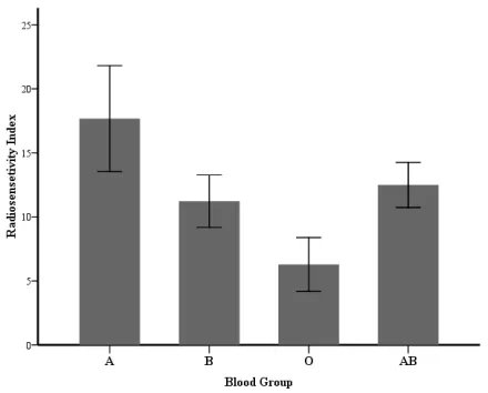
Fig. 1. Mean frequency of micronuclei in control and exposed
groups of different blood groups. Fig. 2.Radiosensitivity index (RI) of different blood groups.
exposed samples in each blood group was
considered as its radiosensitivity index (1, 3). If one
person's cells are more radiosensitive, after taking 2
Gy radiation dose, more DNA breaks (or MNs)
occurs and its proportion to non exposed cells will
be higher than a person with lower radiosensitivity.
Statistical analysis
The statistical analysis was performed using
SPSS 16 by Pair sample t-test between the control
and exposed groups and analysis of variance
(ANOVA) test between the different blood groups.
The p-value < 0.05 was considered statistically
significant.
Results
The mean micronuclei frequencies of the
different blood groups were shown in figure 1. As
the graphs show, the micronuclei frequency in the
exposed samples for all of the blood groups is
significantly higher than the control (P <0.001).
Also, there is a significant difference in the
micronuclei frequencies of O+ and other blood
types in control group (p< 0.001). The difference of
micronuclei frequencies between A+ and O+ in
exposed groups is significant too (P= 0.015).
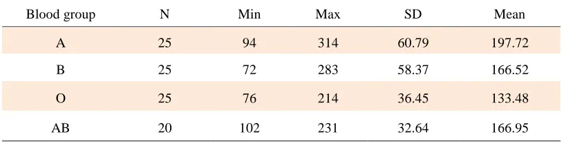
The increase in the number of micronuclei after
exposure to 2 Gy irradiation for all of the four
blood groups are shown in table 1. As quoted in this
table, the highest and lowest increase was seen in A
and O blood groups, respectively. Also this
parameter was significantly different between A
and O blood groups.
Discussion
CBMN assay is the standard technique for
measuring the human population micronuclei and
the estimation of absorbed dose for the prediction
of deterministic and stochastic effects in nuclear
accident (7). It is a cytogenetic method for the
evaluation of cytotoxic effects of chemical
materials and ionizing radiation in mammalian
system too. In vitro cytokinesis blocked
micronuc-leus assay can be used for the prediction of
radiosensitivity of the tumor cells and as an index
for the inter-individual differences in
radio-sensitivity (13).
Our data clearly indicate that the mean
frequency of MN in exposed groups is remarkably
higher than the control groups. These findings are
in agreement with the previous cytogenetic
inves-tigations which have been done by Maffie et al.
(14), Thierens (15) and Khosravifarsani et al. (1).
The recent study performed by
Khosravi-farsani et al. in 2012 showed that radiosensitivity in
left-handed is greater than right-handed breast
cancer women. They also explained that
radiosensitivity in left-handers is higher compared
to right-handers (1). Our data suggest that A+ is the
most radiosensitive and O+ have the lowest
radiosensitivity among the studied blood groups.
The results obtained from Garriga and Ghossein
revealed that O blood group has greater radiation
response than either blood type in carcinoma of
cervix patients (16).
A previous study performed by Dabelsteen et
al. reported that alteration of the cell-surface is a
factor in the development of malignancies (17).
Earlier observations have declared that there is an
association between various cancers and ABO
blood groups (18-20). Stamatakos et al. showed that
the frequency of ductal breast cancer is higher in A
blood group (18). Tursen et al. have investigated
the relationship between blood groups and skin
cancer (19). They reported that although patients
with A and O blood groups have the most and the
lowest occurrence of skin cancer but this difference
is not statistically significant. Wolpin et al.
concluded that the frequency of pancreatic cancer
in patients with O blood group was obviously lower
than other blood groups (20). Doll et al. showed
that the number of gastric ulcer and neoplasm in
patients with A blood group were significantly
higher than the other blood groups (21). You et al.
indicated that the frequency of dysplasia and
metaplasia and gastric atrophy in A blood group
was higher than the blood groups (22). To date, no
report has evaluated the association of blood groups
and radiosensitivity.
The present study addresses more
investiga-tions on the association of ABO blood groups and
radiosensitivity. From a genetic point of view, the
repair of double strand breaks generated by
radiation involves two main mechanisms,
non-homologous end-joining (NHEJ) and non-homologous
recombination (HR). Our data suggest that there
may be an association between some alleles of
ABO blood groups and specific alleles of genes
involved in DNA double strand breaks repair.
Further molecular studies are needed to investigate
candidate loci involved in the DNA repair systems
on chromosome 9 near ABO locus at 9q34. If future
research could prove our findings, blood groups
could be used as a radiosensitivity index for
determining the given dose to radiotherapy patients
and protecting workers against exposure to ionizing
radiation.
Acknowledgment
The authors are thankful to Dr Ebrahim Zabihi for
his valuable technical assistance, Mr. Fazelnezhad
for providing the blood samples and the staff of the
Radiotherapy Department of Shahid Rajaii Hospital
for their cooperating in irradiation of blood
samples.
Conflict of Interest
There is no conflict of interest.
References
1. Khosravifarsani M, Shabestani Monfared A, Elahimanesh F,
Table 1. Micronuclei number increase in blood samples exposed to 2 Gy irradiation
Mean SD
Max Min
N Blood group
197.72 60.79
314 94
25 A
166.52 58.37
283 72
25 B
133.48 36.45
214 76
25 O
166.95 32.64
231 102
20 AB
et al. Is there association between handedness and
radiosensitivity in breast cancer women? Med Oncol
2012;29:2552-5.
2. Hall Eric J, Giaccia Amato J. Radiobiology for the radiologist.
7th ed. Philadelphia: Wolters Kluwer Health/Lippincott
Williams & Wilkins; 2012.
3. Khosravifarsani M, Shabestani Monfared A, Akhavan-Niaki
H, et al. The study of radiosensitivity in left handed compared
to right handed healthy women. BMC Medical Physics
2012;12:1-4.
4. Alsbeih G, El-Sebaie M, Al-Harbi N, et al. Radiosensitivity of
human fibroblasts is associated with amino acid substitution
variants in susceptible genes and correlates with the number of
risk alleles. Int J Radiat Oncol Biol Phys 2007;68:229-35.
5. Chang HW, Kim SY, Yi SL, et al. Expression of Ku80
correlates with sensitivities to radiation in cancer cell lines of the
head and neck. Oral Oncol 2006;42:979-86.
6. Jongmans W, Hall J. Cellular responses to radiation and risk
of breast cancer. Eur J Cancer 1999;35:540-8.
7. Di Giorgio M, Sardi M, Busto E, et al. Assessment of
Individual Radiosensitivity in Human Lymphocytes using
Micronucleus and Microgel Electrophoresis “ Comet ” Assays.
11th International Congress on the International Radiation
Protection Association; Madrid, Spain2004. p. 53-60.
8. Choi JW, Pai SH. Associations between ABO blood groups
and osteoporosis in postmenopausal women. Ann Clin Lab Sci
2004;34:150-3.
9. Woo SM, Joo J, Lee WJ, et al. Risk of pancreatic cancer in
relation to ABO blood group and hepatitis C virus infection in
Korea: a case-control study. J Korean Med Sci 2013;28:247-51.
10. Li Q, Yu CH, Yu JH, et al. ABO blood group and the risk of
hepatocellular carcinoma: a case-control study in patients with
chronic hepatitis B. Plos One 2012;7:e29928.
11. Yuzhalin AE, Kutikhin AG. ABO and Rh blood groups in
relation to ovarian, endometrial and cervical cancer risk among
the population of South-East Siberia. Asian Pac J Cancer Prev
2012;13:5091-6.
12. Fenech M. The cytokinesis-block micronucleus technique: a
detailed description of the method and its application to
genotoxicity studies in human populations. Mutat Res
1993;285:35-44.
13. Fenech M, Chang WP, Kirsch-Volders M, et al. HUMN
project: detailed description of the scoring criteria for the
cytokinesis-block micronucleus assay using isolated human
lymphocyte cultures. Mutat Res 2003;534:65-75.
14. Maffei F, Angelini S, Forti GC, et al. Micronuclei
frequencies in hospital workers occupationally exposed to low
levels of ionizing radiation: influence of smoking status and
other factors. Mutagenesis 2002;17:405-9.
15. Thierens H, Vral A, Morthier R, et al. Cytogenetic
monitoring of hospital workers occupationally exposed to
ionizing radiation using the micronucleus centromere assay.
Mutagenesis 2000;15:245-9.
16. Garriga R, Ghossein NA. The ABO blood groups and their
relation to the radiation response in carcinoma of the cervix.
Cancer 1963;16:170-2.
17. Dabelsteen E, Gao S. ABO blood-group antigens in oral
cancer. J Dent Res 2005;84:21-8.
18. Stamatakos M, Kontzoglou K, Safioleas P, et al. Breast
cancer incidence in Greek women in relation to ABO blood
groups and Rh factor. Int Semin Surg Oncol 2009;6:14.
19. Tursen U, Tiftik EN, Unal S, et al. Relationship between
ABO blood groups and skin cancers. Dermatol Online J
2005;11:44.
20. Wolpin BM, Kraft P, Gross M, et al. Pancreatic cancer risk
and ABO blood group alleles: results from the pancreatic cancer
cohort consortium. Cancer Res 2010;70:1015-23.
21. Doll R, Swynnerton BF, Newell AC. Observations on blood
group distribution in peptic ulcer and gastric cancer. Gut
1960;1:31-5.
22. You WC, Ma JL, Liu W, et al. Blood type and family cancer
history in relation to precancerous gastric lesions. Int J
Epidemiol 2000;29:405-7.