DEVELOPMENT OF AN ELECTRONIC AEROSOL ATOMISATION SYSTEM FOR GENERATING THREE-DIMENSIONAL (3D) CELLS IN
MICROENCAPSULATIONS AND MICROTISSUES CHARACTERISATION
LEONG WAI YEAN
A thesis submitted in
fulfilment of the requirement for the award of the Degree of Master of Electrical Engineering
Faculty of Electrical and Electronic Engineering Universiti Tun Hussien Onn Malaysia
Special dedication with full gratitude on the guidance and encouragement to families who loved, especially my beloved father and mother and not forgotten to my supervisor
iv
ACKNOWLEDGEMENT
First, I would like to extend my deepest appreciation and heartfelt gratitude to my supervisor, Assoc. Prof. Dr. Soon Chin Fhong for her patience, tremendous support, and excellent guidance in terms of knowledge and continuous encouragement through my master studies, research, and thesis work, where ideas and supervision from her is very important in the completion of this project.
My special appreciations to my parents, family members and friends who has encourage, support, love, patience and understanding me throughout my involvement in this research project. Thank you for all the encouragement and affection given.
Finally thanks to my friends and any party involved directly or indirectly in this project which was conducted in Biosensor and Bioengineering Laboratory, Microelectronics and Nanotechnology – Shamsuddin Research Centre (MiNT-SRC), Faculty of Electrical and Electronic Engineering, Universiti Tun Hussein Onn Malaysia (UTHM).
LIST OF ASSOCIATED PUBLICATIONS
Journal
1. Wai Yean Leong, Soon Chuan Wong, Kian Sek Tee, Sok Ching Cheong, Siew Hua Gan, Mansour Youseffi, Chin Fhong Soon, “In vitro growth of human keratinocytes and oral cancer cells into microtissues: an aerosol-based microencapsulation technique”,Biotechnology and Applied Biochemistry. Impact factor: 1.429 (Q3, JCR, ISI Indexed). [In preparation]
2. Wai Yean Leong, Chin Fhong Soon, Soon Chuan Wong, Kian Sek Tee, “Development of an electronic aerosol system for generating microcapsules”,
Journal Teknologi, Volume 78, Issue 5-7, Pages: 79-85, May 2016.
vi
ABSTRACT
ABSTRAK
Pengkapsulan sel adalah teknologi mikro digunakan secara meluas dalam bidang penyelidikan sel dan tisu, pemindahan tisu dan perubatan regeneratif. Pelbagai teknik telah dibangun untuk menghasilkan kapsul mikro untuk membalut sel tetapi memberi ancaman kepada sel disebabkan layanan kasar atau kimia semasa proses pengkapsulan. Dalam kajian ini, sistem pengabusan aerosol elektronik yang mudah dan ekonomi telah dicadang untuk menghasilkan kapsul mikro kalsium alginat. Sistem ini dibangunkan dengan penggabungan pam picagari konvensional, pam udara dan litar pengawal motor. Kapsul mikro dan tisu mikro telah dicirikan. Bagi output sistem, saiz kapsul mikro menunjukkan sedikit peningkatan dengan kadar penyemperitan dan menurun nyata sekali dengan kadar aliran udara. Pada 20 µl/min kadar penyemperitan dan 0.3 l/min kadar aliran udara, kapsul mikro dengan diameter 220 - 270 µm telah dihasilkan. Masa jangkaan polimerisasi kapsul mikro adalah 10 minit selepas rendam dalam larutan kalsium klorida. Kapsul mikro menunjukkan struktur permukaan yang berliang tinggi dalam pengimejan mikroskopi elektron imbasan-emisi medan (FE-SEM). Sel
keratinocytes (HaCaT) dan Oral Squamous Cell Carcinoma (ORL-48) pada kepadatan 3
× 107 dan 9 × 10⁷ sel/ml telah digunakan untuk pengkapsulan dan berjaya tumbuh menjadi tisu mikro selepas 16 hari kultur. Inframerah transformasi Fourier (FTIR) bagi sel 3D menunjukkan peregangan ikatan fosfat dalam tulang belakang asid deoksibonukleik (DNA) dan asid ribonukleik (RNA), lipid dan protein. Sel tisu mikro
HaCaT dan ORL-48 hidup tetapi menunjukkan perbezaan dalam saiz nukleus.
viii
CONTENTS
TITLE i
DECLARATION ii
DEDICATION iii
ACKNOWLEDGEMENT iv
LIST OF ASSOCIATED PUBLICATIONS v
ABSTRACT vi
ABSTRAK vii
CONTENTS viii
LIST OF TABLES xiv
LIST OF FIGURES xv
LIST OF SYMBOLS AND ABBREVIATIONS xxii
LIST OF APPENDICES xxvi
CHAPTER 1 INTRODUCTION 1
1.1 Research background 1
1.2 Problem statement 3
1.3 Aim 5
1.4 Objectives 5
1.5 Scopes 5
1.6 Thesis contribution 6
CHAPTER 2 LITERATURE REVIEW 8
2.1 Introduction 8
2.2 Cells and tissue 8
2.2.1 Extracellular matrix (ECM) and cell 10 adhesion
2.3 Epithelial cells and skin 12
2.3.1 Human keratinocyte cell lines (HaCaT) 12 2.3.2 Oral squamous cell carcinoma cell line 13
(ORL-48)
2.4 Rationale of growing 3D cells 14
2.5 Methods for culturing microtissues 15
2.6 Microencapsulation 16
2.6.1 Application of microencapsulation 18 2.6.2 Technologies for microencapsulation of 20
cells
2.6.2.1 Extrusion, Jet break-up methods 21 and spinning disc
2.6.2.2 Micro nozzle array and vibrating 22 nozzle
2.6.2.3 Microfluidic device 23
2.6.2.4 Electrostatic droplet generation 25
2.6.2.5 Atomisation technique 27
2.6.3 Biopolymers used for cell 30
microencapsulation
2.7 Review on microscopy and spectroscopy 33 techniques
2.7.1 Inverted phase contrast microscopy 33
x
2.7.3 Field emission scanning electron 35 microscopy (FE-SEM)
2.7.4 Fourier transform infrared (FTIR) 37 spectroscopy
2.8 4‟, 6-diamidino-2-phenylindole dihydrochloride 39 (DAPI) staining
2.9 Live/dead viability assay kit 39
2.10 Alginate lyase 40
2.11 Summary 40
CHAPTER 3 METHODOLOGY 41
3.1 Introduction 41
3.2 Development of an electronic aerosol atomisation 45 system
3.2.1 Hardware design of an electronic aerosol 45 atomisation system
3.2.2 Controller circuit design of an electronic 49 aerosol atomisation system
3.2.3 Programming the microcontroller for the 51 air pump
3.2.4 Performance validation of the electronic 56 aerosol atomisation system
3.2.4.1 Verification of the PWM signals 56 generated by the circuit of aerosol
atomisation system
3.2.4.2 Investigate the relationship of 56 potentiometer voltage and PWM
3.2.4.3 Investigate the relationship of PWM 57 signals and output voltage to air pump
3.2.4.4 Investigate the effect of PWM signals 57 to airflow rate
3.2.4.5 Extrusion rate calibration of the 58 commercial syringe pump
3.2.4.6 Airflow rate calibration of the 59 aerosol atomisation system
3.3 Experimental setup of aerosol atomisation system 59 for producing microcapsules
3.3.1 Validation of microcapsules drop distance 61 3.3.2 Determining the size of calcium alginate 63
microcapsules
3.3.3 Spectroscopy analysis of the calcium alginate microcapsules
3.4 Microencapsulation of cells 64
3.4.1 Cell culture and preparation 64
3.4.2 Preparation of cells-alginate suspension 65 3.4.3 Microencapsulation of cells using the 66
developed aerosol atomisation system
3.4.4 3D cell culture and monitoring 67 3.5 Biophysical properties characterisation of the 67
microcapsules and microtissues
3.5.1 Fourier transform infrared (FTIR) 67 spectroscopy measurement
xii
3.5.3 DAPI (4‟, 6-diamidino-2-phenylindole 70 dihydrochloride) staining
3.6 Live and dead cell stainings 70
3.7 Degradation of calcium alginate microcapsules 71 membranes using alginate lyase
3.8 Replating of microtissues 71
3.9 Summary 71
CHAPTER 4 RESULTS AND DISCUSSION 73
4.1 Introduction 73
4.2 The electronic aerosol atomisation system 73 4.2.1 The mechanism and operation of an 74
electronic aerosol atomisation system
4.2.2 System verification 78
4.2.2.1 Duty cycle of the pulse width 78 modulation (PWM)
4.2.2.2 The relationship of potentiometer 79 voltage and pulse width modulation
4.2.2.3 The relationship of pulse width 80 modulation and output voltage to
air pump
4.2.2.4 Airflow rate measurement 81 4.2.2.5 Calibration of the extrusion rate of 82
the syringe pump
4.2.2.6 Airflow rate calibration of the 83 aerosol atomisation system
4.3 The effect of drop distance to the structure of 84 the microcapsules
4.4.1 The effects of different extrusion rates to 87 the size of microcapsules
4.4.2 The effects of different airflow rates to 90 the size of microcapsules
4.5 Polymerisation time of calcium alginate 95 microcapsules based on spectroscopy analysis
4.6 In vitro growth of encapsulated cells (3D cells) 97 into microtissues
4.7 The biophysical properties of the microcapsules 101 and microtissues
4.7.1 FTIR spectrum of calcium alginate 101 encapsulated cells
4.7.2 FE-SEM physical and surface structure 105 scanning
4.7.2.1 Physical structure of calcium 105 alginate microcapsules
4.7.2.2 Physical structure of 3D 106 microtissues
4.7.3 Nucleus distribution of the cells in the 108 microtissues
4.8 Viability of the cells in microtissues 109 4.9 3D microtissues extracted from degraded 110
calcium alginate microcapsules
4.10 The effect of replating the 3D microtissues 111
4.11 Summary 113
CHAPTER 5 CONCLUSION AND FUTURE WORK 114
5.1 Conclusion 114
xiv
REFERENCES 117
APPENDIX A 144
APPENDIX B 147
APPENDIX C 154
LIST OF TABLES
2.1 A summary of 3D cell culture methods for culturing 3D microtissues
15
2.2 Comparison of different microencapsulation technologies for encapsulation of cells
20
2.3 Summary of materials and cell types involved with microfluidic technologies used for cell encapsulation
25
2.4 An overview on biopolymers used for cells
encapsulation, the encapsulated cells type and their applications
30
3.1 The specification of electronic aerosol atomisation system and the parameters used to generate 3D cells
43
3.2 Establishment of experiments 44
4.1 The standard operating procedures of the electronic aerosol atomisation system
xv
LIST OF FIGURES
2.1 The anatomy of human cell 9
2.2 The four basic types of tissue 10
2.3 Cell adhesion to the ECM. (a) Suspended cells adhere to the surface of ECM via integrins (b) The structures of actin cytoskeleton, focal adhesion complexes, integrin receptors, and adhesion proteins to form cross-linked platforms
11
2.4 Phase contrast micrographs of HaCaT cells cultured for 3 days at 1 : 6 dilutions (Scale bar: 100 µm)
13
2.5 Phase contrast micrographs of ORL-48 cells cultured for 3 days at 1 : 6 dilutions (Scale bar: 100 µm)
14
2.6 Different morphology of microcapsules (a) Mono-core, Single-core or reservoir type, (b) Poly-core or Multiple-core, (c) and (d) Matrix type
17
2.7 Principle of immunoisolation by a microcapsule 18 2.8 Schematic diagram of different microencapsulation
processes in forming microcapsules: (a) Extrusion, (b) Jet cutter and (c) Spinning disc
22
2.9 Schematic diagram of microencapsulation processes in alginate: (a) Micro nozzle array and (b) Vibrating nozzle
23
2.10 Illustrations of microfluidics system mechanism for microencapsulation
24
2.11 Illustration of microcapsules fabrication methods based on microfluidics device. (a) Flow-focusing and
(b) T-junction beads formation
2.12 A schematic view of electrostatic droplet generation system
26
2.13 Electric charges distribution when the charged droplet is hanging on the needle tip. Capillary, electrostatic and gravitational forces are exerted on the charged droplet
27
2.14 The coaxial air-flow experiment setup 28
2.15 The illustration of the airflow based on the pressure at two different point
29
2.16 The working principle of aerosol atomisation system 29
2.17 The monomers of alginate 31
2.18 The molecular structure of calcium alginate 32 2.19 (a) Calcium binding site in G-blocks and (b)
“Egg-box” model for alginate gel formation
33
2.20 The working principal of phase contrast microscope 34 2.21 The working principal of fluorescence microscope 35 2.22 The working process of field emission-scanning
electron microscopy
36
2.23 A FE-SEM available in Microelectronic and Nanotechnology-Shamsuddin Research Centre, Universiti Tun Hussein Onn Malaysia
37
2.24 Working principle of FTIR spectroscopy 38
2.25 Fourier transform infrared spectroscope, Perkin Elmer Spectrum 100
38
3.1 Flow chart for the development of electronic aerosol atomisation system and techniques used to
characterise the microcapsules and 3D microtissues
42
3.2 Three major parts of the electronic aerosol atomisation system
45
xvii
aerosol atomisation system
3.4 The control panel of the electronic aerosol atomisation system
47
3.5 The direct current air pump of the electronic aerosol atomisation system
47
3.6 The block diagram of an electronic aerosol atomisation system
48
3.7 The schematic circuit design diagram of the electronic aerosol atomisation system
50
3.8 The PCB layout for the circuit connection 51
3.9 The programming flow of the aerosol atomisation system
52
3.10 Source code for controlling the PWM signal 53
3.11 Source code of start or stop button 53
3.12 Source code of airflow rate function control 55 3.13 The setup for airflow rate measurement of the air
pump
58
3.14 A schematic illustration of the experimental setup of an electronic aerosol atomisation system for
generating calcium alginate microcapsules
60
3.15 An illustration of the aerosol nozzle. (a) The
schematic diagram of the insulin syringe needle head area and (b) the picture of the insulin syringe needle head
61
3.16 (a) The microcapsules drop distance validation setup and (b) the schematic diagram of the dispersed coverage (C), angle (θ) and drop distance (D)
62
3.17 The 96 wells plate containing calcium alginate microcapsules for spectroscopy analysis
64
3.18 Preparation of 1.5 % wt/v cell-alginate suspension 66 3.19 Insulin needle with 100 µl cell-alginate suspension 66 (b)
3.20 The samples on FTIR spectroscopy stage. (a) Sodium alginate powder, (b) calcium alginate microcapsules and (c) calcium alginate encapsulated cells
68
3.21 The mounting stub with microcapsules and 3D microtissues
69
4.1 The overall experiment of an electronic aerosol atomisation system
74
4.2 (a) The electronic circuit boards in the casing and (b) the front panel of the electronic aerosol atomisation system
75
4.3 The airflow rate knob used to select the airflow rate and the LCD displays the selected airflow rate of the electronic aerosol atomisation system
76
4.4 The output signals of PWM: (a) 0 %, (b) 20 %, (c) 40 %, (d) 60 %, (e) 80 % and (f) 100 % duty cycle. T denotes the period for a cycle of pulse
79
4.5 The corresponding value of PWM to the
potentiometer voltages manipulation for controlling the air pump
80
4.6 The corresponding value of output voltage to the duty cycle of PWM signals manipulation
81
4.7 The correspond value PWM to the airflow rate of the aerosol atomisation circuit developed
82
4.8 Calibration results for the extrusion rate of the commercial syringe pump
83
4.9 The airflow rates calibration result of the aerosol atomisation system
84
4.10 The drop distance between needle tip and CaCl2 bath
surface at (a) 3, (c) 6 and (e) 9 cm and the
photomicrographs of polymerised calcium alginate formed at drop distance of (b) 3, (d) 6 and (f) 9 cm
xix
(Scale bar: 200 µm)
4.11 The drop distance between the aerosol nozzle and the CaCl2 solution surface determine the microdroplets
coverage region
86
4.12 Morphological and size distribution of calcium alginate microcapsules with the extrusion rate of (a) 5, (b) 10, (c) 15 and (d) 20 µl/min and a fixed airflow rate of 0.3 l/min (Scale bar: 200 µm)
88
4.13 The effect of different extrusion rates generated by the aerosol atomisation system on average diameter distribution of microcapsules (airflow rate = 0.3 l/min)
89
4.14 The size distribution of calcium alginate microcapsules prepared by aerosol atomisation system with (a) 5, (b) 10, (c) 15 and (d) 20 µl/min extrusion rate and a fixed airflow rate of 0.3 l/min
90
4.15 Morphological and size distribution of calcium alginate microcapsules with the airflow rate of (a) 0.2, (b) 0.3, (c) 0.4 and (d) 0.5 l/min and a fixed extrusion rate of 20µl/min (Scale bar: 200 µm)
92
4.16 The effect of different airflow rates generated by the aerosol atomisation system on average diameter distribution of microcapsules (extrusion rate = 20 µl/min)
93
4.17 The size distribution of calcium alginate microcapsules prepared by aerosol atomisation system with (a) 0.2, (b) 0.3, (c) 0.4 and (d) 0.5 l/min airflow rate and a fixed extrusion rate of 20 µl/min
94
4.18 The effects of extrusion rate and airflow rate to the size of microcapsules
95
upon irradiation at wavelength of 330 nm light 4.20 The microcapsules of calcium alginate (a) before and
(b) after polymerisation in calcium chloride bath
97
4.21 Phase contrast microscopic images of calcium alginate encapsulated 3D HaCaT cells in growth transition for 16 days of culture. (a) Day 0, (b) 2, (c) 4, (d) 6, (e) 8, (f) 10, (g) 12, (h) 14 and (i) 16 (Scale bar: 100 µm)
99
4.22 Phase contrast microscopic images of calcium alginate encapsulated 3D ORL-48 cells in growth transition for 16 days of culture. (a) Day 0, (b) 2, (c) 4, (d) 6, (e) 8, (f) 10, (g) 12, (h) 14 and (i) 16 (Scale bar: 100 µm)
100
4.23 Protrusion of cells starting from Day 8 of cells culture, the dissolved calcium alginate over time and 2D monolayer cells (Scale bar: 100 µm)
101
4.24 FTIR spectra of (a) sodium alginate, (b) calcium alginate microcapsules, calcium alginate
encapsulated with (c) HaCaT and (d) ORL-48 cells
104
4.25 The size, shape and surface structure of the calcium alginate microcapsules at (a) 150 ×, (b) 300 × and (c) 10,000 × magnification
106
4.26 Field emission-scanning electron micrographs of 3D HaCaT microtissue under FE-SEM at (a) 150 ×, (b) 300 × and (c) 1,500 × magnification, respectively
107
4.27 Field emission-scanning electron micrographs of 3D ORL-48 microtissue under FE-SEM at (a) 150 ×, (b) 300 × and (c) 1,500 × magnification, respectively
108
4.28 DAPI staining of cells in the 3D microtissues of (a) HaCaT and (b) ORL-48 after 16 days of culture (Scale bar: 100 µm)
xxi
4.29 Live and dead staining fluorescence microscopic micrographs of calcium alginate encapsulated (a) HaCaT and (b) ORL-48 3D microtissues after 16 days of culture (Scale bar: 100 µm)
110
4.30 Phase contrast microscopic images of the calcium alginate encapsulated (a) HaCaT and (b) ORL-48 microtissues before degradation process, and the extracted (c) HaCaT and (d) ORL-48 microtissues after degradation process at 100 × magnification (Scale bar: 100 µm)
111
4.31 Phase contrast microscopic image of replating the 3D HaCaT microtissues (a) Day 0, (b) Day 1, (c) Day 2 and (d) Day 3 (Scale bar: 100 µm)
112
4.32 Phase contrast microscopy image of replating the 3D ORL-48 microtissues (a) Day 0, (b) Day 1, (c) Day 2 and (d) Day 3 (Scale bar: 100 µm)
LIST OF SYMBOLS AND ABBREVIATIONS
2D - Two-Dimensional
3D - Three-Dimensional
- Alpha
- Beta
o
C - Degree Celsius
< - Lower Than
% - Percent
cells/ml - Cells per Milli Litre
cm - Centimeter
cm2 - Centimeter Square
f - Frequency
F - Force
cm-1 - Reciprocal Centimeter kg/m3 - Kilo Gram per Cubic Meter
kV - Kilo Volt
l/min - Litre per Minute
µg/ml - Micro Gram per Milli Litre
µl - Micro Litre
µl/min - Micro Litre per Minute
µm - Micro Meter
µM - Micro Molar
mA - Milli Ampere
mg/l - Milli Gram per Litre mg/ml - Milli Gram per Milli Litre
xxiii
ml - Milli Litre
mm - Milli Meter
mM - Milli Molar
ms - Milli Second
ms-1 - Milli per Second
nm - Nano Meter
nM - Nano Molar
R2 - Coefficient of Determination
s - Second
units/ml - Units per Milli Litre
v - Velocity
V - Volume
A - Ampere
ARES - Advanced Routing and Editing Software
A-T - Adenine−Thymine
ATR - Attenuated Total Reflection
A-U - Adenine−Uracil
BD - Becton Dickinson
CaCl2 - Calcium Chloride
CLS - Cell Line Services
CO2 - Carbon Dioxide
DAPI - 4‟, 6-Diamidino-2-Phenylindole Dihydrochloride
dc - Direct Current
DI - Deionised
DMEM - Dulbecco‟s Modified Eagle Medium DNA - Deoxyribonucleic Acid
ECM - Extracellular Matrix
ER - Endoplasmic Reticulum
EthD-1 - Ethidium Homodimer ex/em - Excitation/Emission
FBS - Fetal Bovine Serum
FDA - Food and Drug Administration
FE-SEM - Field Emission-Scanning Electron Microscope FTIR - Fourier Transform Infrared
G - Guluronate
HaCaT - Human Keratinocyte Cell Line HBSS - Hank‟s Balanced Salt Solution HTS - High-throughput Screening
Hz - Hertz
I-C - Hypoxanthine−Cytosine
ICF - Inertial Confinement Fusion
IL - Illinois
ISIS - Intelligent Schematic Input System LABE - Low Angle Backscatter Imaging LCD - Liquid Crystal Display
LED - Light Emitting Diode
LEI - Lower Secondary Electron Imaging
M - Mannuronate
MiNT-SRC - Microelectronics and Nanotechnology-Shamsuddin Research Centre
MO - Missouri
N - Newton
Na+ - Sodium
NaCl - Sodium Chloride
NIH - National Institutes of Health
ORL-48 - Oral squamous cell carcinoma (OSCC) cell line OSCC - Oral Squamous Cell Carcinoma
Pa - Pascal
PCB - Printed Circuit Board
Pd - Dynamic Pressure
xxv
PEG - Polyethylene Glycol
PFPE-PEG - Perfluoropolyether - Polyethylene Glycol PVC - Polyvinyl Chloride
PWM - Pulse Width Modulation
Q - Airflow Rate
RNA - Ribonucleic acid
RPM - Revolutions Per Minute
SD - Standard Deviation
SEI - Upper Secondary Electron Imaging
UK - United Kingdom
US - United States
USA - United States of America
UTHM - Universiti Tun Hussein Onn Malaysia
V - Volt
Vdc - Volt Direct Current
LIST OF APPENDICES
APPENDIX TITLE PAGE
A List of hardware used and specification 144
B The Arduino source code of electronic aerosol atomisation system
147
C The mechanism and specification of PVC airflow meter
CHAPTER 1
INTRODUCTION
1.1 Research background
Monolayer cultures in plastic vessels are routinely used in biological studies. However, the use of two-dimensional (2D) cell models for cell biological studies has its limitations [1, 2]. In 2D culture, the proliferation, differentiation, gene and protein expression, functionality and morphology of cells is considerably different from their physiological origin in vivo [3]. By contrast, the three-dimensional (3D) cell culture creates an artificial environment where cells are permitted to grow or interact with its surroundings. 3D cell culture is believed to have a better approximation to the tissue model for cell and tissue research because it restores specific biochemical and morphological features similar to the corresponding tissue in vivo [4]. In 3D cell culture, the connections between cells are more native-like and the behaviour of cells is more reflective of in vivo cellular responses [3, 5].
Regenerative medicine or biotechnology for creating living functional tissues in
vitro is urgently needed for repair or replacement of damaged organs [6], application in
membrane of the microcapsule can facilitate the transportation of proteins, deoxyribonucleic acid (DNA), and drug and allows the diffusion of oxygen, nutrients, therapeutic products and wastes, while blocking the entry of antibodies and immunocytes [15]. In tissue transplantation, microcapsules segregate cells from the surrounding tissue to protect the implanted cells from the recipient‟s immune system [16]. Therefore, cell encapsulation in biocompatible and semipermeable biopolymeric membranes is an effective method to overcome rejection of the implanted organ [17].
There are various types of biopolymer such as agarose, collagen, alginate, chitosan and gelatin that are widely applied for encapsulation of cells [14, 18]. These materials are different in polymerisation process and hence this consideration greatly influences the design of the microencapsulation system. Among them, alginate is the most commonly used biopolymer for encapsulation of living cells because of many advantages it offers [19, 20]. Alginate is a naturally derived polymer, biocompatible in
vitro and in vivo, with excellent biodegradability and provide rapid gelation process in
the presence of divalent cations at room temperature [21]. Indeed, alginate has been employed for encapsulating cells and tissues to be transplanted into human body, as it is biocompatible to both the host and the enclosed cells [22]. Furthermore, alginate has been studied extensively and it is currently recognised as a clinically ready application material by the United States Food and Drug Administration (US FDA) [15, 23].
3
threatening the survival rate of the living organism encapsulated in the microcapsules [8, 28, 42].
Amongst previous methods discussed [26-28, 30-33, 37-39, 43], aerosol atomisation technique is a simple and efficient method to generate microcapsules with well-controlled size and shape without the use of harsh chemicals [19, 36, 44]. In this study, an electronic aerosol atomisation system had been developed for the generation of 3D human keratinocytes (HaCaT) and oral squamous cell carcinoma (OSCC) cells (ORL-48) in microencapsulations of calcium alginate that leads to the growth of 3D microtissues in vitro.
1.2 Problem statement
for application in cell microencapsulation [52]. This is because the oil layer could block the exchange of gas and nutrient to the cells in the microcapsules. Involvement of harsh treatment to remove the oil film causing the cells in the microcapsules exposed more to the divalent ions or solvents which may present threats to the survival rate of the cells [8, 28]. Hence, the simpler the production process(without harsh and post-processing treatment), the less threat to the cells whilst ensuring cells to proliferate in the encapsulations.
5
1.3 Aim
The aim of the research is to develop an electronic aerosol atomisation system to generate calcium alginate microcapsules that are size controllable and able to encapsulate cells that leads to the growth of 3D microtissues.
1.4 Objectives
The following research objectives were established to achieve the aim. The objectives for this research are:
a) To develop an electronic aerosol atomisation system for generating calcium alginate based microcapsules of cells.
b) To encapsulate human Keratinocytes (HaCaT) and OSCC cell lines (ORL-48) using microcapsules of calcium alginate to form 3D cells.
c) To characterise the biophysical properties of calcium alginate microcapsules and the 3D microtissues produced.
1.5 Scopes
The four scopes of the research work are as follows:
a) Development and characterisation of an electronic aerosol atomisation system to generate microcapsules.
b) Synthesis of calcium alginate microcapsules with a diameter ranging from 200 to 300 µm, as the thickness of human epidermis by using an aerosol atomisation system.
d) Encapsulation of HaCaT and OSCC (ORL-48) cells using calcium alginate to form 3D cells, monitor their growth in the encapsulation and investigate their biophysical properties.
1.6 Thesis contribution
The main contributions of this thesis are:
a) Electronic aerosol atomisation system with controllable airflow rate
The aerosol atomisation system has revived previous cell encapsulation techniques with no post-treatment process, no complex fabrication design of nozzle or high voltage requirement that would affect the cell survival rate in the alginate microcapsules [8, 28, 29, 42, 58].
b) Round shape and suitable size of 3D cells generated for the application
The findings obtained from the aerosol atomisation system have contributed to the understanding of how alginate is involved in driving the growth of both HaCaT and ORL-48 microtissues whereby the microcapsules and 3D cells generated were round shape and in consistent size [8, 30].
c) Encapsulate new cell lines of HaCaT and ORL-48
7
1.7 Thesis outline
Chapter 1 introduces the overview of this project with technology and technique of microencapsulation. The problems of the current 3D cell encapsulation technique were discussed, followed by the problem statement, aims, objectives, scopes, thesis contribution and thesis outline.
Chapter 2 consists of the review of the essential background study information in understanding the current body knowledge of microencapsulation and the latest development or technique developed in the field associated with the research topic.
Chapter 3 presents the methodology used to develop the electronic aerosol atomisation system and technique to produce 3D cells based on the calcium alginate microencapsulation. Calcium alginate microencapsulation to generate 3D cell technique, the development of an electronic aerosol atomisation system, the programming of microcontroller of the air pump, the circuit design and simulation of the aerosol atomisation system, the procedure in preparing the cells and calcium alginate for microencapsulation and the biophysical properties characterisation of the microcapsules and microtissues were discussed.
Chapter 4 unveils the performance of the aerosol atomisation system based on the pulse width modulation (PWM), potentiometer voltage and the effects of airflow rate and extrusion rates to the size of microcapsules. The biophysical properties of microcapsules generated were assessed using the optical microscopy, fourier transform infrared spectroscopy (FTIR) and field emission-scanning electron microscopy (FE-SEM). The growth of 3D HaCaT and ORL-48 cells into microtissues were monitored using inverted phase contrast and fluorescence microscopy. Nonetheless, the results of the biophysical properties of the microtissues formed were reported and discussed.
CHAPTER 2
LITERATURE REVIEW
2.1 Introduction
This chapter discussed and explained the background knowledge and information of cells and tissues, types of epithelial cells applied for microencapsulation, rationale of growing 3D cells, microencapsulations, applications of microcapsules, microencapsulation technics, biopolymers used for fabrication of microcapsules for cell microencapsulation and the review on microscopy and spectroscopy techniques applied in this research.
2.2 Cells and tissue
9
Figure 2.1: The anatomy of human cell [60]
Tissue is structural organisation of cells with similar or identical specialised characteristics, contributing to the performance of a specific function. Tissues are parts of organs that provide numerous functions of organs necessary to maintain biological life. In humans, there are four basic types of tissue, which are epithelial, connective, muscular, and nervous tissues (Figure 2.2). Epithelial tissue covers the body surface and forms the lining for most internal cavities. The major function of epithelial tissue includes protection, secretion, absorption, and filtration. The skin is an organ made up of epithelial tissue which protects the body from harmful microbes [61]. Cells of the epithelial tissue have different shapes. Connective tissue is tissue that supports and binds other tissues. It consists of connective tissue cells embedded in a large amount of extracellular matrix.
Mitochondria
Lysosome Centrioles
Microtubules
Golgi apparatus Vesicle
Cytoplasm
Plasma membrane
Microfilaments Smooth ER
Figure 2.2: The four basic types of tissue [62]
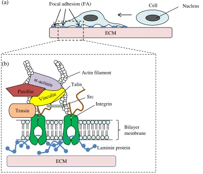
2.2.1 Extracellular matrix (ECM) and cell adhesion
All cells in solid tissue are surrounded by extracellular matrix (ECM). ECM is composed of proteins and polysaccharides. In animal cells, the ECM surrounds cells as fibrils that contact the cells. Cells are linked directly to each other by cell adhesion molecules at the cell surface. ECM provides mechanical support [63], a biochemical barrier [64], a medium for extracellular communication [65], cell matrix adhesion [66], and adhesion matrix for cell migration [67-69] during cell development.
Adhesion of cells to the ECM is key to the regulation of cellular morphology, migration, proliferation, survival, and differentiation [70]. These functions are essential during development, maintenance of tissue architecture and the induction of tissue repair. Integrin are the predominant receptors that mediate cell adhesion to the ECM proteins [71, 72].
11
platform for the recruitment of cellular proteins and signaling proteins to the inner surface of the plasma membrane, where they form structures called focal adhesions (FA) (Figure 2.3 (a)) [74]. The FA provide strong linkages to the actin cytoskeleton mediated by integrins to connect cells firmly to the ECM [75].
Cells adhere to the ECM via integrins that function as a heterodimer that composed of subunits alpha (α) and beta (β) transmembrane linked to cell cytoskeleton actin microfilaments via talin and vinculin [76]. Talin is a main regulator of the initial process of FA assembly [77]. During the initial step of FA formation, the binding of talin to integrin stabilises the ligand-induced clustering by mediating crosslinking of integrins with vinculin and α-actinin (Figure 2.3(b)) [78].
(a)
[image:37.612.110.507.305.646.2](b)
Figure 2.3: Cell adhesion to the ECM. (a) Suspended cells adhere to the surface of ECM via integrins (b) The structures of actin cytoskeleton, focal adhesion complexes, integrin
receptors, and adhesion proteins to form cross-linked platforms
ECM
Cell
Focal adhesion (FA) Nucleus
Talin
Integrin
Bilayer membrane Src
Paxillin
Actin filament
Tensin
Laminin protein
2.3 Epithelial cells and skin
HaCaT and OSCC (ORL-48) are non-cancer and cancer epithelial cells, respectively. The microtissue models for epidermis and oral cancer cell study are scarce. Hence, the growth of both cell types into biomimetic microtissues would provide value in tissue implant [79], pharmacology [80] or even cancer therapeutic drugs study [11].
2.3.1 Human keratinocyte cell lines (HaCaT)
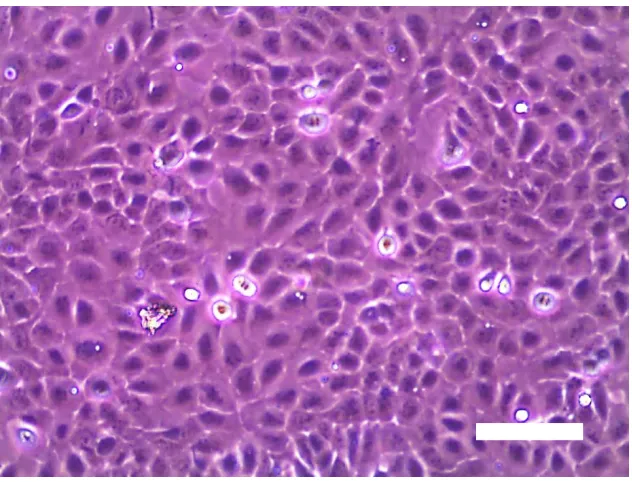
13
Figure 2.4: Phase contrast micrographs of HaCaT cells cultured for 3 days at 1 : 6 dilutions (Scale bar: 100 µm)
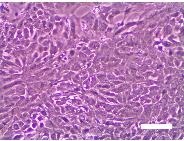
2.3.2 Oral squamous cell carcinoma cell line (ORL-48)
Oral cancer is defined as malignant lesion within oral cavity. Most cancerous oral cells originate from the oral squamous epithelium cell which is the primary surface structure of the lips and mucous membrane of the oral cavity [90]. OSCC has been histologically characterised as irregular nests, columns or malignant epithelial cells [91]. Abnormalities of oral cancerous cells are believed to be associated with several consecutive genetic mutations [92]. By clonal selection of viable cells which have accumulated genetic damages, normal mucosa cells ultimately evolve into malignant mucosa cells over an indefinite period [93].
Figure 2.5: Phase contrast micrographs of ORL-48 cells cultured for 3 days at 1 : 6 dilutions (Scale bar: 100 µm)
2.4 Rationale of growing 3D cells
15
2.5 Methods for culturing microtissues
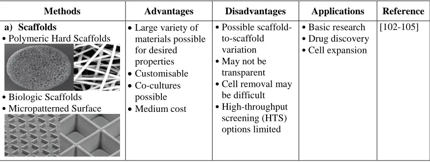
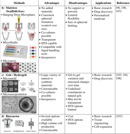
[image:41.612.109.546.484.649.2]3D cell culture methods are commonly accepted as more physiologically relevant methods and are believed to improve prediction of drug development process [95, 96]. There are several methods for culturing cells into 3D microtissues, which involved scaffolds, matrices (scaffold-free), gels or hydrogels and bioreactor as listed in Table 2.1. Scaffold based method is available in variety of materials with different porosities, permeabilities and mechanical characteristics designed to mimic the in vivo ECM of the specific tissues [1]. Whereas, microtissue culture using scaffold-free platforms do not contain added biomaterials or ECM. Cells grown and organised with their own generated ECM [96]. Gels or hydrogels culturing method aim to mimic the ECM and it has a soft tissue-like stiffness [97]. Cells can be cultured directly on the hydrogels (agarose, collagen and alginate) to form microtissues [98-100]. This method can be combined with other methods, such as scaffolds and microchips. The most ideal 3D cell culture method for high volume cell production and in vitro tissue engineering applications are the bioreactors method [101]. Microtissues cultured by using bioreactor method allows circulation of nutrients and removal of wastes within the reactor.
Table 2.1: A summary of 3D cell culture methods for culturing 3D microtissues
Methods Advantages Disadvantages Applications Reference
a) Scaffolds
•Polymeric Hard Scaffolds
•Biologic Scaffolds •Micropatterned Surface
Large variety of materials possible for desired properties
Customisable
Co-cultures possible
Medium cost
•Possible scaffold-to-scaffold variation •May not be
transparent •Cell removal may
be difficult •High-throughput
screening (HTS) options limited
•Basic research •Drug discovery •Cell expansion
Table 2.1 (continued): A summary of 3D cell culture methods for culturing 3D microtissues
Methods Advantages Disadvantages Applications Reference
b) Matrices
(Scaffold-free)
•Hanging Drop Microplates
•Microfluidic
•Microarray
No added materials
Consistent spheroid formation (control over size)
Co-cultures possible
Transparent
HTS capable
Compatible with liquid handling tools
Inexpensive
No support or porosity
Limited flexibility
Size of spheroid limiting
Basic research
Drug discovery
Personalised medicine
[96, 106, 107]
c) Gels / Hydrogels
•Micromold
•Microencapsulation
•Large variety of natural or synthetic materials •Customisable •Co-cultures possible •Inexpensive •Gel-to-gel variation and structural changes over time •Undefined constituents in natural gels •May not be transparent •HTS options
limited
•Basic research •Drug discovery
[103, 104, 108]
d) Bioreactor •Several options
available •High volume cell
production •Customisable
•Cost •HTS options
limited
•Basic research •Tissue
engineering •Cell expansion
[101]
2.6 Microencapsulation
[image:42.612.108.542.120.568.2]17
compound is coated or masked, to present it in the form of multiparticulate system. Microencapsulation process can be classified in terms of the microparticles or microspheres, based on their external morphology and internal structure (homogeneous or solid spheres) in micrometer range diameters [13, 34]. The material inside the microcapsule is referred to as the core, internal phase, or fill, whereas the wall is sometimes called a shell, coating, or membrane. Microcapsules can be classified into three basic categories as mono-core (also called single-core or reservoir type), poly-core (also called multiple-core) and matrix types (Figure 2.6). Mono-core is microcapsule which has a single hollow chamber within the capsule [110]. Poly-core is microcapsule which has a number of different size chambers within the shell [110]. Matrix type is of microparticle that has the active compounds integrated within the matrix of the shell material [110]. However, the morphology of the internal structure of a microparticle depends mainly on the shell materials and the microencapsulation methods that are employed [110].
(a) (b) (c) (d)
(a) (b) (c) (d)
Figure 2.6: Different morphology of microcapsules. (a) Mono-core, Single-core or reservoir type, (b) Poly-core, Multiple-core, (c) and (d) Matrix type [110]
ability [34]. For microencapsulation of cells, the selection of a suitable encapsulating material is critical. The material is required to have appropriate porosity, which can facilitate the transport of nutrients, proteins, DNA, and drug while blocking attack of antibodies and immune cells [6]. The capsules must be mechanically stable and easy to handle. These requirements may be fulfilled by controlling the pore size and the thickness of encapsulating polymer membrane at microscale. Smaller pore size and thicker capsules membrane showed higher mechanical stability [112, 113]. The cell viability and metabolic status must be optimal if the encapsulated cells are in the order of hundreds micron in size [15].
Figure 2.7: Principle of immunoisolation by a microcapsule [15]
2.6.1 Application of microencapsulation
19
micro and nanostructured materials applied to a wide variety of applications. There are various application of microcapsules that already been introduced in the market.
One of the most important applications of microencapsulated products is in the area of crop protection [114, 115]. Polymer microcapsules, such as gelatin, serve as efficient delivery vehicles to deliver pheromone by spraying the capsule dispersion and protect the pheromone from oxidation and light during storage and release [116].
The major applications area of encapsulation technique is pharmaceutical or biomedical for controlled drug delivery [117-120]. Several drug delivery systems are replacement of therapeutic agents, gene therapy and vaccines use. The capsules are engineered to stick tightly to and even penetrate linings in the gastrointestinal track before transferring the drug contents over time into circulatory system or the targeted spot [119, 121]. Other than that, one of the most important medical applications of microencapsulation technology is to serve as a cushion or implant, such as breast implant [110].
Microencapsulation is used to overcome all the challenges in food industry by providing technology to incorporate minerals, vitamins, flavours [122] and essential oils in food [123]. Microencapsulation simplify the food manufacturing process by converting liquids to solid powder, decreasing production cost, help fragile and sensitive materials survive processing and packaging conditions and stabilise the shelf life of the active ingredient [124-126].
Microencapsulation also plays a crucial role in energy generation field. Hollow and multilayered plastic microspheres loaded with gaseous, deuterium, a fusion fuel, are used to harness nuclear fusion for producing electrical energy [127]. This fusion experiment process has been named as inertial confinement fusion (ICF) and it has been in use since 1980s [128].
2.6.2 Technologies for microencapsulation of cells
[image:46.612.111.549.334.642.2]The encapsulation of various materials and living cells inside capsules for different purposes in the pharmaceutical, chemical, food industry, agriculture, tissue engineering, biotechnology and medicine is of great importance. Microencapsulation of cells in hydrocolloid gel matrices is the technique that the cells are entrapped during gel formation, leading to spherical droplets containing cells. Some of the popular microencapsulation technologies generally produce the capsules of micron to millimeter size for microencapsulation of cells were listed in Table 2.2.
Table 2.2: Comparison of different microencapsulation technologies for encapsulation of cells
Micro-encapsulation process
Cost Complex design
High voltage
Material volume
Uniform Size (mm) Post-processing /harsh treatment Reference
Extrusion Low No No Large No 2 - 10 No [129-131]
JetCutter break-up
High Yes Yes Large Yes < 1 No [39, 49, 132-134] Spinning disc High Yes No Large Yes 0.2 - 5 No [135]
Micro nozzle array
High Yes No Large Yes > 0.5 Yes [8, 19]
Vibration nozzle
Low Yes No Medium No 0.1 - 3 No [37, 136,
137] Coacervation/
emulsion method
High Yes No Large Yes 0.02 - 2 Yes [130, 131, 138, 139]
Electrostatic droplet generation
High Yes Yes Medium Yes > 0.1 No [31-33, 133]
Flicking High No No Medium Yes 0.2 - 0.4 No [97]
Air atomisation
Low No No Medium No 0.08 -
0.6
21
2.6.2.1 Extrusion, Jet break-up methods and spinning disc
Extrusion (Figure 2.8 (a)) is the most common methods widely used to produce microcapsules due to its ease, simplicity, low cost, gentle condition and high quantity of encapsulated cells. Jet break-up and spinning disc techniques are also originated from the extrusion method. In a basic extrusion technique, the alginate containing cells are extruded through a syringe needle as droplets into calcium chloride (CaCl2) solution to
be polymerised. The size and shape of the capsules were influenced by the aperture size of the needle, concentration of the CaCl2 solution and the surface tension of the CaCl2
solution. The basic extrusion technique produced capsules size ranging from 2 - 10 mm [129-131].
Jet cutter method is suitable to be used with high viscosity polymer solutions such as poly(vinyl alcohol) solutions [134]. In this technique, the mixture of cells-alginate suspension was forced through a nozzle to form liquid jet and then cut by a rotating cutting wire (Figure 2.8 (b)). The number of cutting wires, rotations speed of cutting tool and the infusion rate manipulates the size of the capsules.
For the spinning disc technique, the capsules are formed by infusing the cells-alginate suspension onto the high velocity spinning disc (Figure 2.8 (c)) due to the centrifugal force at the edge of the spinning disc, the droplets are formed and dropped into the CaCl2 solution to be polymerised. The size of the capsules is controlled by the
(a) (b) (c)
Figure 2.8: Schematic diagram of different microencapsulation processes in forming microcapsules: (a) Extrusion, (b) Jet cutter and (c) Spinning disc [143]
2.6.2.2 Micro nozzle array and vibrating nozzle
Micro nozzle is a developed technique for microencapsulation in year 2000. In this technique, the cell-alginate suspension is flew through silicon micro nozzle array and then cut off by the high stream of oil to form droplets [8]. The gel droplets drop into the oil stream that directs the flow of the droplets to a solution of positive ions (Figure 2.9 (a)). Due to the high flow pressure conditions, micro nozzle array are suitable to be used with high viscosity solution [144]. If this method is to be scaled up for large production, the cost of the oil and its disposability could be the limitations of this technique [145].
For vibration nozzle technique (Figure 2.9 (b)), the microcapsules are formed by oscillating and purging the mixture cells suspension through a nozzle into the hardening bath, resulting in size distribution of capsules as 0.1 - 3.0 mm in diameter [137].
Collecting bath
Liquid jet
Cutting tool with wires
23
[image:49.612.123.535.69.222.2]
(a) (b)
Figure 2.9: Schematic diagram of microencapsulation processes in alginate: (a) Micro nozzle array [8] and (b) Vibrating nozzle [146]
2.6.2.3 Microfluidic device
Microfluidics device has emerged as a powerful platform for the generation of microparticles with tailored structure and properties [147-150]. This technique allows direct integration of different input fluids into the polydimethylsiloxane (PDMS) microfluidic channel as shown in Figure 2.10. The working principle of microfluidic to generate microcapsules is based on the emulsification of alginate solution.
Microcapsule fabrication methods based on microfluidics device may be classified into two major approaches, that are flow-focusing and T-junction capsule formation. The flow-focusing microfluidic approach, as shown in Figure 2.11, forms microcapsules by allowing a core fluid (cell-alginate suspension) to be surrounded by sheath stream (oil) flowing. In contrast, T-junctions microfluidic is designed to form microcapsules by permitting the core fluid to be swept away by one sheath stream in only one direction. A summary of the microfluidic emulsification technologies based on PDMS microfluidic chip design for both flow-focusing and T-junction capsules formation methods, used for the application of cell encapsulation were listed in Table 2.3 [6].
Alginate CaCl2
Microcapsules Vegetable
oil
Figure 2.10: Illustrations of microfluidics system mechanism for microencapsulation [151]
(a) (b)
[image:50.612.161.491.423.569.2]REFERENCES
[1] D. Antoni, H. Burckel, E. Josset, and G. Noel. Three-dimensional cell culture: a breakthrough in vivo. International Journal of Molecular Sciences. 2015. 16(3): 5517 - 5527.
[2] G. R. Souza, J. R. Molina, R. M. Raphael, M. G. Ozawa, D. J. Stark, C. S. Levin, L. F. Bronk, J. S. Ananta, J. Mandelin, M. M. Georgescu, J. A. Bankson, J. G. Gelovani, T. C. Killian, W. Arap, and R. Pasqualini. Three-dimensional tissue culture based on magnetic cell levitation. Nature Nanotechnology. 2010. 5(4): 291 - 296.
[3] R. Edmondson, J. J. Broglie, A. F. Adcock, and L. Yang. Three-dimensional cell culture systems and their applications in drug discovery and cell-based biosensors. Assay and Drug Development Technologies. 2014. 12(4): 207 - 218. [4] L. Kunz-Schughart, J. P. Freyer, F. Hofstaedter, and R. Ebner. The use of 3-D
cultures for high throughput screening: the multicellular spheroid model. Journal
of Biomolecular Screening. 2004. 9(4): 273 - 285.
[5] C. Soon, K. Thong, K. Tee, A. Ismail, M. Denyer, M. Ahmad, Y. Kong, P. Vyomesh, and S. Cheong. A scaffoldless technique for self-generation of three-dimensional keratinospheroids on liquid crystal surfaces. Biotechnic &
Histochemistry. 2016. 91(4): 283 - 295.
[6] A. Kang, J. Park, J. Ju, G. S. Jeong, and S. H. Lee. Cell encapsulation via microtechnologies. Biomaterials. 2014. 35(9): 2651 - 2663.
[7] M. M. Stevens, H. F. Qanadilo, R. Langer, and V. Prasad Shastri. A rapid-curing alginate gel system: utility in periosteum-derived cartilage tissue engineering.
[8] S. Sugiura, T. Oda, Y. Izumida, Y. Aoyagi, M. Satake, A. Ochiai, N. Ohkohchi, and M. Nakajima. Size control of calcium alginate beads containing living cells using micro-nozzle array. Biomaterials. 2005. 26(16): 3327 - 3331.
[9] E. L. Scheller, P. H. Krebsbach, and D. H. Kohn. Tissue engineering: state of the art in oral rehabilitation. Journal of Oral Rehabilitation. 2009. 36(5): 368 - 389. [10] E. L. da Rocha, L. M. Porto, and C. R. Rambo. Nanotechnology meets 3D in
vitro models: tissue engineered tumors and cancer therapies. Materials Science &
Engineering. C, Materials For Biological Applications. 2014. 34: 270 - 279.
[11] C. S. Shin, B. Kwak, B. Han, and K. Park. Development of an in vitro 3D tumor model to study therapeutic efficiency of an anticancer drug. Molecular
Pharmaceutics. 2013. 10(6): 2167 - 2175.
[12] T. M. Chang. Semipermeable Microcapsules. Science. 1964. 146(3643): 524 - 525.
[13] A. M. Sun. Microencapsulation of cells. Medical applications. Annals of The
New York Academy of Sciences. 1997. 831: 271 - 279.
[14] L. Gasperini, J. F. Mano, and R. L. Reis. Natural polymers for the microencapsulation of cells. Journal of The Royal Society Interface. 2014. 11(100): 20140817.
[15] G. A. Paredes Juarez, M. Spasojevic, M. M. Faas, and P. de Vos. Immunological and technical considerations in application of alginate-based microencapsulation systems. Frontiers in Bioengineering and Biotechnology. 2014. 2: 26.
[16] V. Vaithilingam and B. E. Tuch. Islet transplantation and encapsulation: an update on recent developments. The Review of Diabetic Studies. 2011. 8(1): 51 - 67.
[17] J. M. Rabanel, X. Banquy, H. Zouaoui, M. Mokhtar, and P. Hildgen. Progress technology in microencapsulation methods for cell therapy. Biotechnology
Progress. 2009. 25(4): 946 - 963.
[18] N. C. Hunt and L. M. Grover. Cell encapsulation using biopolymer gels for regenerative medicine. Biotechnology Letters. 2010. 32(6): 733 - 742.
119
Microfabricated airflow nozzle for microencapsulation of living cells into 150 micrometer microcapsules. Biomedical Microdevices. 2007. 9(1): 91 - 99.
[20] P. de Vos, C. G. van Hoogmoed, J. van Zanten, S. Netter, J. H. Strubbe, and H. J. Busscher. Long-term biocompatibility, chemistry, and function of microencapsulated pancreatic islets. Biomaterials. 2003. 24(2): 305 - 312.
[21] K. Y. Lee and D. J. Mooney. Alginate: properties and biomedical applications.
Progress In Polymer Science. 2012. 37(1): 106 - 126.
[22] I. Ghidoni, T. Chlapanidas, M. Bucco, F. Crovato, M. Marazzi, D. Vigo, M. L. Torre, and M. Faustini. Alginate cell encapsulation: new advances in reproduction and cartilage regenerative medicine. Cytotechnology. 2008. 58(1): 49 - 56.
[23] P. de Vos, H. A. Lazarjani, D. Poncelet, and M. M. Faas. Polymers in cell encapsulation from an enveloped cell perspective. Advanced Drug Delivery
Reviews. 2014. 67 - 68: 15 - 34.
[24] E. S. Chan, B. B. Lee, P. Ravindra, and D. Poncelet. Prediction models for shape and size of ca-alginate macrobeads produced through extrusion-dripping method.
Journal of Colloid and Interface Science. 2009. 338(1): 63 - 72.
[25] S. Swioklo, P. Ding, A. W. Pacek, and C. J. Connon. Process parameters for the high-scale production of alginate-encapsulated stem cells for storage and distribution throughout the cell therapy supply chain. Process Biochemistry. 2016.
[26] A. Khademhosseini, G. Eng, J. Yeh, J. Fukuda, J. Blumling, R. Langer, and J. A. Burdick. Micromolding of photocrosslinkable hyaluronic acid for cell encapsulation and entrapment. Journal of Biomedical Materials Research Part A. 2006. 79(3): 522 - 532.
[27] W. G. Koh, A. Revzin, and M. V. Pishko. Poly(ethylene glycol) hydrogel microstructures encapsulating living cells. Langmuir. 2002. 18(7): 2459 - 2462. [28] C. J. Martinez, J. W. Kim, C. Ye, I. Ortiz, A. C. Rowat, M. Marquez, and D.
[29] K. S. Huang, M. K. Liu, C. H.Wu, Y. T. Yen, and Y. C. Lin. Calcium alginate microcapsule generation on a microfluidic system fabricated using the optical disk process. Journal of Micromechanics and Microengineering. 2007. 17(8): 1428 - 1434.
[30] Y. Hu, Q. Wang, J. Wang, J. Zhu, H. Wang, and Y. Yang. Shape controllable microgel particles prepared by microfluidic combining external ionic crosslinking. Biomicrofluidics. 2012. 6(2): 026502.
[31] W. Zhang and X. He. Encapsulation of living cells in small (approximately 100 microm) alginate microcapsules by electrostatic spraying: a parametric study.
Journal of Biomechanical Engineering. 2009. 131(7): 074515.
[32] D. Lewinska, J. Bukowski, M. Kozuchowski, A. Kinasiewicz, and A. Werynski. Electrostatic microencapsulation of living cells. Biocybernetics and Biomedical
Engineering. 2008. 28(2): 69 - 84.
[33] N. Li, X. X. Xu, G. W. Sun, X. Guo, Y. Liu, S. J. Wang, Y. Zhang, W. T. Yu, W. Wang, and X. J. Ma. The effect of electrostatic microencapsulation process on biological properties of tumour cells. Journal of Microencapsulation. 2013. 30(6): 530 - 537.
[34] L. Martin-Banderas, A. M. Ganan-Calvo, and M. Fernandez-Arevalo. Making Drops in Microencapsulation Processes. Letters in Drug Design & Discovery. 2010. 7(4): 300 - 309.
[35] J. H. Cui, J. S. Goh, S. Y. Park, P. H. Kim, and B. J. Le. Preparation and physical characterization of alginate microparticles using air atomization method. Drug
Development and Industrial Pharmacy. 2001. 27(4): 309 - 319.
[36] E.P. Herrero, E.M. Mart´ın Del Valle, and M. A. Galan. Development of a new technology for the production of microcapsules based in atomization processes.
Chemical Engineering Journal. 2006. 117(2): 137 - 142.
[37] M. Whelehan and I. W. Marison. Microencapsulation using vibrating technology. Journal of Microencapsulation. 2011. 28(8): 669 - 688.
121
[39] C. Schwinger, S. Koch, U. Jahnz, P. Wittlich, N. G. Rainov, and J. Kressler. High throughput encapsulation of murine fibroblasts in alginate using the JetCutter technology. Journal of Microencapsulation. 2002. 19(3): 273 - 280. [40] Q. Gao, Y. He, J.-z. Fu, J.-j. Qiu, and Y.-a. Jin. Fabrication of shape controllable
alginate microparticles based on drop-on-demand jetting. Journal of Sol-Gel
Science and Technology. 2015. 77(3): 610 - 619.
[41] A. Gautier, B. Carpentier, M. Dufresne, Q. Vu Dinh, P. Paullier, and C. Legallais. Impact of alginate type and bead diameter on mass transfers and the metabolic activities of encapsulated C3A cells in bioartificial liver applications.
European Cells & Materials. 2011. 21: 94 - 106.
[42] J. Wan. Review Microfluidic-based synthesis of hydrogel particles for cell microencapsulation and cell-based drug delivery. Polymers. 2012. 4(2): 1084 - 1108.
[43] U. Prüsse, L. Bilancetti, M. Bučko, B. Bugarski, J. Bukowski, P. Gemeiner, D. Lewińska, V. Manojlovic, B. Massart, C. Nastruzzi, V. Nedovic, D. Poncelet, S. Siebenhaar, L. Tobler, A. Tosi, A. Vikartovská, and K.-D. Vorlop. Comparison of different technologies for alginate beads production. Chemical Papers. 2008. 62(4): 364 - 374.
[44] S. Tendulkar, S. H. Mirmalek-Sani, C. Childers, J. Saul, E. C. Opara, and M. K. Ramasubramanian. A three-dimensional microfluidic approach to scaling up microencapsulation of cells. Biomedical Microdevices. 2012. 14(3): 461 - 469. [45] M. Whelehan and I. W. Marison. Microencapsulation by dripping and jet break
up. Bioencapsulation Innovations. 2011. 1(1): 4 - 10.
[46] A. Dalmoro, A. A. Barba, and M. d'Amore. Analysis of size correlations for microdroplets produced by ultrasonic atomization. The Scientific World Journal. 2013. 2013: 7.
[47] Y. Zhang and M. L. Ma. Microscale Technologies for Cell Engineering. Springer International Publishing: Springer. 2016.
[48] D. Chicheportiche and G. Reach. In vitro kinetics of insulin release by microencapsulated rat islets: effect of the size of the microcapsules.
[49] U. Pruesse, U. Jahnz, P. Wittlich, and K. D. Vorlop. Scale-up of the JetCutter technology. Chemistry & Industry. 2003. 12: 636 - 641.
[50] C. Heinzen, A. Berger, and I. Marison. Use of vibration technology for jet break-up for encapsulation of cells and liquids in monodisperse microcapsules. In: V. Nedović and R. Willaert. Fundamentals of Cell Immobilisation Biotechnology. Dordrecht: Springer Netherlands. 2004. 257 - 275.
[51] M. A. Neves, I. Kobayashi, and M. Nakajima. Development of microchannel emulsification technology for monodispersed soybean and olive oil-in-water emulsions. Journal of Arid Land Studies. 2009. 19(1): 97 - 100.
[52] C. Kim, K. S. Lee, Y. E. Kim, K. J. Lee, S. H. Lee, T. S. Kim, and J. Y. Kang. Rapid exchange of oil-phase in microencapsulation chip to enhance cell viability.
Lab On A Chip. 2009. 9(9): 1294 - 1297.
[53] A. Sohail, M. S. Turner, A. Coombes, T. Bostrom, and B. Bhandari. Survivability of probiotics encapsulated in alginate gel microbeads using a novel impinging aerosols method. International Journal of Food Microbiology. 2011. 145(1): 162 - 168.
[54] S. Ahn, H. Lee, L. J. Bonassar, and G. Kim. Cells (MC3T3-E1)-laden alginate scaffolds fabricated by a modified solid-freeform fabrication process supplemented with an aerosol spraying. Biomacromolecules. 2012. 13(9): 2997 - 3003.
[55] S. Ahn and G. Kim. Cell-encapsulating alginate microsized beads using an air-assisted atomization process to obtain a cell-laden hybrid scaffold. Journal of
Materials Chemistry B. 2015. 3(47): 9132 - 9139.
[56] S. Hamid, K. P. Lim, R. B. Zain, S. M. Ismail, S. H. Lau, W. M. Mustafa, M. T. Abraham, N. A. Nam, S. H. Teo, and S. C. Cheong. Establishment and characterization of Asian oral cancer cell lines as in vitro models to study a disease prevalent in Asia. International Journal of Molecular Medicine. 2007. 19(3): 453 - 460.
[57] C. Scully and S. Porter. ABC of oral health. Oral cancer. BMJ - British Medical
123
[58] W. Chen, J. H. Kim, D. Zhang, K. H. Lee, G. A. Cangelosi, S. D. Soelberg, C. E. Furlong, J. H. Chung, and A. Q. Shen. Microfluidic one-step synthesis of alginate microspheres immobilized with antibodies. Journal of The Royal Society
Interface. 2013. 10(88): 20130566.
[59] E. Bianconi, A. Piovesan, F. Facchin, A. Beraudi, R. Casadei, F. Frabetti, L. Vitale, M. C. Pelleri, S. Tassani, F. Piva, S. Perez-Amodio, P. Strippoli, and S. Canaider. An estimation of the number of cells in the human body. Annals of
Human Biology. 2013. 40(6): 463 - 471.
[60] L. Sherwood. Fundamentals of Human Physiology. 4th Ed. USA: Brooks/Cole Cengage Learning. 2012.
[61] M. Wilson. Microbial inhabitants of Humans: Their Ecology and Role in Health
and Disease. 1st Ed. United Kingdom: Cambridge University Press. 2005.
[62] J. McDowell. Encyclopedia of Human Body Systems Volume 1. USA: Greenwood. 2010.
[63] M. Chiquet, M. Matthisson, M. Koch, M. Tannheimer, and R. Chiquet-Ehrismann. Regulation of extracellular matrix synthesis by mechanical stress.
Biochemistry and Cell Biology-Biochimie ET Biologie Cellulaire. 1996. 74(6):
737 - 744.
[64] R. Londono, V. S. Gorantla, and S. F. Badylak. Emerging implications for extracellular matrix-based technologies in vascularized composite allotransplantation. Stem Cells International. 2016. 2016(1541823): 16.
[65] M. Fujita, D. C. Spray, H. Choi, J. Saez, D. M. Jefferson, E. Hertzberg, L. C. Rosenberg, and L. M. Reid. Extracellular matrix regulation of cell-cell communication and tissue-specific gene expression in primary liver cultures.
Progress in Clinical and Biological Research. 1986. 226(1): 333 - 360.
[66] C. Frantz, K. M. Stewart, and V. M. Weaver. The extracellular matrix at a glance. Journal of Cell Science. 2010. 123(24): 4195 - 4200.
[67] S. H. Kim, J. Turnbull, and S. Guimond. Extracellular matrix and cell signalling: the dynamic cooperation of integrin, proteoglycan and growth factor receptor.
[68] M. P. Sheetz, D. P. Felsenfeld, and C. G. Galbraith. Cell migration: regulation of force on extracellular-matrix-integrin complexes. Trends in Cell Biology. 1998. 8(2): 51 - 54.
[69] K. Wolf, M. Te Lindert, M. Krause, S. Alexander, J. Te Riet, A. L. Willis, R. M. Hoffman, C. G. Figdor, S. J. Weiss, and P. Friedl. Physical limits of cell migration: control by ECM space and nuclear deformation and tuning by proteolysis and traction force. The Journal of Cell Biology. 2013. 201(7): 1069 - 1084.
[70] B. M. Gumbiner. Cell adhesion: the molecular basis of tissue architecture and morphogenesis. Cell. 1996. 84(3): 345 - 357.
[71] H. Truong and E. H. Danen. Integrin switching modulates adhesion dynamics and cell migration. Cell Adhesion & Migration. 2009. 3(2): 179 - 181.
[72] J. T. Parsons, A. R. Horwitz, and M. A. Schwartz. Cell adhesion: integrating cytoskeletal dynamics and cellular tension. Nature Reviews Molecular Cell
Biology. 2010. 11(9): 633 - 643.
[73] M. Nagano, D. Hoshino, N. Koshikawa, T. Akizawa, and M. Seiki. Turnover of focal adhesions and cancer cell migration. International Journal of Cell Biology. 2012. 2012(310616): 10.
[74] V. Petit and J. P. Thiery. Focal adhesions: structure and dynamics. Biology of
The Cell. 2000. 92(7): 477 - 494.
[75] B. Geiger, A. Bershadsky, R. Pankov, and K. M. Yamada. Transmembrane crosstalk between the extracellular matrix--cytoskeleton crosstalk. Nature
Reviews. Molecular Cell Biology. 2001. 2(11): 793 - 805.
[76] B. H. Luo, C. V. Carman, and T. A. Springer. Structural basis of integrin regulation and signaling. Annual Review of Immunology. 2007. 25: 619 - 647. [77] E. Goksoy, Y. Q. Ma, X. Wang, X. Kong, D. Perera, E. F. Plow, and J. Qin.
Structural basis for the autoinhibition of talin in regulating integrin activation.
Molecular Cell. 2008. 31(1): 124 - 133.
![Figure 2.1: The anatomy of human cell [60]](https://thumb-us.123doks.com/thumbv2/123dok_us/8755495.892795/35.612.113.532.71.403/figure-anatomy-human-cell.webp)
![Figure 2.2: The four basic types of tissue [62]](https://thumb-us.123doks.com/thumbv2/123dok_us/8755495.892795/36.612.159.501.67.341/figure-basic-types-tissue.webp)
![Figure 2.7: Principle of immunoisolation by a microcapsule [15]](https://thumb-us.123doks.com/thumbv2/123dok_us/8755495.892795/44.612.152.495.280.539/figure-principle-immunoisolation-microcapsule.webp)
![Figure 2.8: Schematic diagram of different microencapsulation processes in forming microcapsules: (a) Extrusion, (b) Jet cutter and (c) Spinning disc [143]](https://thumb-us.123doks.com/thumbv2/123dok_us/8755495.892795/48.612.143.516.69.247/figure-schematic-different-microencapsulation-processes-microcapsules-extrusion-spinning.webp)
