Sławomir Jeka
1, Paweł Żuchowski
2, Mikołaj Karmowski
3,
Bohdan Gworys
4, Andrzej Karmowski
3Review and Principles of Modern Methods
of Ultrasound Imaging in Rheumatology
Przegląd i podstawy nowoczesnych metod
obrazowania ultrasonograficznego stosowanych w reumatologii
1 Clinical Division of Rheumatology and Connective Tissue Diseases,Dr. J. Biziel’s 2nd University Hospital in Bydgoszcz, Medical College of Nicolaus Copernicus University, Poland 2 NZOZ Nasz Lekarz, Torun, Poland
31st Department and Clinic of Gynecology and Obstetrics, Wroclaw Medical University, Poland 4 Department of Normal Anatomy, Wroclaw Medical University, Poland
Abstract
Ultrasound examinations belong to the group of most easily available visual examinations. They are not burdened with such an amount of contraindications as computed tomography. They are cheaper and faster to perform than magnetic resonance. The progress which has taken place in recent times in ultrasonography has, to a large extent, influenced the increased quality of images, as well as also allowing for new applications in diagnostic imaging. This also concerns rheumatology, including diagnosis of early rheumatoid arthritis. The available options in ultrasound examination allow for an accurate depiction of synovium or erosions. Unfortunately, diagnostic options also have their restrictions and in special cases their improper usage can lead to worsening of the quality of imaging. It is par-ticularly visible in the case of rheumatology, in which great problems appear with establishing diagnostic criteria of illnesses. On that account it is extremely important to have the knowledge in the scope of modern ultrasonography which will enable the correct application of available techniques of imaging, which improves the diagnostic value of this type of examination in rheumatology. That it is required to have a basic knowledge of rules that are used with modern ultrasound options such as SRI, CRI or THI. Thanks to the correct use of these options, based on theoretical knowledge it is possible to obtain very accurate images in the process of an ultrasound examination. Addressing the need to have such knowledge, we can even improve the number of courses organized in this field by ACR or EULAR (Adv Clin Exp Med 2011, 20, 6, 779–785).
Key words: ultrasonography, contrast agents, tissue harmonic imaging, speckle reduction imaging.
Streszczenie
Badanie ultrasonograficzne jest jednym znajbardziej dostępnych badań.Nie jest ono obciążonetaką liczbą
przeciw-wskazań, jak np.tomografia komputerowa.Jest ono również tańsze i szybszedo wykonanianiżrezonans
magnetycz-ny.Postęp, jaki dokonał się w ciągu ostatnich latwultrasonografii w znaczącym stopniuwpłynął napoprawę jakości
obrazowania, stworzyło to możliwość nowych zastosowań w diagnostyce obrazowej. Dotyczy to również reumato-logii, w tym diagnostyki wczesnego reumatoidalnego zapalenia stawów. Dostępne nowe opcje w USG pozwalają na dokładne obrazowanie błony maziowej i nadżerek. Niestety, nowe opcje obrazowania mają również swoje ogranicze-nia, w niektórych wypadkach ich niewłaściwe użycie może prowadzić do pogorszenia jakości obrazu. Jest to szcze-gólnie widoczne w przypadku reumatologii, w której istnieją duże trudności w ustalaniu kryteriów diagnostycznych.
Z tego względujest bardzoważne, aby posiadać odpowiednią wiedzę zzakresu nowoczesnej ultrasonografii, która umożliwia prawidłowe stosowaniedostępnych technikobrazowania,co zwiększawartość diagnostycznątego bada-niawreumatologii. Dotyczy to w szczególności takich opcji badania ultrasonograficznego, jak SRO, CRI czy THI.
Dziękiprawidłowemu wykorzystaniu tych opcji,opartym nawiedzy teoretycznej, jest możliweuzyskanie bardzo
dokładnych obrazówpodczas badaniaUSG.Okonieczności posiadaniatakiej wiedzymożeświadczyćliczbakursów
organizowanychw tej dziedzinie przezACRlubEULAR (Adv Clin Exp Med 2011, 20, 6, 779–785).
Słowa kluczowe: ultrasonografia, środki kontrastowe, obrazowanie w technologii II harmonicznej, redukcja
ziar-nistości obrazu w czasie rzeczywistym.
Adv Clin Exp Med 2011, 20, 6, 779–785 ISSN 1230-025X
REvIEWS
S. Jeka et al.
780
Ultrasound imaging is one of the most acces-sible imaging studies in medicine. It is associated with the low cost of such an examination and the absence of most of the contraindications to its ex-ecution, as are in case of other imaging techniques, especially X-ray and CT. The universality and the availability of this test have contributed to the very dynamic development of ultrasonography.
Modern ultrasound devices can produce high resolution images, thanks to new technical solutions in image analysis, construction of a high frequency head up to 18 Mhz, or by using second generation ultrasound contrast agents. With the improvement of the quality of imaging, the range of ultrasonog-raphy applications is also expanding. In rheumatol-ogy interventions and treatments under ultrasound examination, inter alia, punctures, and administra-tion of drugs have become the standard. Also, the monitoring of changes using ultrasound during the course of rheumatic diseases is an increasingly frequent practice. Modern methods of imaging in ultrasound allow precise tracking of inflammatory changes occurring in the synovial membrane and cartilage pannus, presence of inflammatory granu-lation tissue or detection of erosions, which are not yet visible on a classical X-ray.
This is particularly important in the case of rheumatoid arthritis (RA), in which early diagno-sis is extremely difficult due to a lack of sensitive and specific methods of diagnosis [1, 2].
The majority of rheumatic diseases lead to dis-orders of synovium function. In the presence of ex-cessive amounts of inflammatory fluid in the joints, tendon sheaths and burs synovium become clearly visible by ultrasound. In addition, the swelling or hypertrophy, and the increased visibility of the gran-uloma have caused a growth of the importance of this type of test in the diagnosis and monitoring of rheumatoid arthritis and other rheumatic diseases.
Unfortunately, the opportunities offered by new developments in the field of ultrasound are still not fully used in rheumatological practice. In this state of affairs, rheumatologists lack a suffi-cient level of knowledge of ultrasonography and clear criteria for assessing ultrasound [3]. While the second problem’s solution is not easy, the first problem can be solved quite easily.
Discussion
New Methods Used
in Ultrasound
Increasing computing power used for analyz-ing the signal collected by the ultrasonographic head and increasing the frequency of the
ultra-sound emitted are the most obvious ways to im-prove the quality of images obtained during the test. Of course, such improvements in image qual-ity do not require any medical expertise in the field of ultrasound, because they would not have any effect on how the test is performed. Unfortu-nately, the development of ultrasound devices in this direction has its limits, beyond which it can no longer go to improve image quality. The compu-tational speed of modern processors is already so high that it can quickly convert all signals gathered by the head without any problems. However, a fur-ther increase in the frequency of the ultrasound emitted by the head, increases the amount of de-tail that can be visualized, but reduces the depth of penetration where you can get a picture. The higher the ultrasound frequency the more they are absorbed by the body and in such cases, the organs located inside the tissues cease to be visible.
Tissue Harmonic Imaging – THI
The phenomenon of the generation of har-monic waves was originally observed in optics in the course of an experiment with a ruby laser, whose light passed through a quartz crystal. After passing through the crystal, in addition to receiv-ing the light at the fundamental frequency, the light was also received at twice the frequency compared to the light of the fundamental frequency emitted by the laser [4]. The formation of a wave having twice the frequency – the second harmonic – is as-sociated with the effects of nonlinear wave propa-gation in the medium. In the case of ultrasound, it is related to the vibration of tissue [5–7]. As in the case of optics, so the acoustic wave can generate harmonics of higher orders. However, due to the decrease in the intensity of successive harmonics, in practice it is most common to do imaging using the second harmonic.The advantage of harmonic imaging is the ability to use the heads, which emit ultrasound of relatively low-frequency sounds in relation to the ultrasound which go back to the head and which are used to obtain the image. A head emitting a fre-quency of 5 MHz ultrasound will record the vibra-tions with a frequency of 10 MHz, so the amount of detail and resolution of the resulting image will be much greater than would result from the fre-quency of the head itself.
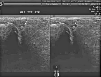
Fig. 1. Image of bone erosion within the joint of the metatarsal-phalangeal third finger of the left foot, longitudinal cross section. Left photograph made without additional options. Right photograph with the THI option. In compari-son to the photograph performed without using additional options, in the photo it is possible to notice poorer imag-ing of deeper situated anatomy structures. White arrows indicate the bone erosion in both photographs
Ryc. 1. Obraz nadżerki stawu śródstopno-paliczkowego, trzeci palec lewej stopy, przekrój podłużny. Lewe zdjęcie wykonane bez dodatkowych opcji. Prawe zdjęcie z opcją THI. W porównaniu do zdjęcia wykonanego bez użycia dodatkowych opcji, na zdjęciu można zauważyć gorsze obrazowanie głębiej położonej struktury anatomicznej. Białe strzałki wskazują nadżerkę na obu zdjęciach
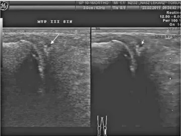
Fig. 2. Image of bone erosion within the joint of the metatarsal-phalangeal third finger of the left foot, longitudinal cross section. Left photograph made without additional options. Right photograph with the SRI option. White arrows indicate the bone erosion in both photographs
Ryc. 2. Obraznadżerkistawu śródstopno-paliczkowego, trzeci palec lewej stopy, przekrój podłużny. Lewe zdjęcie
S. Jeka et al.
782
Fig. 3. Image of bone erosion within the joint of the metatarsal-phalangeal third finger of the left foot, longitudinal cross section. Left photograph made without additional options. Right photograph with the CRI option. White arrows indicate the bone erosion in both photographs
Ryc. 3.Obraznadżerkistawu śródstopno-paliczkowego, trzeci palec lewej stopy, przekrój podłużny. Lewe zdjęcie
wykonanobezdodatkowych opcji.Prawe zdjęcie z opcjąCRI.Białestrzałkiwskazująnadżerkęna obuzdjęciach
Fig. 4. Image of bone erosion within the joint of the metatarsal-phalangeal third finger of the left foot, longitudinal cross section. Left photograph made without additional options. Right photograph with the SRI and CRI options. White arrows indicate the bone erosion in both photographs
Ryc. 4.Obraznadżerkistawu śródstopno-paliczkowego, trzeci palec lewej stopy, przekrój podłużny. Lewe zdjęcie
wykonanobezdodatkowych opcji.Prawe zdjęcie z opcjamiSRI i CRI.Białestrzałkiwskazująnanadżerkęna obu
A harmonic wave of lower intensity also al-lows you to get a significantly “cleaner” image – especially in the case of imaging structures such as cysts. In the case of harmonic imaging, there is a reduction of artifacts due to the fact that the sec-ond harmonic has a much smaller echo than the wave generated by the head.
SRI/XVIEW
At the beginning of this article the authors mentioned that one of the most obvious ways of improving the quality of imaging is to increase the computing power of processors used for analyz-ing the recorded signal. Processors installed in the latest ultrasonographic machines actually already have the computing power to fully cover the needs of signal analysis. Its power is large enough to be used for additional analysis without sacrificing the speed of the projecting image. One possible appli-cation of high performance computing is a digital analysis of the processed signal before it is dis-played on the monitor.
This option is the SRI or Xview – the name changes depending on the manufacturer of the ultrasonographic machine. However, the basic algorithm of this method is the same. During the
study, one of the most common causes of poor im-age quality was a very high amount of noise that hampers giving an accurate diagnosis during the study. This can be seen by both too little contrast between structures in the studied tissue and small amounts of detail shown.
Initially, the pixels of the image of which it is built are segregated into two groups – the pixels showing the tissue, and pixels which are noise. In-clusion of a pixel into one of these two groups is based on an analysis of the pixels that lie directly in its environment. Evaluation is done on a deter-mination of the pixel gray level in comparison to neighboring pixels. If the shades vary randomly, the pixel is added to the noise group. Conversely, if the transition is smooth, then the pixel is counted as a structural element of the test tissue.
In the case that the pixel is classified as noise, the algorithm adjusts its tint to the surrounding pixels. Conversely, if the pixel at the beginning has been classified as part of the tissue, the algorithm for analyzing the remaining pixels in the surround-ing area is trysurround-ing to keep this trend [7, 8].
Thanks to this algorithm, the sharp edges of structures are depicted, which in the absence of such analysis is difficult to obtain due to the scatter-ing of the ultrasound at the borders of structures.
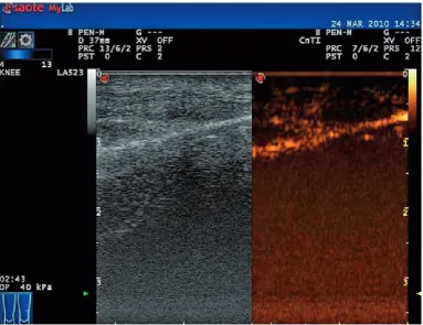
Fig. 5. Longitudinal section through the knee joint. Image on the left was obtained using traditional ultrasonography. Image on the right was obtained using sulphur hexafluoride intravenous contrast medium
S. Jeka et al.
784
CrossXBeam CRI
This option is an attempt to improve the qual-ity of imaging from a different side than in the case of the previously-discussed techniques. In both of these cases, one of the advantages was to remove noise – once by reducing the signal strength and increasing the frequency wavelength registered by the head, and the second time by digital image analysis.
In the case study using the CRI option, ultra-sound beams are emitted at different angles from the head. The appearance of noise and artifacts will vary depending on the angle of the ultrasound beam. The image is formed on the basis of a signal from multiple beams – even 11 beams can be used in this technique nowadays. Improvement in the picture quality is achieved by a clearer distinction between the tissues in this study, and greater vis-ibility of the edges contained in these structures.
In addition, this option can be flawlessly com-bined with harmonic imaging and the noise reduc-tion opreduc-tion – SRI.
Contrast Agents
The use of contrast has long been common in imaging studies – even in studies with such very high image quality as computed tomography or magnetic resonance imaging. For many reasons, among others because of the manner of obtaining the image and the availability of ultrasound, con-trast agents used in ultrasound must be completely different than the specificity of contrast agents used for example in computer tomography and magnetic resonance imaging.
The development of contrast agents proceeded quite differently and for a long time they did not have great use in ultrasonography. This was mainly due to the short duration of the maintenance of the contrast agents in the bloodstream which did not allow for accurate performance testing.
Only second-generation contrast agents made it possible to carry out a thorough ultrasound examination [7, 9]. Compared to the first con-trast agents, which were nothing more than gas bubbles, the second-generation contrast agents are the product of advanced chemistry laborato-ries. The gas bubbles that were used during the 60s of the twentieth century have been replaced by phospholipid membranes, with the gas placed
inside these membranes. Such a structure of bubbles, whose dimensions are about 3 microns, significantly improves their stability. They are much more resistant to environmental factors, like changes in pressure in the circulatory system. This has resulted in an increase in the time you can do the test with their use to several minutes. There is no longer insufficient time to perform a thorough ultrasound examination – including an assessment of blood flow within the synovial membrane, which, as already mentioned at the beginning, is often in the early stages of develop-ment of rheumatic diseases [10].
The resistance of the new generation of con-trast agents to mechanical damage, as a result of pressure changes caused by the passage of ultra-sound or a change in blood pressure, also allows for wider use of them. When properly selected, ultrasonic frequencies emitted by the head can have non-linear oscillations (vibrations) forced on them, which causes them to become a source of the second harmonic wave which we mentioned ear-lier. This allows for even better picture quality.
Conclusions
The development of ultrasound techniques have certainly contributed to the growth of appli-cations of this type of research in different fields of medicine. This implies, however, the need for the continuous upgrading of knowledge by the doctors performing the tests, in order to use the options that are best for the type of test. Even harmonic imaging is a very good option for imaging of su-perficial structures, parts of organs such as the nip-ples, testicles, salivary glands or thyroid. However, it is no longer the best choice for deep-lying organs such as kidneys, spleen and bladder.
References
[1] Jeka S, Murawska A: Ultrasonografia błony maziowej w chorobach reumatycznych. Reumatologia 2009, 47, 6, 339–343.
[2] Korkosz M, Wojciechowski W, Kapuścińska K et al.: Niskopolowy rezonans magnetyczny i ultrasonografia wy-sokiej rozdzielczości nadgarstka, stawów śródręczno-paliczkowych i międzypaliczkowych bliższych rąk oraz prze-ciwciała antycytrulinowe i czynniki reumatoidalne w rozpoznaniu reumatoidalnego zapalenia stawów u pacjentów z niezróżnicowanym zapaleniem wielostawowym. Reumatologia 2009, 47, 51–59.
[3] Filippucci E, Meenagh G, Epis O et al.: Ultrasound imaging for the rheumatologist. XIII. New trends. Three-dimensional ultrasonography. Clin Exp Rheumatol 2008, 26(1), 1–4.
[4] Kielich S: Molekularna optyka nieliniowa. PWN, Warszawa–Poznań 1977.
[5] Berry M, Howdhury V, Suri S: Diagnostic Radiology – Advances in Imaging Technology, Jaypee Brothers Medical Publishers, New Delhi 2005.
[6] Słapa R: Nowoczesne techniki ultrasonograficzne w badaniach tarczycy. Ultrasonografia 2009, 38(9).
[7] Jeka S, Sokólska E, Ignaczak P, Dura M: Nowoczesne techniki ultrasonograficzne obrazowania błony maziowej w chorobach reumatycznych. Roczn Pom Akad Med Szczecin 2010, 56, suppl. 1, 16–24.
[8] Mikowski A, Yadong L, Becker D, Ishark SO: Speckle Reduction Imaging, GE Medical Systems Ultrasound.
[9] Jakubowski W: Postępy w ultrasonograficznych środkach kontrastujących (UŚK). Ultrasonografia 2004, 15.
[10] Madej T, Kolarz B, Stępniak C et al.: Ultrasonograficzne środki kontrastujące w ocenie aktywności procesu za-palnego w obrębie stawów i pochewek ścięgnistych rąk u chorych na reumatoidalne zapalenie stawów. Ultrasono-grafia 2007, 31, 85–89.
[11] Kane D, Grassi W, Sturrock R, Balint PV et al.: A brief history of musculoskeletal ultrasound: From bats and ships to babies and hips. Rheumatology 2004, 43, 931–933.
Address for correspondence:
Sławomir Jeka
Clinical Division of Rheumatology and Connective Tissue Diseases Dr. J. Biziel 2nd University Hospital in Bydgoszcz
Medical College of Nicolaus Copernicus University Ujejskiego 75
85-168 Bydgoszcz Poland
E-mail: [email protected]
Conflict of interest: None declared