Cochlear hair cell fate determination and
differentiation
in vitro
JUSTIN NEIL MACDONALD WEIR
115 Pembroke Road,
Bristol, BS8 3EU.
BSc, MB BS, FRCS
Charing Cross and Westminster Medical School
ProQuest Number: U642221
All rights reserved
INFORMATION TO ALL USERS
The quality of this reproduction is dependent upon the quality of the copy submitted. In the unlikely event that the author did not send a complete manuscript and there are missing pages, these will be noted. Also, if material had to be removed,
a note will indicate the deletion.
uest.
ProQuest U642221
Published by ProQuest LLC(2015). Copyright of the Dissertation is held by the Author.
All rights reserved.
This work is protected against unauthorized copying under Title 17, United States Code. Microform Edition © ProQuest LLC.
ProQuest LLC
789 East Eisenhower Parkway P.O. Box 1346
SUMMARY
Mammalian cochlear hair cells are relatively inaccessible and few in number. This
hampers any research on their fate determination and differentiation. The production
of conditionally immortal cell lines from the H2K^/5À58 transgenic mouse should
overcome these difficulties.
The aims of the present study were threefold. Firstly, to establish that the cell lines
provide a viable in vitro system, by examining the pattern o f molecular expression in
the cochlear hair cell line UB/OC-1. Secondly, to examine differentiation by using
clonal derivatives from the heterogeneous cell line UB/OC-1. Thirdly, to explore the
process of lateral specification in the determination o f cell fate and to explain the
differentiation o f hair cells and supporting cells from a common precursor.
The methods used were cell culture, immunocytochemistry, reverse transcription
polymerase chain reaction and western blot. The results demonstrated that firstly; the
temporal expression pattern of Brn3.1, an essential transcription factor required for
hair cell differentiation, and the a9 subunit of the nicotinic acetylcholine receptor,
followed a similar pattern to that during normal development. Secondly, epithelial cell
markers such as, vimentin, cytokeratin, actin and cadherin, and specific hair cell
markers such as myosinVIIA and fimbrin were expressed when the hair cells
differentiated. The pattern o f expression suggested parallel pathways of gene
expression during differentiation of hair cells. Thirdly, from the expression o f
Numblike, Notch 1, Jagged 1 and jagged!, factors which are known to be involved in
The results also demonstrate the muph greater experimental flexibility offered by cell
lines in understanding hair cell development. Future studies will focus on functional
A C K N O W L E D G E M E N T S
I wish to thank Dr. Matthew Holley for giving me the opportunity for performing this
research in his laboratory at the Department of Physiology, Bristol University and for
his constant supervision and inspiration. I also wish to thank Dr. Marcelo Rivolta and
Dr. Patrick Lawlor for showing me the techniques o f cell culture, RT-PCR, western
blot and film development and particularly for putting up with my constant
questioning. Professor Tony Wright for his supervision of the writing of the thesis,
and to all those who kindly donated antibodies. Finally to my wife, Carrie, for her
constant encouragement and support over the last eighteen months.
I performed all the experimental procedures used in this study, of cell culture, mRNA
and protein extraction, cDNA synthesis, RT-PCR, western blot and black and white
C O N TEN T S
Page
Summary 2
Acknowledgements 4
Contents 5
Figures 7
Chapter One. Introduction
1.1 Deafness; the Problem 9
1.2 Structure of the mammalian cochlea 11
1.3 Hair cell contact 14
1.4 Hair cell structure 15
1.5 Supporting cell structure 17
1.6 Molecular development of the murine cochlea 18
1.7 Hair cell fate determination 21
1.8 Hair cell differentiation 26
1.9 a 9 acetylcholine receptor subunit 28
1.10 Regeneration of hair cells 29
1.11 Cochlear preparations 30
1.12 Production o f cell lines 32
Chapter Two. Methods
2.1 Cell culture 38
2.2 Immunocytochemistry 39
2.3 Reverse Transcription Polymerase Chain Reaction 40
2.4 Western Blot 44
Chapter Three. Results
3.1 Temporal expression of Brn3.1 and a9AChR in UB/OC-1 47
3.1.1 Expression o f BrnS. 1 47
3.1.2 Expression of a9AChR 47
3.2 Cloning UB/OC-1 to test for clonality and generate Brn3.1 negative 49
cell lines
3.3 Characterisation o f clones #14, #15 and #24 54
3.3.1 Epithelial characteristics 54
3.3.2 Hair cell characteristics 59
3.4 Characterisation o f the clones in relation to the mechanism of 64
lateral specification
Chapter Four. Discussion 70
4.1 Bm 3.1 and a9AChR expression under differentiating conditions 71
4.2 Cloning UB/OC-1 to test for clonality and generate Bm 3.1 negative 73
cell lines
4.3 Characterisation of clones #14, #15 and #24 74
4.4 Characterisation o f the clones in relation to the mechanism of lateral 80
4.5 Critical analysis o f the use of cell lines in determining cochlea 84
hair cell differentiation
4.6 Conclusion 86
References 88
FIGURES
Chapter One
1.1 Gross anatomy of the mammalian inner ear 10
1.2 The adult organ of Corti 12
1.3 Epithelial junctions 13
1.4 Development of the mouse inner ear from E. 10.75 to E. 17 19
1.5 Experimental Design 3 5
Chapter Two
2.1 Concentration of reagents used in RT-PCR reactions 42
2.2 Conditions o f each RT-PCR reaction 43
Chapter Three
3 .1 BrnS.l and o9AChR RT-PCR reactions 48
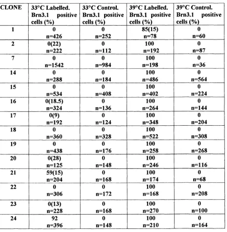
3.2 Table of clones and labelling with Bm3.1 50
3.3 Phase contrast pictures o f clones #14, #15 and #24 at 33°C and 39°C 51
3.4 Brn3.1 immunolabelling of #14, #15 and #24 at 33°C and 39°C 52
3.5 Vimentin, actin, pan-cadherin and pan-cytokeratin immunolabelling of 55
#14, #15 and #24 at 33°C and 39°C
3.7 Brn3.1 western blot 61
3.8 Myosin VIIA western blot 62
3.9 Fimbrin western blot 63
3.10 Notch 1 western blot 65
3.11 Jagged 1 western blot 66
3.12 Numblike western blot 67
3.13 OCP-2 western blot 68
3.14 Summary table of protein and gene expression in #14, #15 and #24 69
Chapter Four
4.1 In vivo and in vitro expression of genes and proteins 70
analysed in this study
CHAPTER ONE
INTRODUCTION
1.1 Deafness; the Problem
Deafness remains one of the great challenges of the century. The morbidity
associated with sensorineural hearing loss ranges from minor annoyance to the
impairment of learning, social interaction and safety. Causative factors responsible for
this affliction are varied, and include genetic defects, drugs, toxins, infectious agents
and the normal ageing process. With approximately 10% o f the global population
affected (Rubel, 1997) and the knowledge that every second person over the age of 75
will to some degree be deaf (U.S. Department of Health and Human Services, 1994),
the need to understand the ontogeny of the auditory system is paramount. For this will
provide a basis to understanding the necessary biological mechanisms that will one
day ensure that deafness is defeated.
In order to place this work in context in understanding the molecular biology o f
development within the cochlea it is necessary to give a brief overview o f the
structure o f the mammalian cochlea and the specialised characteristics of sensory hair
cells. This is followed by a review of cochlear development and in particular hair cell
fate determination and differentiation. The issue of hair cell regeneration is briefly
discussed, as are current methodologies for in vitro preparations. Finally, the use of
1.2 Structure of the mammalian cochlea.
The vertebrate inner ear is a complex sensory organ responsible for the senses of
balance and hearing (Fig. 1.1). It is composed o f a bony labyrinth, the otic capsule
that encases the membranous labyrinth, which contains the cellular structures. These
structures include a closed epithelial layer that is diversified into specific regions
containing the sensory-transducing cells, the sensory hair cells, as well as supporting
and secretory cell types. Hair cells are specialised mechano-receptors organised in a
sophisticated topographical pattern. Within the cochlea they are surrounded by
perilymph and covered at their apex by endolymph. The potassium-rich endolymph,
produced by the stria vascularis, is essential for the process of transduction by the hair
cells. The endolymph contains ions, which provide the necessary voltage and ionic
gradient that can change the resting potential o f the hair cell. This in turn results in
transmitter release from the hair cell and excitation o f the afferent nerve fibres. The
perilymph, within the scala vestibuli and scala tympani, is separated from the
endolymph, within the scala media, by tight junctions between adjacent epithelial
cells. The organ o f Corti, which contains the specialised epithelial cells; the hair cells
and supporting cells, spirals along the basilar membrane (BM), between the apex and
the base o f the cochlea (Fig. 1.2).
In the human/mouse there are approximately 16000/3000 cochlea hair cells
respectively (Echteler et a l 1994) in each ear, which are separated from one another
by various types of supporting cells. The organ of Corti contains one row of inner hair
cells (IHCs) medially and three rows of outer hair cells (OHCs) laterally. Hair cells
and their stereocilia (Echteler et a l 1994) increase in length between the base and the
FIGURE.1.1
Gross anatomy of the mammalian inner ear. Depicted in detail is the membranous
labyrinth, a connected system of ducts and pouches that contains the vestibular and
auditory end organs. Surrounding the membranous labyrinth is the bony labyrinth.
The arrows indicate the direction of flow of perilymph following inward direction of
the oval window.
Subdural sp a c e Subaracbnold sp a c e
Endolymphatic sa c Endolymphatic duct
Perilymphatic d uct Saccule
Helicotrem a
S cala vestibuli
S cala m edia S cala tympani
D uctus reuniens
C ochlea
m eatus Auditory tube
(Source; Echteler, et al. 1994. Structure of the mammalian cochlea. In Comparative
FIGURE.1.2
The adult organ of Corti. The inner hair cells (IHC) and three rows of outer hair cells
(OHC), along with the inner pillar (IP), outer pillar (OP), Deiters (D), Hensen (H),
and Claudius (C) cells rest on the basilar membrane (BM). The tectorial membrane
(TM) covers the apical surface of the sensory and supporting cells. The scala vestibuli
(SV), and scala tympani (ST) are filled with perilymph. The scala media contains
endolymph and its boundaries include the reticular lamina of the organ of Corti,
Reissner’s membrane (RM), and the lateral wall made up of the stria vascularis
(St.V), the spiral ligament (SpL), and the spiral prominence (SP).
(Source; Slepecky, N. 1996. Structure of the mammalian cochlea. In: The Cochlea.
These morphological characteristics, in particular the stiffness of the BM, relate to the
tuning capacities and sensitivities that enable such a diverse frequency range to be
detected from high frequencies in the base to low frequencies in the apex. Tight
junctions between hair cells and supporting cells enable them to separate the
endolymph from the perilymph. Other junctions include zonula adherens junctions,
desmosomes and gap junctions (Slepecky N. 1996). Tight junctions and zonula
adherens junctions are present at the apical surface of the cells, whilst desmosomes
and gap junctions are found along the basolateral surfaces (Fig. 1.3).
FIGURE.1.3
Diagram showing the four principal types of intercellular junction that occur between
two simple epithelial cells.
Source; Garrod and Collins. 1992. Intercellular junctions and cell adhesion in epithelial cells. In: Epithelial organisation and Development. Fleming (ed). 1-35. Cliapman&Hall, London.
Apical surface
Tight junction
Zonula adherens
_ Desmosome
Gap junction
Intercellular junctions play an integral part in determining the polarity o f epithelial
cells. In turn, epithelial polarity influences not only membrane topography and
composition, but also the internal organisation of the cell, including that of the
membrane skeleton and the cytoskeleton.
Tight junctions (zonula occludens) have two major functions: firstly, they create a
barrier to the diffusion o f membrane molecules in the plane o f the lipid bilayer, and
secondly, they seal the intercellular space preventing the passage of electrolytes and
macromolecules between the apical and basolateral surfaces o f the epithelium. In the
organ o f Corti they create a barrier between endolymph and perilymph. They are
found between the hair cells and supporting cells and between supporting cells. A
number of proteins associated with the cytoplasmic aspect of tight junctions have
been identified in the organ of Corti, namely, Zonula Occludens 1 (ZO-1) (Stevenson
et a l, 1986), Cingulin (Citi et a l, 1988, Raphael and Altschuler 1991).
Adherens junctions are found in a sub-apical ring beneath the tight junctions
physically binding the cells together and mediating intercellular communication. They
have three domains, cytoplasmic, intramembrane, and extracellular. At the
cytoplasmic domain, the junction is linked to the intracellular actin cytoskeleton,
which forms a circumferential ring within the cell, by vinculin (Geiger et al., 1985)
and a-actinin (Lazarides and Burridge, 1975). The intramembrane and extracellular
domains are composed of adhesive molecules from the cadherin family o f calcium-
dependent glycoproteins (Takeichi, 1988, 1990). This family includes E-cadherin
(uvomorulin) (Yoshida and Takeichi, 1982), N-cadherin (Hatta et al., 1987), P-
cadherin (Nose et a l, 1987), and M-cadherin (Donalies et al., 1991). E-cadherin is
expressed in mature supporting cell/supporting cell adherens junctions (Whitlon,
cells and supporting cells and in supporting cell/supporting cell junctions (Raphael et
a l, 1988).
In the organ of Corti, desmosomes (macula adherens) are not found between hair cells
and supporting cells (Gulley and Reese, 1976). Likewise, gap junctions, which are
composed of connexins, are not found between mature hair cells and supporting cells.
Although present in the otocyst, they are subsequently lost prior to development of
hair cells in the chick (Forge, 1997). However, gap junction-like structures have been
noted at the lower boundaries o f the tight junctions between hair cells and supporting
cells (Nadol, 1978). Recently, the gene encoding the gap-junction protein connexin
26 has been liked to the autosomal recessive non-syndromic sensorineural deafness
(DFNBl) (Kelsell e ta l, 1997).
1,4 Hair Cell Structure
Intermediate filaments (IF), microfilaments and microtubules form an intracellular
structural framework called the cytoskeleton. IF have been studied extensively and
provide useful markers for cell differentiation as their expression pattern tends to be
tissue specific. The diameter of an IF is 8-12nm and falls between that of the smaller
actin filaments and larger microtubules (Steinert et a l, 1984). IF have been divided
into six classes based on differences in their solubility, antigenicity and
electrophoretic mobility (Lazarides, 1982; Wang et a l, 1985). The currently
recognised classes of IF are desmin, vimentin, cytokeratins, neurofilaments, nestin
and glial filaments. Within the inner ear, in particular the hair cells, the expression of
IF’s cytokeratin and vimentin is variable. Kuijpers et a l (1992) demonstrated that in
the rat inner ear the expression of vimentin decreased in hair cells from embryonic
cells and mesenchyme with trace amounts present in the adult hair cells. Similarly, the
limited expression o f cytokeratin in the developing sehsory epithelia decreases after
E l 2.5 (Kuijpers et a l, 1991b). The transient co-expression o f vimentin and
cytokeratin within the developing epithelia may play a role in differentiation and can
be used as an early marker of differentiation (Viebahn, et a l, 1997). Glial filaments
and desmin have not been observed at any point in the development o f hair cells in the
guinea pig, rat or human (Raphael, 1987; Kuijpers et a l, 1991b; Oesterle et a l, 1990).
More than most other cells, hair cells rely on actin. Actin is present in at least four
regions of the hair cell (Flock and Cheung, 1977; Slepecky and Chamberlin, 1982;
Drenckhahn et a l, 1991). The majority of actin is found in the stereocilia, where it is
present as bundles of parallel filaments that extend into the cuticular plate. Apically, it
forms a circumferential ring of filaments associated with the adherens junctions at the
reticular lamina. It is also a component of the cortical lattice just inside the cell
membrane along the lateral wall and it is found in the cytoplasm (Holley et a l, 1992).
Each o f these regions has different actin-binding proteins which include profilin
(Flock et al., 1982), tropomyosin (Slepecky and Chamberlin, 1985b), spectrin (Holley
and Ashmore, 1990; Ylikoski et a l, 1990), a-actinin (Slepecky and Savage, 1994),
and fimbrin (Matsudaira et a l, 1983). Co-localisation studies show that fimbrin cross
links the highly organised parallel actin filaments in the stereocilia o f both IHC’s and
OHC’s (Drenckhahn et a l, 1991). This cross-linking contributes to the stiffness o f the
stereocilia. In the rat, fimbrin is first noted at E l 8 in IHCs and by birth it is present in
OHCs (Zine et a l, 1995). It is an essential part of the formation and ontogenesis of
stereocilia.
The isoenzyme myosin VIIA, an unconventional myosin, is another important protein
maintains the structural integrity of the hair bundles by cross-linking between
adjacent stereocilia rootlets within the cuticular plate and is also found in the
pericuticular necklace and cell body (Hasson et al., 1997). If myosin VIIA is non
functional as in the shaker-1 mutant mouse (Gibson et a l, 1995) then the stereocilia
are disrupted and deafness and vestibular dysfunction ensues. In the human, mutations
o f the myosin VIIA gene cause Usher syndrome IB (Weil et a l, 1995) and the non-
syndromic deafnesses DFNB2 and DFNAl 1 (Weil et a l, 1997; Lui et a l, 1997c). A
developmental study o f myosin VIIA showed that its’ mRNA is first expressed at E9
in the otic vesicle (Sahly et a l, 1997). At this stage, the presumptive sensory hair cells
and supporting cells, both of which develop microvilli, cannot be distinguished. As
cell differentiation proceeds, myosin VIIA expression is restricted to the cochlea and
vestibular hair cells. It can, along with fimbrin, be used as a specific hair cell marker
as it is not found in supporting cells.
1.5 Supporting Cell Structure
In supporting cells, organised bundles o f microtubules and actin filaments pass from
the reticular lamina to the BM. These are thought to play a role in mechanical support
and coupling o f the BM to the reticular lamina. Both cytokeratin and vimentin have
been shown to be present in adult supporting cells (Raphael et a l, 1987; Bauwens et
a l, 1991; Kuijpers et a l, 1991b). This is unusual as it is rare to find these two IF in
the same cell and vimentin is normally only found in cells o f mesenchymal origin.
Specific proteins that are found only in supporting cells include the calcium binding
protein S-100 (Foster et a l, 1994; Pack and Slepecky, 1995), and organ o f Corti
regulate in some manner the intracellular or transcellular flux in the supporting
cells.
1.6 Molecular development of the murine cochlea
The inner ear develops from a thickened patch o f ectoderm called the otic placode,
which invaginates by E9 to form the otic vesicle or otocyst (Noden and Van De
Water, 1986) (Fig. 1.4). The epithelium o f the otic placode also gives rise to the
primary neurons of the statoacoustic ganglion or what is later termed the
otic/cochleovestibular ganglion. This can be watched by time lapse videomicroscopy
in the transparent zebrafrsh embryo where the neuroblasts crawl out individually from
the ventral region o f the otocyst epithelium into the connective tissue below (Haddon
and Lewis, 1996). It is thought that placodes originally evolved from primordial
primary sensory cells into primordia. Primordia contain both sensory cells and ganglia
that connect these axonless sensory cells to the brain (Fritzsch, 1993). Development
o f the otocyst depends on gene expression in the rhombencephalon (the hindbrain),
the nonneural portion o f the otocyst, the neuronal precursors derived from the placode
and the neuronal, glial, schwann sheath and melanocyte precursors derived from the
neural crest (Fritzsch et a l, 1997). Many o f these genes are required for axis
specification, segmentation, and body plan patterning (Keynes and Krumlauf, 1994).
Such a bewildering array o f genes include the important homeobox genes which are
thought to play a determinant role in the development o f the hindbrain, branchial
arches, vertebrae, and limbs (Keynes and Krumlauf, 1994). They are so named as they
encode transcription factors which contain a highly conserved sixty amino acid DNA-
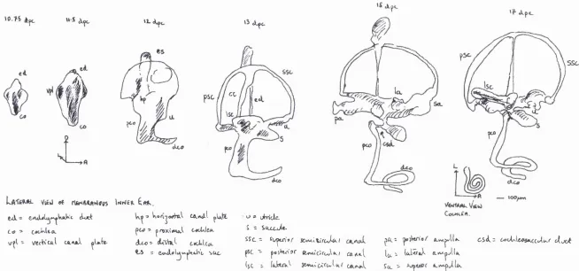
FIGURE. 1.4
Schematic representation of the developing inner ear of the mouse from E. 10.75 to E. 17. (Source; Morsli et a i, 1998).
u s
I
11. ck
es
134 '
ftjL " t Auct WoK^oWfO.^ CA.*»<rl pW lt
C<o •> ■> ^ n 9 X ( M A .l
~ V&fKc&l ocAJii dLco"
e.3 : sue.
Ct
psc
'■> » cAn'cJlc- S 2 S o x c jU
-SSC 2 Sv/ytn'*/" iCvM-iiircvli*-f
- poiW isr awcirovVfc.f (CftAo-i
SSC-la.
| ) R . 2 d L v ^ | » v U ^ csif = 6ooUUo5<ka«/(<^/’ o(o>o^
Homeobox (Hox) genes that are involved in the development of the otocyst include;
H oxal (Lufkin et a i, 1991; Chisaka et a l, 1992); distal-less {dlx-5) (Ekker et a l,
1992); those related to Drosophila muscle segment homeobox genes (Msx-1, Msx-2)
(Suzuki et a l, 1991; Davidson, 1995); NK- related homeobox genes (NkxS.l, Nkx5.2)
(Rinkwitz-Brandt et a l, 1995); Drosophila orthdenticle related genes {Otxl and Otx2
(Simeone et a l, 1993) and paired-box transcription factors {Pax2 and Pax2) (Nomes
et a l, 1990; Hoth et a l, 1993; reviewed in Rivolta, 1997).
Secreted factors such as wingless-int (Wnt) (Parr et al, 1993), fibroblast growth
factors (fgfS) (Wilkinson et a l, 1989), and bone morphogenetic protein (Bmp) (Oh et
a l, 1996) are also expressed at the otocyst stage. Similarly, the zinc finger genes;
Msal, the mouse homologue of the spalt gene from Drosophila (Ott et a l, 1996),
GATA-3 (George et a l, 1994) and the thyroid hormone receptors (TRal, TRa2,TRpl
and TRp2) (Bradley et a l, 1994) are all expressed in the otocyst. The combinatory
action of all these genes is to determine the axis orientation {dlx-3, fgf3), proliferation
ifgf3), formation of the inner ear {Hoxal, Pax2, Msx-1, Msx-2, Otxl, and Nkx5.1) and
cranial nerves VII,
Vni
{Hoxal), and migration o f neural crest precursors {Pax3).Axis orientation in the anteroposterior, dorsoventral and medial lateral planes relies
on the close spatial proximity o f the hindbrain and the subsequent gene expression as
described above. The cells within the otocyst then undergo proliferation and
segregation o f the sensory epithelia. The cochlea and saccule are derived from the
ventral half o f the otocyst at E l l , whilst the semicircular canals and utricle are
derived from the dorsal half (Li et a l, 1978) (Fig. 1.4). Only after E l 1.5 is the otocyst
completely autonomous in its differentiation (Van De Water, 1983). It appears that
from studies conducted by Li and McPhee (1979) and Van De Water (1983) that the
such as insulin and insulin-like growth factors (IGFs) derived from the mesenchyme
cause mitogenesis in the epithelium (Leon et al., 1995b). In particular IGF-1 appears
to act on restricted areas of the epithelium, which may account for the observed
regional differences in growth rate. Associated with the mitogenic activity o f IGF-1 is
the rapid induction of c-fos (Leon et al., 1995a). c-fos is an essential element in the
propagation o f mitogenic signals regulating the expression of secondary genes, which
eventually lead to cell division (Morgan and Curran, 1991). In contrast, low
concentrations o f retinoic acid (RA) have been shown to inhibit cell proliferation and
stimulate sensory and secretory cell differentiation (Represa el a l, 1990). Kelley et al.
(1993) provided further supporting evidence for this role. They demonstrated that
exogenous RA added to the otocyst could produce supemumary hair cells.
Importantly, if the RA was added at the time o f terminal mitosis then more hair cells
and supporting cells were generated.
1.7 Hair cell fate determination
Relatively little is known about the exact nature of events that leads to a particular cell
fate in the inner ear. However, recent research is beginning to unravel this complex
story. The initial steps of axis orientation and compartmentalisation in the otocyst
appear to be crucial for subsequent cell fate specification (Fekete, 1996). Studies in
mutant zebrafish, such as dog-eared, colourless and van gogh (Whitfield et a l, 1996)
have shown that hair cells are able to form in grossly malformed ears that lack any
obvious morphological compartments. This suggests that sensory patch specification
does occur before gross morphogenesis. In the otocyst, the differential expression of
genes at the compartment boundaries probably accounts for the distribution o f the
become the cristae, whereas those in the ventral half will become the maculae and
cochlear duct.
As yet in the mammalian inner ear, there is no data on how the fate o f the sensory cell
progenitors is determined. However, research on neurogenesis (Henrique et al., 1997;
Lewis, 1996 for review), myogenesis (Kopan et a l, 1994) and haematopoesis (Li et
a l, 1998) has led to an understanding o f some of the mechanisms involved. In
neurogenesis of Drosophila, the first step to take place is the switching on of
proneural genes in a cluster o f cells. These genes form the achaete-scute complex
(Ghysen and Dambly-Chaudiere, 1988) and endow the cells that express them with
the potential to form neural precursors. The achaete-scute complex, termed Mash
{mammalian achaete-scute homolog) in mammals (Johnson et a l, 1990), encode
transcriptional regulators of the basic helix-loop-helix (bHLH) type. Another
important bHLH gene is atonal {Math, mammalian atonal homolog, in mammals
(Akazawa et al., 1995)). In Drosophila, achaete-scute and atonal do not only act as
proneural genes for their respective lineages but they also help to determine the type
o f sensory organ developed ie; either an external sensory organ (sensory bristle) or
photoreceptor (Jarman et a l, 1993b). Once the proneural cluster has been determined
and the particular bHLH genes switched on then the process by which one cell within
the cluster is singled out to follow a particular fate depends on both extrinsic and
intrinsic mechanisms.
The intrinsic mechanism, at least in part, relies on the gene numb (Uemura et a l,
1989; Rhyu et al, 1994). Numb protein is membrane associated and asymmetrically
localised in the shape of a crescent in the sensory organ precursor of Drosophila prior
to cell division. Upon division. Numb protein is preferentially segregated into one of
mutants have shown that cell fate can be altered by changing the level of expression
o f numb. Furthermore, numb is able to generate asymmetry in the subsequent division
of the daughter cell. This appears to be a general feature o f asymmetric cell divisions
in the fly nervous system. In the mouse, Zhong et a i, (1996) have found a numb gene
that shares a high degree of sequence homology with fly numb. Mouse Numb protein
is also membrane associated prior to cell division, but unlike fly Numb it can be
distributed either symmetrically or asymmetrically in the daughter cells. This
difference may reflect the different strategies the fly and mouse have adopted during
evolution to fulfil neurogenesis. In the fly, except in the eye, all the neural precursors
undergo asymmetric cell divisions to generate neurons. In the mouse, neural
precursors undergo symmetric cell division to increase the size of the precursor pool
before undergoing asymmetric cell division to generate neurons (Anderson and Jan,
1997). Mouse numblike, which also has significant sequence similarity to Drosophila
numb has been found to be symmetrically distributed in dividing neural precursors
(Zhong et al., 1997). Nevertheless, its action is similar to that of numb which is to
antagonise Notch signalling activity.
The extrinsic mechanism relies on the interactions between members o f the Notch and
Delta/Serrate/Lag family of proteins (Hartenstein and Posakony, 1990; Goriely et a l,
1991; Heitzler et al, 1991). Notch 1-4 are cell surface receptors that are activated
through direct cell surface contact with a ligand expressing cell.
Deltal,2,3/Serrate 1,2/Jagged 1,2 are the ligands (Dunwoodie et a l, 1997; Lindsell et
al, 1995). Notch/ligand (eg. Jagged) are the mediators o f “lateral
inhibition/specification" (Greenwald, 1998). This is a critical signalling system that
enables one cell to follow a particular fate, whilst its neighbours follow a different
differentiation within that cell. It appears to be a universal system in possibly all
multicellular organisms in a variety o f developmental processes (Chitnis et al., 1995).
Once Notch has been activated its intracellular domain is cleaved by a protease
(Schroeter et a l, 1998). This domain then interacts with a transcription factor named
Suppressor o f Hairless (Su[H]) in Drosophila and RBP-Jk/KBF2/CBF1 in mammals;
collectively known as the CSL proteins ( Tamura et al., 1995; Jzirriault et al., 1995).
Prior to induction by Notch, the CSL proteins interact with a corepressor complex
containing SMRT (Silencing Mediator of Retinoid and Thyroid hormone receptors)
and the hi stone deacetylase HDAC-1. Notch disrupts the formation of the repressor
complex such that the CSL protein now becomes an activator o f transcription (Kao et
a l, 1998). The Notch/CSL protein complex drives the transcription o f a group of
related genes known as Enhancer o f split (E[spl]) in Drosophila and Hes (Homolog
enhancer o f split) in mammals (Bailey and Posakony, 1995; Lecourtois and
Schweisguth, 1995). These genes encode bHLH transcription factors which combine
with a protein called Groucho to create a repressor (Paroush et a l, 1994). Finally, the
JTgjbHLH/Groucho protein complex represses genes of the achaete-scute complex
(Oellers et a l, 1994; Heitzler et a l, 1996) thereby blocking differentiation. As the Ac-
Sc proteins activate the expression o f Delta (Kunisch et ah, 1994), this feedback loop
operates to repress Delta expression in the cell in which Notch has been activated.
Thus, lateral specification appears to be regulated through the Notch —> RBP-J -^HES
-> Math pathway. Furthermore, Notch signalling through the expression o f HES can
also upregulate the expression o f Notch itself (Weinmaster, 1998). This positive
regulation o f Notch would serve to reinforce the cell’s responsiveness to Notch
Taken together with the activity of numb, Jan and Jan (1995) have proposed that
Notch mediated signalling is influenced by numb at each successive step in the
sensory organ precursor (SOP) lineage. Essentially by antagonising Notch, numb
creates a bias between sister cells that signal each other. They suggest this as a
mechanism to increase the reliability o f Notch-mediated signalling in situations
requiring rapid decision making, when there may be insufficient time to activate
transcription-based feedback mechanisms. The mechanism by which Numb
antagonises Notch is not yet known, although one possibility is that Numb interferes
with the interaction o f Notch with RBP-J (Frise et a l, 1996).
Other proteins also modulate the activity of Notch and its ligands. This has been
found during wing formation in Drosophila where activation of the ligands. Serrate
and Delta, is modulated by Fringe (Irvine and Wieschaus, 1994). Fringe can inhibit
Serrate signalling through Notch yet enhance Delta activated Notch in a cell
autonomous fashion (Panin et a l, 1997). This differential signalling through Notch is
crucial in establishing the dorsal-ventral boundary o f the wing margin, which is
required for morphogenesis and patterning o f the wing. The mammalian homologues
of fringe; lunatic, manic and radical fringe have recently been isolated (Johnston et
a l, 1997; Cohen et a l, 1997). Lunatic fringe is required for boundary formation
(Zhang and Gridley, 1998; Evrard et a l 1998) and symmetric patterning. In the murine
inner ear, lunatic fringe was first expressed at E9.5 in the anteroventral aspect o f the
otocyst (Morsli et a l, 1998). By postnatal day 1 (PI) lunatic fringe transcripts were
restricted to the supporting cells underneath the inner and outer hair cells. Morsli et a l
(1998) suggested that fringe plays a similar role in the inner ear by specifying
The mRNA expression of notchl, jaggedl and jagged2 in the cochlea has been
determined by Lanford et al. (1998). At E l 3, notchl was distributed widely
throughout the cochlear epithelium, was also expressed at E13 whilst jagged
2 was not expressed until E l 5. Their expression was localised to a narrow band that
ran from the base to the apex o f the cochlea. This band o f expression migrated from
the neural edge of the epithelium to the abneural edge. By P3, jaggedl was restricted
to supporting cells whilst jaggedl was found only in hair cells. It would appear that
the mechanism o f lateral specification is critical for hair cell determination and
depends, as it does in neural development, on the activity o f Notch, its ligands and
their modulators.
1.8 Hair cell difTerentiation
Once the fate of a sensory precursor cell within the epithelium has been determined it
then undergoes differentiation. Initiation o f differentiation in the cochlea begins at
around E13 in the base and progresses towards the apex by E l 5 (Ruben, 1967;
Nishida et a l, 1998). It occurs after terminal mitosis o f the entire population of
sensory precursor cells, which begins at E 12 in the apex and finishes at around E14 in
the base (Ruben 1967; Kelley et a l, 1993). One gene, B m S .l, is critical for hair cell
differentiation. It is a member of the POU-domain transcription factor family. This
family o f transcription factors was first found to play a critical role in neural
development (Treacy and Rosenfeld, 1992; Wegner et a l, 1993). The POU-domain
functions as a bipartite DNA-binding domain that contains a POU-specific domain o f
about 70 amino acids and a POU-homeodomain o f about 60 amino acids, joined by a
variable linker. It was first identified in the mammalian pituitary-specific Pit-1, the
al., 1988). The POU-domain family is divided into four classes. The class III factor
Brn-4/POU3F4 is expressed in the rat otic vesicle (Le Moine and Young, 1992;
Mathis et a l, 1992), and mutations in the corresponding human gene cause X-linked
progressive sensorineural deafness and stapes fixation (DFN3) (de Kok et al, 1995).
The class IV Ï2iCXor B m S.l/B m Sc/P 0U 4F 3 has been noted in sensory cell epithelium
at E13 (Erkman et al, 1996; Xiang et al, 1997; Ryan, 1997; Xiang et a l, 1998). A
null mutation of the gene leads to loss of only cochlear and vestibular hair cells,
leading to profound deafness and vestibular dysfunction. In the human, a small
deletion o f the Brn3.l coding region has been linked to the autosomal dominant
hearing loss DFNA15 (Vahava et a l, 1998).
Originally, it was thought that Bm3.1 was critical for the initial differentiation of the
hair cell phenotype (Ryan, 1997). However, recent work by Xiang et a l (1998) has
shown that in Brn3. FT mutants, hair cells undergo differentiation before degenerating
via apoptosis. Their data indicates that Brn3.1 is required for the maturation, survival
and proper positioning of hair cells, whereas it appears to play virtually no role in the
commitment and initial differentiation of hair cells. The downstream target of B m 3.1
is an important factor to determine, and as yet there has been no conclusive evidence
to suggest such a factor. However, Brn3 genes are known to have a regulatory effect
on the promoter o f some acetylcholine genes (Milton et a l, 1996). They demonstrated
that the regulatory region o f the nicotinic acetylcholine receptor a 2 subunit gene is
activated by Brn3.2 but not by Brn3.0 or Brn3.L Therefore, a good candidate for a
downstream target of B m 3.1 is the a 9 acetycholine receptor subunit found in IHC and
1.9 a9 acetycholine receptor subunit
The efferent innervation of the cochlea arises from neurons in the superior olivary
complex nuclei (Rasmussen, 1946). Lateral olivocochlear neurons project to the
ipsilateral cochlea and terminate on spiral ganglion cell dendrites under the IHCs.
Medial olivocochlear neurons project to the contralateral cochlea and terminate on the
bases o f OHCs (Warr, 1992 for review). Several neurotransmitters are associated with
efferent terminals including acetylcholine, y-aminobutyric acid (GABA), enkephalin,
dopamine, calcitonin gene related peptide (CGRP) and dynorphin (Hunter, 1992).
Acetylcholine is the predominant neurotransmitter for both medial and lateral
efferents (Housley and Ashmore, 1991; Eybalin, 1993).
Activation o f the acetylcholine receptor (AChR) mediates a fast influx of Ca^^ into
the OHC, which then activates an outwardly-directed Ca^^-dependant channel
(Sewell, 1997). The AChR is classified as a subunit of the neuronal nicotinic
receptors with mixed nicotinic-muscarinic pharmacology (Housley et al., 1992).
Elgoyhen et al. (1994) isolated the subunit from the rat and named it a9.
Recombinant DNA technology had already resulted in the identification of the
vertebrate muscle nAChR subunits a l , p i, y, 5, and 8, and the neuronal subunits a2,
a3, a4, a5, a6, a7, a8, P2, p3, and P4 (Housley and Ryan, 1997).
a9nAChR mRNA has been found by in situ hybridisation and single cell reverse
transcription-polymerase chain reaction in both OHC and IHC (Park et al., 1997;
Glowatzki, 1997). It first appears in the presumptive cochlear hair cells in the rat at
E18 and peaks at PIO (Luo et al., 1998). Its expression in IHC is not readily
explained, as they do not receive direct efferent innervation. However, transient
efferent innervation has been reported to occur during development (Lenoir et al.,
concentrated at the base of the hair cell (Park et a l, 1997). The a9nAChR is specific
for hair cells and can be used as a marker of hair cell differentiation.
1.10 Regeneration of hair cells
In mammals, cochlear hair cells are generated during embryogenesis, as described,
and must remain viable throughout the lifetime o f the organism to maintain normal
auditory function. Critically, they are not replaced when they are damaged or die
(Kelley et al., 1995; Corwin and Oberholtzer, 1997; Forge et al., 1998; Stone et al,
1998). In contrast to mammals, continuous post-embryonic hair cell production occurs
in fish, amphibians and birds (Corwin, 1985; Roberson et a l, 1992). Furthermore in
birds, hair cells within the cochlea were regenerated following aminoglycoside and
acoustic trauma (Cruz et a l, 1987; Cotanche, 1987). The reinitiation o f mitosis among
the supporting cells within the sensory epithelium gives rise to new hair cells (Corwin
and Cotanche, 1988; Ryals and Rubel 1988; Stone and Cotanche, 1994).
If it were feasible to regenerate or prevent degeneration o f mammalian hair cells then
an enormous amount of morbidity would be alleviated. This possibility was given
added impetus by the discovery of hair cell replacement in the vestibular sensory
epithelia o f mature mammals (Forge et al, 1993; Warchol et a l, 1993). Forge et a l
demonstrated the appearance of immature stereociliary bundles in situ in the
vestibular epithelia o f adult guinea pigs, which increased in number several weeks
after aminoglycoside induced hair cell loss. Complementary studies performed in
vitro with organotypic explants of utricular macula from mature guinea pigs
suggested that sensory epithelial cells can be induced to divide following
aminoglycoside induced damage (Warchol et a l, 1993). However, the exact
In non-mammalian species, hair cells have arisen directly from supporting cells
without an intervening mitotic event (Baird et al., 1993; Baird et al., 1996; Roberson
et al., 1996; Adler et al., 1997; Steyger et al., 1997). In mice, Kelley et a l (1995)
found that embryonic cochlear hair cells, and on rare occasions neonatal hair cells,
were replaced after laser microbeam irradiation. They suggested that supporting cells
were able to change their pre-existing fate in response to hair cell damage. This
supports the concept of lateral specification, for as the supporting cells are released
from this mechanism they become hair cells. In the adult guinea pig. Forge et a l
(1998) showed that spontaneous limited recovery did occur in vestibular hair cells
following aminoglycoside damage. They, too, propose that the new hair cells were
derived, without having to undergo mitosis, from existing supporting cells.
Evidence supporting the role o f repair in hair cells that have been subjected to sub-
lethal damage, has come from work on the developing organ of Corti in vitro
(Sobkowicz et a l, 1997). Furthermore, microvillar tufts resembling immature
stereociliary bundles were detected in young rats after drug induced damage (Romand
et a l, 1996; Lenoir et a l, 1997), suggesting that cochlear hair cells do attempt to
regenerate but fail to differentiate and eventually die.
1.11 Cochlear preparations
In order to study the current complexities surrounding hair cell regeneration,
recovery, repair and maintenance as well as the normal development of hair cells it is
necessary to have a suitable cochlear preparation. For many years tissue cultures of
(Sobkowicz et a l, 1993). In particular, studies o f the ultrastructural development of
the sensory and supporting elements o f the inner ear, the development and
maintenance of the afferent innervation, and the regeneration o f the organ of Corti,
have all been analysed using tissue culture (Sobkowicz et a l, 1975; Sobkowicz et a l,
1984; Sobkowicz et a l, 1992). However, one of the major disadvantages associated
with organ tissue culture is that the cells do not survive for longer than a couple of
weeks. Moreover, they have only limited potential for analysing gene fiinction. The
more traditional method of identifying genes and understanding their function has
been the use of randomly generated mutations. In the mouse, many mutants are
known, such as kreisler (Cordes and Barsh, 1994), that show an ear phenotype and
represent good models for human deafness (Petit, 1996). The zebrafish mutants have
also become a rich source for identifying novel genes in ear development (Whitfield
et a l, 1997). Other methods used to find new genes include subtractive hybridisation
(Robertson et a l, 1994), gene trapping (Yang et al., 1998) and the use o f specific
probes to isolate the gene encoding a particular protein. More recently, with progress
in the genome project there is an opportunity to isolate a large number of genes
responsible for normal cochlear function and deafness using a scanning genome array
(Chen et a l, 1998). However, without a suitable in vitro system to explore and
characterise these genes more easily then the pace of subsequent progress will be
slow. Furthermore, the relative inaccessibility o f the cochlea and the small number of
hair cells do hamper any research into the molecular genetics of the inner ear.
The need for an in vitro system that has the advantages o f tissue culture and the ability
to fulfil the dual entities o f identification and functional analysis of genes is
paramount. Cell lines offer the opportunity to study the mechanisms o f hair cell
unlimited source for the biochemical isolation o f low abundance molecules. That such
a system is theoretically possible comes from work performed on other cell types. The
production o f cell lines that have been genetically manipulated to create an immortal
and, importantly, stable culture of cells has made such a scenario possible.
1.12 Production of ceil lines
In tissue culture, differentiated somatic cells have a limited life span. They divide a
finite number o f times before they arrest (senesce). Even if they are given forced
signals, eg. growth factors, to undergo continued division they will still eventually die
(crisis). The current model to explain this phenomenon implicates the critical role of
telomerase in this process o f ageing (Greider, 1994). As telomeres shorten with each
successive round o f mitosis, at some critical length, a genetic program of senescence
is switched on. By introducing specific viral oncogenes into dividing mortal cells,
cellular programs for limiting the rate and the number of replications are inhibited and
the proteins involved in senescence are inactivated (Hopfer et al., 1996). The cells are
now deemed to be immortal. It is important that the transfected cell line retains the
differentiated characteristics o f the mortal cell if it is to have any benefit as an
experimental model. This has proven to be the case in immortalised cell lines used to
study cystic fibrosis (Gruenert et a l, 1995).
Recently, several viral oncogenes have been identified which increase the probability
of successful immortalisation o f primary tissue culture cells. These include; simian
virus 40 (SV40) large T antigen (TAg), polyoma TAg, human papillomavirus E6 and
E7 and adenovirus E la (Rassoulzadegan et a l, 1982; Jat et al., 1986). The ideal
animal to facilitate cell line generation would be one where the immortalising gene is
activated by the investigator, ie., in tissue culture. Conditional activation is an
important feature not only as a means o f preventing tumour formation but also as a
means o f regulating experimental conditions. If the activation mechanism were
generally applicable to all cell types, then a wide variety of cell lines could be derived
which had similar expression of the oncogene. The oncogene could then be switched
off in vitro so that differentiation o f the tissue could be studied.
The H2k*’/5A58 transgenic mouse (Immortomouse) fufills all these criteria (Jat et al,
1991). It contains a temperature sensitive immortalizing oncogene {tsA5%) derived
from a mutant SV40 and can be used to establish cell lines from the most challenging
material. The ubiquitous major histocompatibility complex H2k*’ class-1 promoter
controls its expression, which can be upregulated in the prescence o f y-interferon
(ylFN) (Kimura et al., 1986; Baldwin et a l, 1987). The gene codes for a thermo labile
variant of TAg that is stable at 33°C but is rapidly degraded in vivo at mouse body
temperature (37°C-40°C) (Jat et al, 1989). Thus every cell within the mouse should
possess a copy o f an immortalising gene which can be conditionally expressed by the
control o f both temperature and ylFN. The health of the animals is not impaired, as
the oncogene is inactive at body temperature. So far the only abnormality noted is
hyperplasia of the thymus (Jat et a l, 1991). Experimentally, in vitro, the cells
proliferate at 33°C when TAg is stabilised and upregulated by ylFN. But when the
TAg is switched off at 39°C in the absence o f ylFN the cells subsequently
differentiate (Fig. 1.5). The oncogene perpetuates cell division and prevents terminal
differentiation by abrogating both the p53 and retinoblastoma tumor suppressor
pathways (Parkinson, 1996). The Immortomouse has been used widely in other fields
to investigate cells in vitro. These include rat embryo fibroblasts (Gonos et a l, 1996),
mammary epithelia (Zavizion et a l, 1996), glial cells (Bernard et a l, 1994), cells
from the otocyst (Barald et a l, 1997) and vestibular epithelial cells (Holley et a l,
1997).
Mammalian cochlear hair cells undergo terminal mitosis by embryonic day 14 (E l4)
prior to terminal differentiation (Ruben, 1967). The cell line UB/OC-1 (University -
Bristol/Organ o f Corti-1) was established from epithelial cells derived from the
embryonic cochlea at E l 3, thus providing the first known established in vitro
embryonic cochlear hair cell lines (UB/OC-1 and UB/OC-2) (Rivolta et a l, 1998).
E l 3 organ of Corti cultures from the Immortomouse were grown at 33°C in the
presence of ylFN. The cultures were then dissociated after the initial population
expansion and the process o f selective adhesion was used to enrich the epithelial cells.
The epithelial cells were then cloned by limiting dilution to produce homogeneous
(UB/OC-2) and heterogeneous (UB/OC-1) cell lines (Grix, PhD thesis, 1999).
UB/OC-1 was characterised and found to contain exclusively hair cells which
expressed B m 3.1, a9AChR, Myo7a, and fimbrin at 39°C (Rivolta et al., 1998). At
33°C, there were trace amounts of B m S .l, Myo7a, fimbrin but no a9AChR
Electrophysiological studies showed that the a9AChR was functionally active in 30%
FIGURE.1.5
Experimental Design. Cell lines were derived prior to terminal mitosis and the TAg
was switched on at 33°C in the prescence of ylFN. TAg was switched off at 39°C in
the absence of ylFN allowing differentiation to occur. At 33°C cells were harvested
after 2 days for all experimental procedures (equivalent to day 0). At 39°C cells were
harvested after 14 days for all experimental procedures unless otherwise specified.
KMBKYONIC DEVELOPMENT
0 E5 ElO E I5
BIRTH POSTNATAL DEVELOPMENT E20 F5 PIO P15
50 —
PROLIFERATION
Hatr cells labelled with tritiated thymidine
(Ruben» 1967)
l>l FFEREN riA T I ON
PROLIFERATION BV T ANTIGEN
A A
* y l F
1.13 Aims and outline of the present study
The purpose of this study was to examine a number of features of cochlear hair cell
fate determination and differentiation. By looking at the pattern o f expression of some
of the genes involved in these processes it was anticipated that a model could be
proposed that might explain our current understanding o f the mechanism of lateral
specification. This knowledge is important to our understanding of the wider issues of
hair cell survival and regeneration. The long-term goal will always be the cure and
prevention o f sensorineural deafness.
The cell line UB/OC-1 could provide an ideal experimental tool with which to explore
some of these fundamental issues. To establish this important role it not only has to
provide a viable in vitro system but it also must be able to relate to hair cell
development in vivo.
The study has been divided into four separate experiments. At the time o f the first two
experiments the evidence suggested that BrnS. 1 was the critical factor necessary for
the initial stage of hair cell differentiation. Indeed, it had been postulated that B m 3.1
was the factor involved in hair cell commitment. Furthermore, there was little
information as to what might be the downstream effects o f B m S .l. There was the
possibility o f a causal link between the expression of BrnS. 1 and the a9AChR.
The first experiment involved looking at the expression of BrnS. I and the a9AChR at
selected time points under differentiating conditions. Particularly, this would establish
whether or not there was the potential for a link between the two genes. It would also
provide some information about whether the timing and sequence o f expression of
these genes in this in vitro system is similar to that of normal development and
Secondly, the aim was to derive clonal cell lines from UB/OC-1, which would not
express Brn3.1 under proliferating conditions. This would then provide the
opportunity to alter the fate of the cells and convert them to supporting cells. It would
also provide an opportunity to study the early differentiation o f hair cells.
The remaining two sets of experiments involved the characterisation of a selected
subset o f clones derived from UB/OC-1. The hypotheses were;
1. Would the clones continue to express epithelial and hair cell specific
characteristics under differentiating conditions and if so would this provide
information as to the differentiation of hair cells?
2. Would the clones express the receptors and ligands involved in lateral
specification in a manner consistent with that of normal development and could
the pattern of expression help in forming a model that could explain the
mechanism involved in hair cell fate determination?
The principle methods of investigation employed for these experiments were cell
culture, immunocytochemistry, reverse transcription polyînerase chain reaction (RT-
CHAPTER TWO
METHODS
2.1 Cell Culture.
The cell line UB/OC-1 and its sub-clones were cultured in Minimal Essential Medium
(MEM) with Earle’s salts and Glutamax (Gibco BRL), and 10% fetal calf serum
(FCS) (Gibco BRL) in a 75cm^ polystyrene tissue cultured flask (Corning). Under
proliferating conditions at 33°C, 50U/ml y-interferon (ylFN) (Sigma) was added.
When the cells reached confluence they were washed with modified Hanks Balanced
Salt Solution (BBSS) (Sigma) containing lOmM Hepes (Sigma) and trypsinized with
2.5mg/ml trypsin (Sigma) diluted in HBSS. When the cells had rounded up the trypsin
was inactivated with an equal volume of culture medium. The suspension was
centrifuged at 180g for 3 minutes and the pellet was then resuspended in 5 ml of
medium. lOp.1 aliquots were placed in a haemocytometer (Weber) and the cells
counted. The cell cultures were split as appropriate. Cells for storage were frozen at a
concentration o f 5x10^ cells in 0.5ml o f 10% dimethyl sulphoxide (DMSO) (Sigma)
and 90% FCS and stored in liquid nitrogen. For reculturing, the cells were thawed
rapidly at 37®C for 3 minutes, washed with MEM, centrifuged at 180g for 5 minutes
and resuspended in culture medium. Under differentiating conditions at 39^C cells
were cultured in MEM and 10% FCS in the absence o f ylFN. (From now on "33% ”
means 33% and ylFN, whilst “39®C” means 39®C in the absence of ylFN). At both
33% and 39% the cells were fed twice weekly and incubated in 5% CO2 (RSBiotech
incubators).
Sub-clones were derived by limiting dilution (Freshney 1994) from single cell
cell suspension was diluted to obtain 96 cells in 10ml medium and lOOpl of the
diluted suspension was added to each well (1 cell/well). Plates were incubated at 33°C
for 3 weeks and fed with SOpl of 50% filtered conditioned medium from UB/OC-1 at
33°C and 50% normal medium until they had reached confluence. 24 wells produced
viable cells which were then trypsinized and placed in two 12 well plates (Costar) and
subsequently into 75cm^ flasks.
For immunolabelling and RT-PCR cells were seeded at a density of 83/mm^ at 33°C
in a 35mm dish (Falcon) and 104-208/mm^ in a 35mm diameter dish at 39°C. For
Western Blot cells were seeded at a density o f 6667/cm^ at 33°C and 13,333/cm^ at
39°C in 75cm^ flasks.
2.2 Immunocytochemistry.
Cultures were rinsed in MEM and fixed for 10 minutes with either 4%
paraformaldehyde in phosphate buffered saline (PBS) (pH7.5), or 1:1
Acetone/Methanol depending on the antibody used. Paraformaldehyde-fixed cells
were rinsed with Tris-Buffered Saline (TBS) (137mM NaCl, 2mM KCl, 11.5mM
Tris, 38mM Trizma, 3mM Sodium Azide, pH 7.6) and permeabilized with 0.5%
Triton X-100 (Sigma), whilst the acetone/methanol cells were air dried for 20
minutes. A blocking solution of 5% goat serum in TBS was used for 20 minutes
before the primary antibody was added. The primary antibodies were as follows:-
Bm3.1 antibody (dilution 1:250) is a rabbit polyclonal (Xiang et al 1995), obtained
from BAbCo, California. The monoclonal anti-pan cytokeratin antibody (dilution
1:50), the polyclonal anti-pan cadherin antibody (dilution 1:50), and the monoclonal
anti-vimentin antibody (dilution 1:200) were from Sigma (C-2562, C-3678 and V-
(Molecular Probes). Mouse ascites, rabbit serum or TBS were used as the appropriate
controls. The primary antibodies were left on overnight at 4°C. Bm 3.1 was visualised
with an Enzyme-Labelled Fluorescence Amplification kit (ELF-AP) (Molecular
Probes, Oregon). This utilises an alkaline phosphatase substrate that yields a
fluorescent precipitate. The biotinylated alkaline phosphatase is linked by streptavidin
to the biotinylated secondary antibody ensuring adequate amplification o f the signal
allowing detection of low abundance molecules. Cytokeratin, cadherin, and vimentin
were visualised with an FITC-conjugated secondary antibody (dilution 1:200). The
secondary antibody was washed with TBS before 4,6-Diamidino-2-phenylindole
(DAPI) (dilution Ijig/ml) was added to label the nuclei. One final wash with TBS was
performed before mounting with Vectashield (Vector Laboratories) and a 32mm
glass coverslip. Phase contrast pictures were taken on an inverted DM IRB Leica
microscope using Ilford FP4 125 black and white film. Immunofluoresence pictures
were taken using a Nikon Optiphot-2 microscope using Kodak Tmax 400 black and
white film or Agfa RSX 200 colour film. The negatives were developed
commercially. The black and white pictures were printed on Multigrade IV RC de
luxe paper (Ilford) from the negatives using a Durst Laborator 1000 enlarger, 1:9
solution of Ilford Phenisol film developer and 1:9 solution of Ilford Hypam fixer.
2.3 Reverse Transcription Polymerase Chain Reaction.
Total RNA was extracted from UB/OC-1 and the clones at 33°C and 39°C using
RNeasy preparation (Qiagen). The concentration o f RNA was determined using a
spectrophotometer. 2|ig of total RNA was used to synthesise cDNA. Genomic DNA
contamination was removed by incubating the samples with 1.5U of RQ l DNase
(Stratagene) in buffer of 40mM Tris, 8mM MgCb, 50mM NaCl (pH8) at 37°C for 15
minutes. RQl DNase was inactivated by a 1:1 phenol/chloroform extraction and then
the RNA was ethanol precipitated. For every 50|l i1reaction o f cDNA synthesis, 300ng
OligodT was needed to anneal to the RNA at 65°C for 5 minutes. This took place
before reverse transcription occurred at 37°C for 90 minutes in the presence of 4mM
dNTPs, 40U RNasin, Moloney Murine Leukaemia Virus (MMLV) buffer (50mM
Tris-HCl, 75mM KCl, 3mM MgCl:, pH8.3) and 50U MMLV-RT (Stratagene). The
cDNA was incubated at 90°C for 5 minutes to inactivate the MMLV and then stored
at -80°C.
Primers (Gibco) employed for the detection o f the different transcripts correspond to
mouse sequences whilst that of a9 comes from the rat. gapdh is a common ‘house
keeping’ gene which is used as a normalising control. The primers were: gapdh,
position 248 (5’ AACGGGAAGCCCATCACC 3’) and 672 (S’
CAGCCTTGGCAGCACCAG 3’); B m S J (A) position 205 (5’
CCATGCGCCGACTTTGTCTCC 3’) and 639 (5’
CTCCACATCGCTGAGACACGC 3’) and (B) position 444 (5’
GCGGTACATCAGGGCCTC 3’) and 656 (5’ CACATCGCTGAGACACGC 3’); aP,
position 754 (5’ CCTTACCCAGATGTCACCTTCACTC 3’) and 1466 (5’
AACACCATAGCAAAGAAAATCCACA 3’); Myo 7a, positions 468 (5’
GCTGTATTATCTGCGGGGAG 3’Xahd 856 (5’ CTGGTGATGCACTTCCCCATG
3’); Jag2 (Forward/Upper 5’ TGCATGGATGGCTGGATG 3’) and (Reverse/Lower
5’GTGCTGTGATGGAGGGGTCA 3’). Jag2 sequences were kindly supplied by
Dr.Matthew Kelly, Georgetown University, Washington DC, USA. PCR reactions
were set up according to a standardised protocol (Innis et a l, 1990). Different