IJSRR, 8(2) April. – June., 2019 Page 4359
Research article Available online www.ijsrr.org ISSN: 2279–0543
International Journal of Scientific Research and Reviews
Herbal extract mediated green synthesis of nanoparticles, its
characterization and study of its property
Tejas Joshi and Sunetra Chaudhari*
Dept. Of Chemistry, KishinchandChellaram College, 123, DinshawVacha Road, Vidyasagar Prin, K. M Kundnani Chowk, Churchgate, Mumbai, Maharashtra, 400020
ABSTRACT
Green synthesis of nanoparticles is ecofriendly also more effective in action of their properties.
In the present study, Aconitum heterophyllum was used in the synthesis of nanoparticles.
Characterization of nanoparticles was done by UV spectrophotometer, nano-tracking analysis and
XRD. Nanoparticles were successfully prepared and size was found to be around 35nm and XRD
confirmed the FCC structure of silver crystal. Antimicrobial activity was established by Ditch-plate
method against Staphylococcus aureus, B. Subtilis, E. coli, P. aeruginosa. There was considerable inhibition against tested Staphylococcus aureus, B. subtilis, E. coli, P. aeruginosa but lower compared to the synthetically prepared nanoparticles.
KEYWORDS:
Green synthesis, Aconitum, antibacterial activity, silver nanoparticles.*Correspondence author
Dr. Sunetra Chaudhari
Associate Professor, Department of Chemistry
Kishinchand Chellaram College. Churchgate, Mumbai, 400020
Maharashtra, India
Email: [email protected], [email protected]
IJSRR, 8(2) April. – June., 2019 Page 4360
INTRODUCTION:
The environment we live in has become extremely damaged by the tremendous production of
toxic chemicals and gases. This damage caused by the boom in industrialization and urbanization
shows no sign of slowing down, and hence we need to turn to nature itself to find out how to create and
proliferate the use of green products. This has led us to the discovery and subsequent advancements in
the synthesis of nanoparticles, its characterization and also to study its antimicrobial activities.
Amongst their varied properties, nanoparticles have some peculiar characteristics which make them
highly suitable for biological use. The biological molecules undergo rigidly controlled operations
which make them suitable for synthesis of stable and environment friendly metallic nanoparticles1.
Using plant extracts is one of the most commonly used methods of synthesizing nanoparticles. This has
a huge advantage as plants are not only available everywhere, but they are safer to use and also contain
several metabolites and other useful chemicals which need not be processed separately for carrying out
the synthesis. This not only enhances the efficiency and eco-friendly character of the synthesis but also
cuts down on the time and money spent on other operations2. For all the various kinds of nanoparticles
in use today, metallic nanoparticles show the most potential. They contain a significantly large surface
area to volume ratio which gives them their notable antibacterial properties. This trait is in particular
demand by researchers as there is a rise in the microbial resistance against antibiotics, metal ions and
most importantly, the increase in the number of resistant strains3. The traditional methods of
nanoparticles synthesis require a copious volume of harmful and expensive chemicals which in turn
produce a huge amount of dangerous and toxic byproducts. This compounds the need for green
synthesis and other biological processes which are eco-friendlier. Plant extracts as starting materials
are more advantageous over others like microbial cultures, as they are simple to obtain andincorporate
in the modified procedures. Metal nanoparticles have garnered immense interest in the recent years
owing to their unique characteristics’ good conductivity, chemical stability and catalytic activity. They
also show antibacterial and antifungal properties which is why they are used in superconducting
materials, electric components, cosmetic industries etc4,5.There is a growing interest in nanoparticles of
Gold and Argentum as they present highly beneficial materials. Silver nanoparticles have a bigger
surface area and are thus able to provide amazing catalytic activity, biochemical reactivity and atomic
behavior when compared to bigger particles having the same chemical composition6.Many types of
organic as well as inorganic reducing agents in aqueous or non-aqueous solvents are used to reduce Ag
IJSRR, 8(2) April. – June., 2019 Page 4361 essential hydrogen, N,N-dimethyl formamide [DMF], and sodium borohydride [NaBH4]7,8,9. A type of
component called capping agents are used for stabilizing the size and properties of the nanoparticles.
One advantage of this method is that a huge amount of nanoparticles can be synthesized with as little
waste as possible. Development of nanoparticles by this procedure involves production of byproducts
that are significantly less toxic than the ones produced by the old methods and thus, are eco-friendlier
and greener in nature. This is why production of Silver Nanoparticles using plant extracts and other
similar biomass raw materials is being touted as a key branch of nanotechnology. Therefore, the use of
natural products as raw materials is a better alternative and more preferred than the toxic chemicals
that are traditionally used and the byproducts which pose a potential threat to our physiology10.Atvish
[Aconitum Heterophyllum] is an Ayurvedic herb and is considered very effective for the human body,
especially the digestive system. The roots are tuberous, paired and create a broad spectrum of
alkaloids. Puri has worked extensively on A. heterophyllum and notes that the plant is used internally
as well as externally for ailments. Its stem and underground parts i.e. roots are most extensively used
for clinical use is the stem and the underground roots11.
Using cotton pellets induced granuloma in rats7, Verma et al12. demonstrated the
anti-inflammatory activity of ethanaloicroot extract of A. heterophyllum [225, 450 and 900 mg/kg p.o.].
The diminish of weight of the cotton pellet is an evidence of reduction in inflammation. The results
manifested that the effects and the anti-inflammatory properties of the extract were analogous to
diclofenac sodium, a standard non-steroidal, anti-inflammatory drug.
A. heterophyllum is an effective diuretic and a very impactful aphrodisiac. It is a potent remedy for
gastroenteric fevers, particularly amongst infants and children13. A. heterophyllum has also been
recorded to have anti-fungal, cytotoxic, antiviral and immune-stimulant properties 14-17.
Phytochemical constituents:
A. heterophyllum has been found to possess phytochemicals that have medicinal values and are
used for providing relief from various ailments. Chromatographic separation techniques were used to
isolate and characterize composites of A. heterophyllum such as alkaloids, amide alkaloids, flavonoids,
flavanol glycosides, diterpenoid and norditerpenoid compounds. Their structures were explained using
Nuclear Magnetic Resonance techniques. These composites were the principal focus of medicinal
researchers as they show duality in nature, medicinal as well as toxic. A thorough examination of the
basal constituents of the roots of A. heterophyllum has led to the isolation of seven new diterpene
IJSRR, 8(2) April. – June., 2019 Page 4362 Heterophyllisine, Heterophylline and Heterophyllidine. These are lactone alkaloids and have relative
of Heteratisine. The strong base fraction led to the production of two new alkaloids [Atidine and
F-dihydroatisine]. The very strong base fraction yielded alkaloids that were designated as hetidine and
hetisinone. The latter had been discovered previously as a chemically derivative product of hetisine.18
MATERIALS AND METHODS:
Chemicals and reagents:
Biological sample: roots ofAconitum heterophyllum,
Microbial cultures: Staphylococcus aureus, B. Subtilis, E. coli, P. aeruginosa.
Chemicals: Silver Nitrate solution (Merck), nutrient agar [Hi-media], DPPH[Hi media],
Ampicillin[Hi-media], Ascorbic Acid, Methanol
Green synthesis of silver nanoparticles using A. heterophyllum
The plant sample is washed and rinsed with distilled water to remove external dust and dried in
a muslin cloth for a period of 15 days to remove moisture. A dry powder is made by grinding. The dry
powder is boiled in distilled powder until the solvent is reduced to half. The sample is subjected to
vacuum filtration to obtain the supernatant. 6.5ml of the supernatant was taken and treated with 50ml
of 1mM AgNo3 solution. It was then heated in a microwave for 1 minutes. The formation of yellow
colored solution indicates the presence or formation of AgNPs. The solution is the subjected to
centrifugation at 10000 rpm for 10minutes. The supernatant is discarded and the pellet obtained is
washed several times with distilled water. The pellet now obtained is brownish red in color.
Characterization of plant mediated AgNPs
NanoparticleTracking Analysis: is done for visualization and analysis of particles.
UV Spectrometry analysis: The sample obtained was subjected to UV spectrometry analysis to
characterize the nanoparticles synthesized. A spectrum scan was run from 300-800nm to determine the
absorption maxima.
XRD Analysis: The obtained nanoparticles were characterized using X-ray diffraction,performed
using a Rigaku Smart Lab equipment, operating at45 kV and 200 mA, Cu Kα radiation (1.54059 Å),
IJSRR, 8(2) April. – June., 2019 Page 4363
Evaluation of properties and activities of the silver nanoparticles
Antimicrobial activity:
Bacterial maintenance: Staphylococcus aureus, B. Subtilis, E. coli, P. aeruginosa were maintained on Nutrient agar slants and incubated at 37ºC.
Ditch plate technique: Antimicrobial susceptibility of the organisms towards the crude extract
was observed by using the ditch plate technique. 50% [v/v] of the sample mixed with molten agar was
poured into ditch created of 1.5cm in width and loopful of bacteria was streaked across. The plates
were left overnight to incubate at 37ºC. The Zone of Inhibition was measured
Antioxidant activity: Antioxidant activity of plant extract was assessed based on its, scavenging effect
against 0.002% DPPH. Ascorbic acid was used as a standard. Aliquots of sample were mixed in the
ratio of 1:1 and incubated in the dark for 30 minutes. The mixture was read at 517nm19.
RESULTS:
Nanoparticle tracking analysis
The size of the nanoparticle was found to be 34nm + 21nm with a drift velocity of 1931nm/s.
The total concentration to be 1.30E8 particles/ml. [Fig 1]
Fig1: Graphical representation of NTA
UV spectrometry analysis:
The absorption maxima of the green synthesis of AgNPs was found to be at 412 which is nearer
IJSRR, 8(2) April. – June., 2019 Page 4364 Fig 2. Graphical representation UV spec analysis
X-ray Diffraction:
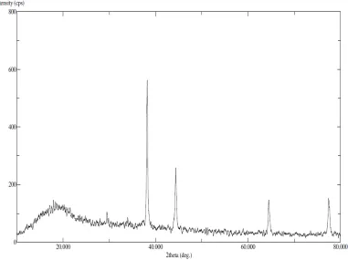
The diffraction spectrum of green synthesized AgNps have shown Bragg Peaks (angle 2θ) at
29.5º, 34º, 38.2º, 44.38º, 54.98º, 59º, 64.44º, 77.46º which corresponds to 210, 122, 111, 200, 142, 241,
220 and 331 miller indices that suggest FCC structure.
IJSRR, 8(2) April. – June., 2019 Page 4365
Antimicrobial Susceptibility testing
:Green nanoparticles showed weaker inhibition in comparison to synthetically prepared nanoparticles.
The aconitum nanoparticles showed strong inhibition against S. aureus and B. subtilis however weaker inhibition against E. coli and Pseudomonas aeruginosa. Ampicillin was used as a control.
Antioxidant activity:
The natural antioxidants of the Aconitum contribute to its antioxidant activity. In comparison
the activity is slightly lesser than ascorbic acid.
Fig5: Graphical representation of antioxidant assay performed by DPPH assay Fig4. AST plates [left to right] of Green AgNPs, synthetic NPs, and control [Ampicillin]
Sr. no Organism
[mm] [mm] [mm] [mm] [mm] [mm] [mm] [mm] [mm]
1 S. aureus 21 24 23 28 27 25 22 24 24
2 B. subtilis 21 22 21 15 18 17 25 23 27
3 E. coli 18 16 17 25 23 27 21 21 19
4 P. aeruginosa 17 16 18 14 15 15 19 20 21
Control Synthetic Nanoparticles Green Nanoparticles
Zone of Inhibition
IJSRR, 8(2) April. – June., 2019 Page 4366
DISCUSSION:
Atvish has been commonly associated with the treatment of intestinal and digestive ailments.
Traditionally, in wester Himalayan region it has often been used in Ayurvedic for relieving flatulence
and also advised for a regular bowel regimen. Traditional Chinese practitioners also use aconite for
pain relief caused by trigeminal and intercostal neuralgia. Aconite is also used as a mild diaphoretic
and to slow pulse rate by its effect on brainstem centers. Due to its association with several clinical
applications, Aconitum and its phytoconstituents have been studied for its properties and extraction.
Nanoparticles have been known to enhance the biological activities of the plants. Therefore, silver
nanoparticles were made to study its antimicrobial activity especially towards enteric bacteria. The
silver nanoparticles were synthesized chemically as well as by green synthesis to compare the activity
and efficiency. It can be noted that while the antimicrobial activity of G-AgNPsappears to be lesser, by
statistically analysis by T test at 0.05 significance level, the activities of chemical synthesis and green
synthesis is comparable. Since green synthesis is also economically and ecologically feasible, it is a
better option.
CONCLUSION:
The green synthesis of silver nanoparticles was carried out by using Aconitum heterophyllum.
The physical characteristics of the nanoparticles were determined. The size of the synthesized AgNPs
was determined using a nanoparticle tracking system, the mean size was found to be 34nm. The
chemically and biologically synthesized nanoparticles were studied for their antimicrobial activity. The
activities of nanoparticles by both syntheses were found to be statistically comparable. They both
showed good activity towards S. aureus and B. subtilis and lesser activity towards E. coli and
Pseudomonas aeruginosa. We can hence conclude that Aconitum can be used as a good candidate to treat enteric infections and be explored in clinical research and since nanoparticles increase catalytic
activity and efficacy, using nanoparticles as a drug delivery system could work out well.
REFERENCES
1. Harekrishna B, Dipak KB, GobindaSP, Priyanka S, Sankar PD. Green synthesis of
IJSRR, 8(2) April. – June., 2019 Page 4367 2. Ankamwar B, Damle C, Ahmad A, Sastry M. Biosynthesis of gold and silver nanoparticles
using Emblica Officinalis Fruit extract and their Phase Transfer and Transmetallation in an Organic Solution. J nanosciNanotechnol 2005;5(10):1665-1671.
3. K.A. Khalil, H. Fouad, T. Elsarnagawy, F.N. Almajhdi Preparation and characterization of
electrospun PLGA/silver composite nanofibers for biomedical applications.Int J Electrochem
Sci 2013; 8: 3483–3493
4. Ahmad, P. Mukherjee, S. Senapati, D. Mandal, M.I. Khan, R. Kumar, M. Sastry. Extracellular
biosynthesis of silver nanoparticles using the fungus Fusarium oxysporum. Colloids Surf B: Biointerfaces, 2003; 28(4) :313–318
5. Klaus-J, R. Joerger, E. Olsson, C. Granqvist. Bacteria as workers in the living factory: metal
accumulating bacteria and their potential for materials science.Trends Biotechnol, 2001; 19 (1)
:15–20
6. Xu ZP, Zeng QH, Lu GQ, Yu AB. Inorganic nanoparticles as carriers for efficient cellular
delivery. Chem Eng Sci. 2006; 61:1027–1040.
7. Tran, Q. H., Nguyen, V. Q., & Le, A.-T.. Silver nanoparticles: synthesis, properties, toxicology,
applications and perspectives. Advances in Natural Sciences: Nanoscience and
Nanotechnology, 2013; 4(3): 033001. doi:10.1088/2043-6262/4/3/033001
8. Guzman MG, Dille J, Godet S. Synthesis of silver nanoparticles by chemical reduction method
and their antibacterial activity. Int J Chem Biol Eng. 2009; 2:104–111
9. Chou KS, Ren CY. Synthesis of nanosized silver particles by chemical reduction method.
Mater Chem Phys. 2000; 64:241–246.
10.Reddy G, Joy J, Mitra T, Shabnam S, Shilpa T. Nanosilver review. Int J Adv Pharm. 2012; 2:
9–15
11.D Ukani, M.K Mehta, N.DNanavati, D. Aconitum heterophyllum (Ativisha) in Ayurveda.
Ancient science of life. 1996; 16: 166-71.
12.Verma S, Ojha S, Raish M. Anti-inflammatory activity of Aconitum heterophyllum on cotton
pellet-induced granuloma in rats. J Med Plants Res 2010; 4(15): 1566-9.
13.Shastri, M.R V. Sachittar Ayurveda 1964; 19: 134
14.Pandey H, Nandi SK, Kumar A, Palni UT, Chandra B, Palni LMS. In vitro propagation of
IJSRR, 8(2) April. – June., 2019 Page 4368 15.Liu FX, Sun S, Cui ZZ. Analysis of immunological enhancement of immunosuppressed
chickens by Chinese herbal extracts. JEthnopharmacol 2010; 127: 251-6.
16.Gupta A, Jagtap RB, Chaphalkar SR. Anti-viral activity of Azadirachtaindicaleaves against Newcastle disease virus: a study by in vitro and in vivo immunological approach. Int J Curr Trends Pharm Res 2014; 2(6): 494-501.
17.Atal CK, Sharma ML, Kaul A, Khajuria A. Immuno modulating agents of plant origin.
I:preliminary screening. J Ethnopharmacol 1986; 18: 133-41.
18.Anwar S, Ahmad B, Sultan M, Gul W, Islam N. Biological and pharmacological properties of
Aconitumchasmanthum. J Biol Sci 2003; 3: 989-93.
19.Nooman A. K, Ashok K.S, A. Al-Othman, Zaha El-Agbar, Husni F. Turk J Biol 32: 51-55
20.Manivasagan P, Venkatesan J, Senthilkumar K, Sivakumar K. and Kim SK. Biosynthesis,