Received 7 April 2015 Accepted 22 May 2015
Edited by M. Gdaniec, Adam Mickiewicz University, Poland
†
Keywords:crystal structure; indolinone deriva-tives; hydrogen bonding; intermolecular inter-actions; dipole moment
CCDC reference:1028641
Supporting information:this article has supporting information at journals.iucr.org/e
Crystal structure of 1-ethyl-5-iodoindolin-2-one
Man Zhang,aYu-Xiang Shen,bQi Fang,b* Lei Wangaand Da-Zhi Lic
aSchool of Chemistry and Chemical Engineering, Shandong University, Jinan 250100, People’s Republic of China,bState
Key Laboratory of Crystal Materials, Shandong University, Jinan 250100, People’s Republic of China, andcBinzhou Key
Laboratory of Material Chemistry, Department of Chemical Engineering, Binzhou University, Binzhou 256603, People’s Republic of China. *Correspondence e-mail: [email protected]
In the title indolinone derivative, C10H10INO, all the non-H atoms, except the
terminal methyl C atom, are almost coplanar. The molecules are arranged into columns extending along thea-axis direction and interact with the molecules in adjacent columnsviaC—H O hydrogen bonds [H O distance = 2.57 (3) A˚ ] and I I short contacts of 3.8986 (3) A˚ . A one-dimensional zigzag iodine chain along theaaxis is apparent between two neighbouring columns.
1. Chemical context
Indolinone derivatives play an important role in the pharmaceutical industry and some of them show anti-neoplastic (Caneet al., 2000), antibacterial (Kumaret al., 2013) and anti-inflammatory (Mammoneet al., 2006) activities. The indolinone skeleton can be also found in many known bioactive drugs, such as horsfiline (Murphy et al., 2005), rhynchophylline (Deiters et al., 2006) and the gelsemium alkaloids (Kitajimaet al., 2006). In addition, indolinone deri-vatives are widely used in the spice industry and agriculture, as functional materials (Jiet al., 2010) and dye intermediates.
In recent years, the synthesis and crystal structures of many indolinone derivatives have been reported including 6-chloro-5-(2-chloroethyl)oxindole (Nadkarni & Hallissey, 2008). We have recently synthesized and reported the crystal structures of several indolin-2-one derivatives including 1-phenyl-indolin-2-one (Wang et al., 2015). As a continuation of our work in this field, we report here the synthesis and crystal structure of the title compound, 1-ethyl-5-iodoindolin-2-one.
2. Structural commentary
The title molecule is shown in Fig. 1. The non-H atoms of the indoline core are virtually coplanar [mean deviation is 0.011 (3) A˚ with a maximum deviation of 0.023 (3) A˚ for C1]. The atoms C9, O1 and I1 are essentially co-planar with the indoline core, with deviations of 0.019 (4) A˚ for C9,
0.070 (3) A˚ for O1, and 0.127 (1) A˚ for I1. The sum of valence angles around N1 is 360.0, indicating ansp2
hybridization of this atom. The two C—N bonds in the five-membered ring have a partial double-bond character [N1. . .C1 1.370 (4) A˚ ; N1. . .C8 1.400 (3) A˚ ], indicating conjugation of the-electrons of the NC=O group with the-electrons of the benzene ring.
3. Supramolecular features
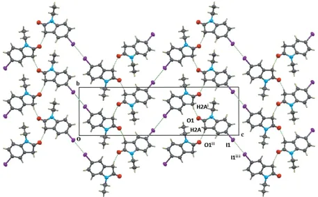
The crystal packing in the title compound is shown in Figs. 2 and 3. The molecules are face-to-face parallel-packed forming a column along the a axis with – interactions centroid–
centroid distances = 4.130 (2) and 4.462 (2) A˚ ]. Molecules from neighbouring columns are connected by a C—H O hydrogen bond (Table 1) with the formation of a layer-type aggregate parallel to (001). There is an I I contact shorter than the sum (3.96 A˚ ) of the van der Waals radii [I Ii 3.8986 (3) A˚ , C—I Ii173.3 (3); symmetry code: (i) x 1
2,
y1
2,z+ 2] joining the columns of molecules in adjacent
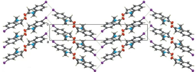
layers and forming a kind of 1-D zigzag chain along thea-axis direction (see Fig. 3). An important feature of the columns is that they are polar, i.e. all molecular dipole moments in the same column point in the same direction.
DFT/b3lyp/genecp calculations were carried out, which took the pseudopotential basis set LanL2DZ for the iodine atom and the 6–311g(d) basis set for the other atoms, to optimize the molecular geometry and calculate the dipole moment using theGAUSSIAN03program (Frischet al., 2003). The dipole moment of the title molecule (1.707 D) is much smaller than that of its precursor molecule, 1-ethyl-5-iodo-indolin-2,3-dione (5.432 D). This difference may partly explain the non-centrosymmetry of the title crystal (space group
P212121) and the centrosymmetry of the crystal of the
research communications
Acta Cryst.(2015).E71, 712–715 Zhanget al. C
[image:2.610.88.259.68.219.2]10H10INO
713
Table 1Hydrogen-bond geometry (A˚ ,).
D—H A D—H H A D A D—H A
C2—H2A O1i 0.99 (3) 2.57 (4) 3.554 (4) 169 (3)
Symmetry code: (i)xþ3;y1 2;zþ
[image:2.610.76.533.438.721.2]3 2.
Figure 2
The view of the structure along the aaxis, showing the C—H O hydrogen bond between columns and the I I interactions between columns. [Symmetry codes: (i)x+ 3,y+1
2,z+ 3
2; (ii)x+ 3,y 1 2,z+
3 2; (iii)x
1 2,y
1 2,z+ 2.]
Figure 1
precursor (Wang et al.,2014). On the other hand, the non-centrosymmetry of the title crystal may be better explained by the I I intermolecular interactions, for there are no I I short contacts in the above centrosymmetric precursor crystal.
4. Database survey
A search of the Cambridge Structural Database (WebCSD, Version 5.36; last update April 2015; Groom & Allen, 2014) for 5-iodoindolin-2-one derivatives gave 15 hits. Of these 16 structures (with the title structure included), the number of
non-centrosymmetric structures (9) is slightly greater than the number of centrosymmetric structures (7). In these 16 struc-tures, there are four structures which exhibit I I short intermolecular contacts and all the four structures are non-centrosymmetric (three of them belong to theP212121space
group and the other one belongs to the P63 space group;
Takahashiet al., 2014). Therefore, the I I contacts seem to promote non-centrosymmetric packing in this kind of compound.
5. Synthesis and crystallization
The title compound was synthesized by reduction of the precursor with an 80% hydrazine hydrate (see reaction scheme) . 1-Ethyl-5-iodoindolin-2,3-dione precursor (1.714 g, 5.69 mmol) and 80% NH2NH2H2O (19.0 mL) were added
into a 50 mL flask and the mixture was stirred under reflux. The reaction progress was tracked by TLC. After 4.5 h, the reaction mixture was cooled down and poured into 100 mL water with precipitation of yellow solid. Then the mixture was extracted with CH2Cl2, the organic phase was washed with
water and dried with MgSO4. The solvent was removed under
reduced pressure and the crude product was purified by silica gel column chromatography with CHCl3 as eluent. The title
compound was obtained as a colorless solid (1.509 g, yield 92.3%). m.p. 403–404 K. Crystals suitable for X-ray diffraction were obtained by slow evaporation of a CHCl3solution.
6. Refinement
[image:3.610.99.508.72.223.2]Crystal data, data collection and structure refinement details are summarized in Table 2. H atoms bound to aromatic C atoms and methylene C atoms were located in difference maps
Figure 3
[image:3.610.326.553.597.679.2]The view of the structure along thebaxis, showing the one-dimensional columnar structure and the zigzag iodine chains along theaaxis.
Table 2
Experimental details.
Crystal data
Chemical formula C10H10INO
Mr 287.09
Crystal system, space group Orthorhombic,P212121
Temperature (K) 295
a,b,c(A˚ ) 4.4622 (1), 8.2664 (2), 27.4400 (5)
V(A˚3) 1012.16 (4)
Z 4
Radiation type MoK
(mm1) 3.12
Crystal size (mm) 0.420.320.16
Data collection
Diffractometer Bruker APEXII CCD Absorption correction Multi-scan (SADABS; Bruker,
2005) Tmin,Tmax 0.354, 0.635
No. of measured, independent and observed [I> 2(I)] reflections
12175, 2938, 2878
Rint 0.020
(sin/)max(A˚
1) 0.704
Refinement
R[F2> 2(F2)],wR(F2),S 0.021, 0.050, 1.21
No. of reflections 2938 No. of parameters 148
H-atom treatment H atoms treated by a mixture of independent and constrained refinement
max,min(e A˚
3) 0.47,0.69
Absolute structure Flack (1983), 1183 Friedel pairs Absolute structure parameter 0.02 (2)
Computer programs:APEX2andSAINT(Bruker, 2005),SHELXS97,SHELXL97and
and freely refined, leading to C—H distances of 0.91 to 1.02 A˚ . The three H atoms bound to methyl C atoms could also be located in difference maps but they were placed at calculated positions and treated using a riding-model approximation with C—H = 0.96 A˚ andUiso(H) = 1.5Ueq(C).
Acknowledgements
This work was supported by the National Science Foundation of China (grant Nos. 21472116 and 20972089).
References
Bruker (2005). APEX2, SAINT and SADABS. Bruker AXS Inc., Madison, Wisconsin, USA.
Cane, A., Tournaire, M.-C., Barritault, D. & Crumeyrolle-Arias, M. (2000).Biochem. Biophys. Res. Commun.276, 379–384.
Deiters, A., Pettersson, M. & Martin, S. F. (2006).J. Org. Chem.71, 6547–6561.
Flack, H. D. (1983).Acta Cryst.A39, 876–881.
Frisch, M. J.,et al.(2003).GAUSSIAN03.Gaussian Inc., Pittsburgh, PA, USA.
Groom, C. R. & Allen, F. H. (2014).Angew. Chem. Int. Ed.53, 662– 671.
Ji, L., Fang, Q., Yuan, M. S., Liu, Z. Q., Shen, Y. X. & Chen, H. F. (2010).Org. Lett.12, 5192–5195.
Kitajima, M., Nakamura, T., Kogure, N., Ogawa, M., Mitsuno, Y., Ono, K., Yano, S., Aimi, N. & Takayama, H. (2006).J. Nat. Prod.69, 715–718.
Kumar, P. K. P., Priyadarshini, C. V., Arulmozhi, S. & Krishna Moorthy, P. (2013).Int. J. Clin. Pharm-Net.5, 5240–5256. Mammone, T., A˚ kesson, C., Gan, D., Giampapa, V. & Pero, W. R.
(2006).Phytother. Res.20, 178–183.
Murphy, J. A., Tripoli, R., Khan, T. A. & Mali, U. W. (2005).Org. Lett. 7, 3287–3289.
Nadkarni, D. V. & Hallissey, J. F.(2008).Org. Process Res. Dev.12, 1142–1145.
Sheldrick, G. M. (2008).Acta Cryst.A64, 112–122.
Takahashi, M., Murata, Y., Yagishita, F., Sakamoto, M., Sengoku, T. & Yoda, H. (2014).Chem. Eur. J.20, 11091–11100.
Wang, L., Shen, Y.-X., Dong, J.-T., Zhang, M. & Fang, Q. (2014).Acta Cryst.E70, o67.
Wang, L., Zhang, M., Jin, Y.-Y., Lu, Q. & Fang, Q. (2015).Acta Cryst. C71, 69–74.
research communications
Acta Cryst.(2015).E71, 712–715 Zhanget al. C
sup-1
Acta Cryst. (2015). E71, 712-715supporting information
Acta Cryst. (2015). E71, 712-715 [doi:10.1107/S2056989015009937]
Crystal structure of 1-ethyl-5-iodoindolin-2-one
Man Zhang, Yu-Xiang Shen, Qi Fang, Lei Wang and Da-Zhi Li
Computing details
Data collection: APEX2 (Bruker, 2005); cell refinement: SAINT (Bruker, 2005); data reduction: SAINT (Bruker, 2005);
program(s) used to solve structure: SHELXS97 (Sheldrick, 2008); program(s) used to refine structure: SHELXL97
(Sheldrick, 2008); molecular graphics: SHELXTL (Sheldrick, 2008); software used to prepare material for publication:
SHELXTL (Sheldrick, 2008).
1-Ethyl-5-iodoindolin-2-one
Crystal data
C10H10INO
Mr = 287.09
Orthorhombic, P212121
Hall symbol: P 2ac 2ab a = 4.4622 (1) Å b = 8.2664 (2) Å c = 27.4400 (5) Å
V = 1012.16 (4) Å3
Z = 4 F(000) = 552
Dx = 1.884 Mg m−3
Melting point = 403–404 K Mo Kα radiation, λ = 0.71073 Å Cell parameters from 9948 reflections θ = 2.5–30.0°
µ = 3.12 mm−1
T = 295 K
Parallelepiped, orange 0.42 × 0.32 × 0.16 mm
Data collection
Bruker APEXII CCD diffractometer
Radiation source: fine-focus sealed tube Graphite monochromator
Detector resolution: 8.3 pixels mm-1
ω scans
Absorption correction: multi-scan (SADABS; Bruker, 2005) Tmin = 0.354, Tmax = 0.635
12175 measured reflections 2938 independent reflections 2878 reflections with I > 2σ(I) Rint = 0.020
θmax = 30.0°, θmin = 2.6°
h = −6→6 k = −11→9 l = −37→33
Refinement
Refinement on F2
Least-squares matrix: full R[F2 > 2σ(F2)] = 0.021
wR(F2) = 0.050
S = 1.21 2938 reflections 148 parameters 0 restraints
Primary atom site location: structure-invariant direct methods
Secondary atom site location: difference Fourier map
Hydrogen site location: mixed
H atoms treated by a mixture of independent and constrained refinement
w = 1/[σ2(F
o2) + (0.0144P)2 + 0.4592P]
where P = (Fo2 + 2Fc2)/3
(Δ/σ)max = 0.002
Δρmax = 0.47 e Å−3
supporting information
sup-2
Acta Cryst. (2015). E71, 712-715Extinction correction: SHELXL97 (Sheldrick, 2008), Fc*=kFc[1+0.001xFc2λ3/sin(2θ)]-1/4
Extinction coefficient: 0.0014 (3)
Absolute structure: Flack (1983), 1183 Friedel pairs
Absolute structure parameter: 0.02 (2)
Special details
Experimental. Scan width 0.4° ω, Crystal to detector distance 6.20 cm, exposure time 20 s, 17 h for data collection Geometry. All e.s.d.'s (except the e.s.d. in the dihedral angle between two l.s. planes) are estimated using the full covariance matrix. The cell e.s.d.'s are taken into account individually in the estimation of e.s.d.'s in distances, angles and torsion angles; correlations between e.s.d.'s in cell parameters are only used when they are defined by crystal symmetry. An approximate (isotropic) treatment of cell e.s.d.'s is used for estimating e.s.d.'s involving l.s. planes.
Refinement. Refinement of F2 against ALL reflections. The weighted R-factor wR and goodness of fit S are based on F2,
conventional R-factors R are based on F, with F set to zero for negative F2. The threshold expression of F2 > σ(F2) is used
only for calculating R-factors(gt) etc. and is not relevant to the choice of reflections for refinement. R-factors based on F2
are statistically about twice as large as those based on F, and R- factors based on ALL data will be even larger.
Fractional atomic coordinates and isotropic or equivalent isotropic displacement parameters (Å2)
x y z Uiso*/Ueq
I1 0.57349 (4) −0.10015 (2) 0.963176 (7) 0.05126 (7) C5 0.7961 (6) 0.0703 (3) 0.91955 (9) 0.0404 (5) O1 1.3877 (6) 0.3521 (3) 0.74792 (8) 0.0644 (7) C4 0.8354 (6) 0.0383 (3) 0.87003 (10) 0.0410 (5) C3 0.9948 (5) 0.1476 (3) 0.84294 (9) 0.0386 (6) C8 1.1096 (6) 0.2881 (3) 0.86420 (9) 0.0394 (5) N1 1.2652 (5) 0.3781 (3) 0.82916 (8) 0.0429 (5) C9 1.4198 (8) 0.5309 (4) 0.83826 (12) 0.0505 (6) C10 1.2102 (8) 0.6717 (4) 0.84543 (12) 0.0539 (7) H10A 1.0772 0.6795 0.8180 0.081* H10B 1.3242 0.7698 0.8482 0.081* H10C 1.0955 0.6557 0.8746 0.081* C1 1.2634 (7) 0.3027 (3) 0.78470 (10) 0.0472 (6) C2 1.0825 (9) 0.1476 (3) 0.78985 (10) 0.0488 (6) C7 1.0700 (8) 0.3218 (3) 0.91288 (10) 0.0456 (5) C6 0.9096 (8) 0.2096 (4) 0.94064 (9) 0.0472 (6) H2A 1.216 (8) 0.054 (4) 0.7827 (11) 0.053 (9)* H2B 0.899 (12) 0.165 (5) 0.7685 (17) 0.103 (16)* H4 0.754 (8) −0.062 (4) 0.8534 (12) 0.058 (9)* H6 0.884 (8) 0.234 (4) 0.9737 (11) 0.051 (9)* H7 1.143 (7) 0.419 (4) 0.9266 (11) 0.053 (9)* H9A 1.548 (9) 0.555 (4) 0.8089 (13) 0.067 (11)* H9B 1.528 (10) 0.520 (5) 0.8662 (14) 0.075 (12)*
Atomic displacement parameters (Å2)
U11 U22 U33 U12 U13 U23
sup-3
Acta Cryst. (2015). E71, 712-715C3 0.0434 (14) 0.0350 (12) 0.0374 (12) 0.0044 (8) 0.0043 (9) −0.0006 (9) C8 0.0386 (12) 0.0356 (11) 0.0440 (12) 0.0026 (10) 0.0029 (10) 0.0009 (10) N1 0.0479 (11) 0.0335 (11) 0.0473 (11) −0.0035 (10) 0.0100 (10) −0.0003 (9) C9 0.0432 (13) 0.0488 (15) 0.0596 (17) −0.0113 (14) 0.0002 (15) 0.0038 (12) C10 0.0615 (18) 0.0403 (15) 0.0598 (17) −0.0119 (14) −0.0008 (15) −0.0031 (13) C1 0.0537 (16) 0.0393 (13) 0.0486 (15) 0.0047 (13) 0.0125 (13) 0.0039 (12) C2 0.0664 (17) 0.0389 (13) 0.0412 (13) −0.0003 (15) 0.0159 (15) −0.0040 (10) C7 0.0526 (14) 0.0414 (13) 0.0428 (13) −0.0052 (14) −0.0023 (13) −0.0048 (10) C6 0.0530 (14) 0.0538 (15) 0.0349 (12) −0.0018 (15) 0.0015 (13) 0.0009 (11)
Geometric parameters (Å, º)
I1—C5 2.099 (3) C9—C10 1.506 (5) C5—C6 1.384 (4) C9—H9A 1.01 (4) C5—C4 1.395 (4) C9—H9B 0.91 (4) O1—C1 1.222 (3) C10—H10A 0.9600 C4—C3 1.369 (4) C10—H10B 0.9600 C4—H4 1.02 (3) C10—H10C 0.9600 C3—C8 1.397 (4) C1—C2 1.521 (4) C3—C2 1.508 (4) C2—H2A 0.99 (3) C8—C7 1.376 (4) C2—H2B 1.02 (5) C8—N1 1.400 (3) C7—C6 1.398 (4) N1—C1 1.370 (4) C7—H7 0.95 (3) N1—C9 1.461 (4) C6—H6 0.94 (3)
C6—C5—C4 121.3 (2) C9—C10—H10A 109.5 C6—C5—I1 119.53 (19) C9—C10—H10B 109.5 C4—C5—I1 119.2 (2) H10A—C10—H10B 109.5 C3—C4—C5 118.0 (3) C9—C10—H10C 109.5 C3—C4—H4 118.8 (19) H10A—C10—H10C 109.5 C5—C4—H4 123.3 (19) H10B—C10—H10C 109.5 C4—C3—C8 120.8 (2) O1—C1—N1 125.5 (3) C4—C3—C2 131.2 (3) O1—C1—C2 126.8 (3) C8—C3—C2 108.0 (2) N1—C1—C2 107.7 (2) C7—C8—C3 121.8 (3) C3—C2—C1 103.1 (2) C7—C8—N1 128.5 (3) C3—C2—H2A 110.2 (18) C3—C8—N1 109.7 (2) C1—C2—H2A 108 (2) C1—N1—C8 111.5 (2) C3—C2—H2B 110 (3) C1—N1—C9 123.3 (2) C1—C2—H2B 105 (3) C8—N1—C9 125.2 (2) H2A—C2—H2B 118 (3) N1—C9—C10 113.4 (3) C8—C7—C6 117.4 (3) N1—C9—H9A 107 (2) C8—C7—H7 121 (2) C10—C9—H9A 108 (2) C6—C7—H7 121.6 (19) N1—C9—H9B 108 (3) C5—C6—C7 120.8 (2) C10—C9—H9B 107 (3) C5—C6—H6 122 (2) H9A—C9—H9B 113 (3) C7—C6—H6 117 (2)
supporting information
sup-4
Acta Cryst. (2015). E71, 712-715I1—C5—C4—C3 −176.70 (19) C9—N1—C1—O1 0.1 (5) C5—C4—C3—C8 −1.1 (4) C8—N1—C1—C2 1.3 (3) C5—C4—C3—C2 178.5 (3) C9—N1—C1—C2 179.6 (3) C4—C3—C8—C7 0.5 (4) C4—C3—C2—C1 −178.6 (3) C2—C3—C8—C7 −179.1 (3) C8—C3—C2—C1 1.0 (3) C4—C3—C8—N1 179.4 (2) O1—C1—C2—C3 178.1 (3) C2—C3—C8—N1 −0.3 (3) N1—C1—C2—C3 −1.4 (3) C7—C8—N1—C1 178.0 (3) C3—C8—C7—C6 0.0 (4) C3—C8—N1—C1 −0.7 (3) N1—C8—C7—C6 −178.6 (3) C7—C8—N1—C9 −0.2 (5) C4—C5—C6—C7 −0.7 (5) C3—C8—N1—C9 −178.9 (3) I1—C5—C6—C7 177.2 (2) C1—N1—C9—C10 108.7 (3) C8—C7—C6—C5 0.0 (5) C8—N1—C9—C10 −73.2 (4)
Hydrogen-bond geometry (Å, º)
D—H···A D—H H···A D···A D—H···A
C2—H2A···O1i 0.99 (3) 2.57 (4) 3.554 (4) 169 (3)