A
Multi
‐
Site
Prospective
Study
of
the
Adverse
Events
and
Complications
Associated
with
Mohs
Micrographic
Surgery
By
Nicole
Y.
Lee
A
Master’s
Paper
submitted
to
the
faculty
of
the
University
of
North
Carolina
at
Chapel
Hill
in
partial
fulfillment
of
the
requirements
for
the
degree
of
Master
of
Public
Health
in
the
Public
Health
Leadership
Program.
Chapel
Hill
2011
_______________________________
First
Reader:
_______________________________
Date
_________________________________
Second
Reader:
_________________________________
Table
of
Contents
Abstract………..……2
Original
Manuscript..……….………..…3
Systematic
Review….………..……….……21
ABSTRACT
Non‐melanoma skin cancer (NMSC) is the leading cause of cancer worldwide with an incidence of more than 1 million new cases each year, occurring at a rate of 3% annually. As the incidence of skin cancer continues to rise, it becomes increasingly important to ensure safe and effective ways to manage cutaneous malignancies. Treatment options for NMSC include traditional excision, curettage and electrodessication, cryosurgery, external beam radiation, topical chemotherapeutic agents or Mohs micrographic surgery (MMS). There has been ongoing debate over the past decade about the safety of outpatient surgery, with increasing scrutiny on complications associated with outpatient procedures. Much of the alarm over outpatient‐based surgical procedures stems from serious adverse events occurring in patients having procedures performed under general anesthesia in an ambulatory surgical facility. Serious adverse events during and following outpatient based cutaneous surgery, however, are rare.
MMS has proven to be effective in the treatment of skin cancer. The cost‐effectiveness of MMS has also been well‐established. Complications and serious adverse events associated with MMS have been evaluated by single‐surgeon studies in both the outpatient clinic and hospital‐based settings. Retrospective studies have also examined the safety of MMS. While these studies provide evidence for the safety of the procedure, a prospective, multicenter study would afford a higher level of clinical evidence, and definitively establish the safety profile of MMS and reconstruction in the ambulatory setting.
This paper is divided into two sections. In the first section, we report the results of an original, prospective, multi‐site research study that evaluated the rate of serious adverse events, post‐operative complications, and post‐operative pain associated with the treatment of skin cancer using MMS in 1550 patients with 1792 tumors. The study resulted in a complication rate of 2.6% (44/1709), with a
breakdown of 21 cases of active bleeding, 2 hematomas, 16 infections, and 5 flap or wound edge necrosis. There were 10 cases of secondary complications. When compared to patients without complications, those who developed a complication were older (73.5±13 vs 69±13, p=0.024), had larger tumors (1.6±1.11 vs 1.12±0.75 cm, p=0.0001), and had larger post‐operative defects (2.41±1.52 vs 1.88±1.2 cm, p=0.0032). Furthermore, patients with a complication reported higher mean pain scores than those who did not develop complications (3.08±2.92 vs 1.96±2.38, p=0.0014). In conclusion, MMS is proven as a highly effective, cost‐effective, well‐tolerated, and extremely safe procedure for the treatment of skin cancer. This prospective, multicenter study helps confirm the high degree of safety of MMS within the United States.
INTRODUCTION:
Non‐melanoma skin cancer (NMSC), which includes basal cell carcinoma and squamous cell carcinoma, is the leading cause of cancer worldwide with an incidence of more than 1 million new cases each year. As the incidence of skin cancer continues to rise, it becomes increasingly important to ensure safe and effective ways to manage cutaneous malignancies. There has been ongoing debate over the past decade about the safety of outpatient surgery, with increasing scrutiny on complications associated with outpatient procedures. Prospective data collection with adequate follow‐up is important to definitively establish the safety of Mohs micrographic surgery (MMS) performed in the outpatient setting.
MMS has proven to be effective in the treatment of skin cancer. 1 The cost‐effectiveness of MMS has also been well‐established.2‐4 Complications and serious adverse events associated with MMS have been evaluated by single‐surgeon studies in both the outpatient clinic and hospital‐based
settings.6,7 Retrospective studies have also examined the safety of MMS. 8 While these studies provide evidence for the safety of the procedure, prospective, multicenter studies offer more substantial clinical evidence, and definitively establish the safety profile of MMS surgery and reconstruction in the
ambulatory setting.
We report the results of a 13 site, 13 surgeon, nationwide prospective cohort study evaluating the rate of serious adverse events, post‐operative complications, and post‐operative pain associated with the treatment of skin cancer using MMS in 1550 patients with 1792 tumors in both private practice and university‐based settings.
METHODS:
Thirteen fellowship‐trained Mohs surgeons belonging to the American College of Mohs Surgeons agreed to participate in a multicenter, prospective, four‐week study tracking the adverse events, complications, and peak post‐operative pain levels associated with MMS and reconstruction. The Institute of Research Board approval was obtained for all sites. Surgical approaches varied from office to office, as did the use of intraincisional antibiotics, post‐operative prophylactic antibiotics, and analgesics, all of which were documented.
Population:
All English speaking patients at least 18‐years‐old undergoing MMS by one of the 13 study surgeons were invited to participate. Patient accrual was planned for 20 consecutive work days in each of the 13 Mohs surgeons’ practices, including private practice and academic settings. Informed consent was obtained for all patients.
Variables:
respiratory distress, hypertensive urgency or emergency, dysrhythmia, pacemaker or defibrillator malfunction, or any event leading to deviation of standard practice or transfer to the emergency department or hospital.
The second phase of the study involved the collection of follow‐up data related to complications after surgery, including infection, bleeding requiring health care provider intervention, significant flap/graft/skin edge necrosis (10% or greater), dehiscence (greater than 2mm by 2mm), and serious adverse events including chest pain, shortness of breath, symptoms of TIA or CVA, or any event leading to the use of emergency medical services or hospitalization. Chronology was used to determine the primary complication. For example, if a patient experienced active bleeding followed by dehiscence or necrosis, active bleeding was considered the primary complication. In patients with treatment for infection and dehiscence or necrosis, infection was considered the primary complication.
Evidence of infection was defined as the presence of erythema, edema, purulence, and increased pain. To minimize the potential for underreporting of infections, any postoperative site
treated for infection was counted as an infection, regardless of whether or not a culture was performed.
Dehiscence was defined as the separation of any area of sutured skin greater than 2 mm by 2 mm in the absence of other complications. Necrosis was defined as devitalized tissue greater than 10% of a flap, graft, or skin edge.
Peak post‐operative pain levels were also recorded at follow‐up. Peak post‐operative pain was recorded using a 0‐10 analog scale for patients returning to the Mohs surgeon’s office and those completing the mail‐in survey.
For patients following up in person, all information was collected at a follow‐up visit within two weeks of the procedure. Those patients not following up in person were given a pre‐addressed, postage paid envelope with a brief survey to complete and mail back to the primary research center within 1‐2 weeks after surgery. Patients who did not complete and return the survey were contacted by telephone for a scripted interview to document post‐operative adverse events, complications, and pain levels.
Analysis:
Mean values, standard deviations and percentiles were used to describe patient characteristics and tumors treated. Bivariable association between the outcomes bleeding, hematoma, infection, flap/edge/graft necrosis or any complication and exposure of interest were calculated using Pearson’s Chi‐Square. Participant satisfaction with pain management based on use of pain medication was
evaluated with Pearson’s Chi‐Square. A 2 –Sample T‐Test was used to compare mean differences for the outcome of complications (active bleeding, hematoma, infection, edge/flap/graft necrosis, and all complications) and age, number of stages, tumor size, and defect size. A 2‐Sample T‐Test was also used to evaluate for any association of reported pain levels and sex, age (<65 years old vs ≥ 65 years old), numbers of stages performed, numbers of lesions removed, anticoagulants, intraincisional antibiotics, tumor size, defect size, type of follow‐up, past medical history, repair type, tumor location, and type of complication. All reported p‐values were two‐sided and statistical significance was set at values of p<0.05. Stata 11.0 was used to conduct all statistical analysis.
RESULTS:
information, follow‐up was obtained in‐office for 69%, by mail‐in survey for 26%, and by telephone interview for 5%.
Patient and Tumor characteristics:
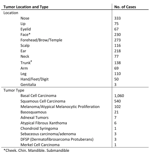
The mean (±SD) age of patients in the study was 69 (±13) years old. Of the tumors treated, 1094 were in men and 698 were in women (Table 1). Most patients were nonsmokers (93%), and over half had a history of prior skin cancer treatment (64.6%). A wide variety of neoplasms were treated, with the majority consisting of basal cell carcinoma (61%) and squamous cell carcinoma (31%) (Table 2). As for location, 77% of the tumors were located on the head and neck with the overall stratification by location shown in Table 2. The mean pre‐operative tumor size was 1.14 (±0.76) cm while the average post‐ operative defect size was 1.89 (±1.2) cm. The average number of stages per tumor was 1.6 (±0.86), with a range from 1‐8.
Wound management:
Eighty‐two percent of the defects resulting from MMS were reconstructed (Table 3), while 18% were managed using second intention healing. Of the 82% of defects that were reconstructed, 92% were repaired by the Mohs surgeon, 8% were referred to another surgeon for reconstruction (typically plastic surgeons working in one of the practices, or oculoplastic surgery colleagues in outside practices).
Types of reconstruction:
Of the defects reconstructed by Mohs surgeons, 72% were repaired with primary linear closure (Table 3, Figure 1); 11% were repaired with a single‐stage transposition flap, V‐Y advancement flap, Burow’s advancement flap, mucosal advancement flap, or a rotation flap; and 15% were repaired with a skin graft. Four interpolation flaps were performed and eight combined repairs carried out (utilization of primary linear repair or a flap in addition to a skin graft). The methods of reconstruction used for the patients who were referred to other surgeons are unknown.
Serious adverse events:
No deaths occurred during treatment or in the post‐operative period. No serious adverse events occurred during MMS or reconstruction. Two adverse events with an unclear relationship to the procedure and without serious sequelae were reported by patients after surgery. One patient, who underwent treatment of a basal cell carcinoma on the alar crease with 3 stages of MMS resulting in a 2.6 cm defect reconstructed using a single‐stage nasolabial transposition flap, was hospitalized on post‐ operative day two for weakness and disorientation but was discharged the next day in his usual health with no clear diagnosis. He had experienced bleeding on post‐operative day one that required returning to the Mohs surgeon’s office, incision of a portion of the flap, and electrodessication along with
resuturing for hemostasis. He had no further bleeding and had no long‐term complications.
Another patient, treated for two basal cell carcinomas on the nose during one visit with two stages of MMS resulting in defects of 1.1 cm and 2.4 cm that were repaired with full thickness skin grafts, experienced syncope at 4 am the morning after surgery. He had an unremarkable evaluation in the emergency department and was discharged home in good condition. He had also experienced post‐ operative bleeding on the day of surgery that required returning to the Mohs surgeon’s office where the bleeding was managed with pressure.
A total of 44 (2.6%) primary post‐operative complications occurred among the 1709 tumors for which we had follow‐up information (Table 4). There were also 10 secondary complications, cases in which a patient with one complication experienced an additional complication. There were two patients with active bleeding requiring physician intervention who were treated empirically with delayed post‐ operative antibiotics. For completeness, these patients are included in the analysis of patients treated for infection, but in these cases, empiric treatment for infection is not considered a primary
complication
Of the 44 complications, 37 were treated for one tumor on the day of treatment while six were treated for two. When compared to patients without complications, those who developed a
complication were older (73.5±13 vs 69±13, p=0.024), had larger tumors (1.6±1.11 vs 1.12±0.75 cm, p=0.0001), and had larger post‐operative defects (2.41±1.52 vs 1.88±1.2 cm, p=0.0032). Patients who underwent primary linear repair were less likely to have a complication while those who underwent repair with a graft, interpolation flap, composite graft, cartilage graft with a flap, or combined repair were more likely to have a complication (1.32% vs 4.20%; p<0.0001). Furthermore, patients with a complication reported higher mean pain scores than those who did not develop complications (3.08±2.92 vs 1.96±2.38, p=0.0014).
Active bleeding requiring physician intervention:
Active bleeding requiring physician intervention was the most common complication encountered during the study, occurring in 21 patients (1.2%). These patients were older (78±11 vs 69±13, p=0.002). Additionally, their tumors tended to be larger (1.69±1.2 cm vs 1.14±0.76 cm,
p=0.0008), and their mean defect sizes also tended to be larger (2.61±1.55 cm vs 1.89±1.2 cm, p=0.006). There was no association between bleeding and location of the tumor.
Of the 21 patients who developed active bleeding requiring physician intervention, five of the post‐operative defects had been managed by second intention healing, five had undergone primary linear closure, four had been reconstructed with a flap, five with a skin graft, and two with a combined closure utilizing a primary linear repair or a flap in addition to a graft. Overall, bleeding occurred at a lower rate in primary linear closures than with second intention healing (0.55% vs 1.75%, p=0.05). When compared to all other repair types, however, (FTSG/STSG, single‐stage flaps, interpolation flaps), patients who healed by second intention did not experience a higher rate of bleeding.
Thirteen of the twenty‐one patients who experienced active bleeding requiring physician intervention were taking an anticoagulant. Bleeding occurred at a higher percentage in patients who were on an anticoagulant than those who were not, but this difference was not statistically significant (1.67% vs 0.73%, p=0.08). Of note, none of 21 patients taking an NSAID for whom we had follow up information available developed post‐operative bleeding.
Of the twenty‐one patients with active bleeding requiring physician intervention, twelve were managed at the Mohs surgeon’s office, four at the emergency department, three at another physician’s office, and one at both the treating physician’s office and another provider’s office. In addition, in one case the Mohs surgeon went to the patient’s home to manage an actively bleeding interpolation flap. In the eleven cases where the method used to control bleeding was reported, pressure alone was effective in eight of the patients while pressure and electrodessication were required in three patients. The mean reported maximum pain score in patients with active bleeding as a complication was minimally higher (2.2±2.5) compared to those without this complication (1.99±2.4), but this difference was not
Active bleeding and secondary complications:
Five of the patients who developed active bleeding requiring physician intervention also developed a minor secondary complication. One patient developed a 2 mm dehiscence of a portion of their skin graft, one patient developed a focal 10% necrosis of the skin edge of a linear repair, and one patient developed a hematoma under a skin graft used to repair an ear defect. Two patients with active bleeding were also given delayed post‐operative empiric antibiotics for increased edema and erythema concerning for a developing infection. Neither patient demonstrated purulence nor had a culture performed.
Infection:
A total of 18 patients were treated for infection, including the two patients discussed above with active bleeding who were given delayed post‐operative antibiotics after bleeding. Both patients
displayed increased edema and erythema but neither demonstrated purulence. Cultures were not performed. These patients are included in the overall analysis of infections, but are not considered primary wound infections. Of the 18 patients treated for infection, 14 were treated by their Mohs surgeon, and 4 by outside physicians.
Infection was confirmed by culture in nine of the 18 patients treated for infection. Two patients developed wound infection with oxacillin sensitive Staphylococcus aureus, four patients with methicillin resistant Staphylococcus aureus, one patient with Enterobacter aerogenes, one patient with Klebsiella pneumonia, and one patient with Serratia marcescens. The overall rate of post‐surgical sites treated for infection was 1% (18/1709). Of the patients who were treated for an infection, 13 of the 18 received intraincisional antibiotics during surgery. Also of note, none of the second intention healing sites treated for infection demonstrated growth of Staphylococcus aureus.
There was no association between anatomic location and the incidence of infection. Of the nine patients with a culture confirmed infection, three defects had been repaired with primary linear closure, three managed by second intention, two with an interpolation flap, and one with an FTSG. Two patients who underwent paramedian forehead flap reconstruction of nasal defects developed a post‐operative infection at the forehead site. Overall, the incidence of infection was higher in patients who healed by second intention (2.11% vs 0.73%, p=0.03) and those whose defects were reconstructed with
interpolation flaps (50% vs 0.85%, p<0.0001) while there was a lower incidence in those who underwent primary linear repair (0.55% vs 1.49%, p=0.05).
Infection and secondary complications:
Four of the eighteen patients treated for post‐operative infection also developed wound dehiscence greater than 2 mm by 2 mm. All four of these patients had culture confirmed infections. One patient developed dehiscence of the forehead site after undergoing a paramedian forehead flap, another developed dehiscence of a supraclavicular donor site after FTSG repair of a large foot defect, another patient developed dehiscence after primary linear repair of a 3.1 cm arm defect, and another developed dehiscence after primary linear repair of a 6.5 cm defect on the back. Of interest, all of these patients had wound infections with Staphylococcus aureus, three of the four with a methicillin resistant strain.
Patients who developed a post‐operative wound infection reported a statistically higher mean pain score (4.9±3) compared to those without an infection (1.96±2.4) (p<0.0001).
Use of post‐operative antibiotics:
A total of 126 patients were given post‐operative prophylactic antibiotics. Of patients whose defects were managed with primary repair, 5% were given post‐operative prophylactic antibiotics, 23% of patients whose defects were reconstructed with a flap were given antibiotics, and 22% of patients whose defects were managed with a skin graft were given antibiotics. Only 2% of patients managed with second intention healing were given post‐operative prophylactic antibiotics. None of the patients undergoing reconstruction with interpolation flaps were given post‐operative antibiotics. Also of note, none of the patients given post‐operative prophylactic antibiotics developed an infection.
Wound dehiscence:
Primary dehiscence
There were no cases of primary dehiscence during the study period.
Secondary dehiscence
There were six cases of secondary dehiscence. Four occurred in patients treated for wound infection. One occurred in an infected forehead wound after nasal reconstruction with a paramedian forehead flap, the second occurred in an infected supraclavicular donor site for an FTSG, the third occurred in an infected primary linear closure of a 3.1 cm arm defect, and the fourth occurred in an infected 6.5 cm wound on the back repaired by linear closure. Of patients who developed an infection following repair of a defect (the group excluding patients with infection of a second intention site because these patients cannot develop wound dehiscence), 33% subsequently developed wound dehiscence.
Two additional cases of secondary dehiscence occurred. One patient developed dehiscence after active bleeding following skin graft repair of an ear defect. Another patient developed dehiscence after hematoma formation following linear closure of a 1.5 cm eyelid defect
Hematoma:
The incidence of primary hematoma formation was extremely low at a rate of 0.12%. There were two primary hematomas during the study. One slowly expansile hematoma was reported, which was managed with partial incision of the wound and aspiration. This hematoma occurred after linear repair of a 4.3 cm wound on the cheek of a patient taking aspirin. A non‐expanding hematoma developed after primary linear repair of a 1.5 cm eyelid defect in a patient not taking anticoagulants. The hematoma was small and therefore not drained. This patient subsequently developed a 2 mm by 2 mm wound dehiscence.
One secondary expansile hematoma developed in a patient who also experienced active bleeding that required physician intervention after FTSG repair of an ear defect.
Necrosis:
necrosis was low at 2.6% (5/196). None of the patients developing graft necrosis were smokers or diabetic. Three skin grafts used for defects on the nose developed necrosis, two developed 30%
necrosis and another greater than 50% necrosis. The residual defect after graft necrosis in this case was subsequently reconstructed utilizing a melolabial interpolation flap. An FTSG on a 4 cm ear defect developed 20% necrosis, but the resulting wound healed well by second intention. One patient developed a slightly larger than 10% secondary necrosis of a primary linear repair on the cheek after developing active bleeding requiring physician intervention. The patient was a smoker.
Location of management of post‐operative complications/adverse events:
Thirty of the forty‐four (69%) primary complications were managed by the Mohs surgeon. Six patients were treated in the emergency department for a complication. Four of these patients were treated in the emergency department for active bleeding following surgery, and two were treated as discussed in the adverse event section, one for syncope and another for weakness and fatigue. Ten patients utilized another physician for assistance in management of post‐operative complications, typically because these patients lived a greater distance from the Mohs surgeon’s office compared to the patient’s primary care provider or general dermatologist.
Post‐operative Pain:
The peak post‐operative pain level was recorded for 1640 tumors treated (92%). The overall average pain score was 1.99. The mean pain score for those without complications was 1.96±2.38, which was statistically lower than the mean pain score for those who had complications (3.08±2.92) (p=0.0014) (Table 5). This difference is mostly attributable to the significantly increased reported pain levels by patients with infection. Patients who developed a post‐operative wound infection reported a statistically higher mean pain score (4.9±3) compared to those without an infection (1.96±2.4)
(p<0.0001).
In summary, men and patients over 65 years old tended report lower pain levels than women and younger patients. Also, post‐operative pain varied by method of wound management as well as anatomic location of the tumor treated. Patients with wounds managed by second intention reported the lowest pain level (1.29±2.04); patients with wounds managed with interpolation flaps reported the highest pain level (6.5±3.04). Tumors treated on the genitalia led to the highest average pain level by location (6±5.29). Tumors treated on the nose and lip led to the highest average pain levels by location on the face (2.57±2.61 and 2.49±2.74, respectively). Patient satisfaction with pain control was also assessed. 1617 responses were recorded and 91% of these patients reported satisfaction with pain control.
DISCUSSION:
As the use of health care dollars becomes increasingly scrutinized, it will be important to have solid evidence of the effectiveness and safety of medical procedures. MMS is both efficacious and cost‐ effective in the treatment of cutaneous malignancies. The safety of MMS and reconstruction in the ambulatory setting has been reported in single‐surgeon studies but generalization of the data from these studies is difficult. 6,7 This 13 surgeon, 13 site prospective study tracking the treatment of patients across a wide geographic range in the US confirms the low rate of minor complications as well as the paucity of serious adverse events associated with MMS.
were rare, and occurred at a rate of 2.6%. Active bleeding was the most frequent primary complication, followed by infection, necrosis, and hematoma. Dehiscence did not occur as a primary complication, but occurred in association with other complications, especially post‐operative wound infection, with 33% of patients with infection after reconstruction also experiencing some degree of dehiscence. On average, patients expressed low levels of post‐operative pain (1.99 on a scale of 0‐10), and 91% of patients reported satisfaction with their post‐operative pain control.
Several conclusions can be reached from this data. MMS and reconstruction in the ambulatory setting is performed with a very high degree of patient safety across multiple practices throughout the US. While two patients in this study developed events reported as serious within the week following surgery, both requiring evaluation in the emergency department, the relationship of these events to treatment is not clear and neither patient suffered any significant short or long‐term morbidity. There were no cases of pacemaker or defibrillator malfunction and no serious adverse events occurring during MMS or reconstruction. Data from this study suggest that active bleeding is more likely to occur in larger defects as well as those managed with closure methods other than primary linear repair.
Patients taking anticoagulants may also be at higher risk for developing active bleeding following surgery,9 though statistical significance was not reached in our study.
Wound infection occurs in less than 1% of tumors treated. For patients with post‐operative wound infection in our study, there was a high correlation with an increased degree of pain, suggesting that increased pain is a useful marker for post‐operative wound infection. Efforts to reduce post‐ operative infection vary, and intraincisional antibiotics have been shown in randomized trials to reduce the incidence of infection10, though in the present study there was no difference in the rate of infections between patients given intraincisional antibiotics and those not. Post‐operative antibiotics are also given to reduce the risk of infection, and in our study, none of the patients given a prophylactic post‐ operative oral antibiotic developed an infection. Given the <1% incidence of infection as well as the concern for increasing antibiotic resistance, post‐operative prophylactic antibiotics should continue to be given selectively.11,12 Patients who undergo reconstruction with an interpolation flap might benefit from prophylactic antibiotics, but a randomized trial would be helpful in determining which patients are most likely to benefit from the use of post‐operative prophylactic antibiotics.
Flap, graft, or skin edge necrosis are rare complications, occurring most commonly in skin grafts. In this study, necrosis occurred in only 2.6% of the skin grafts performed. Hematomas and wound dehiscence also occur rarely, the latter is more likely to develop in patients with a post‐operative wound infection. Patients experiencing one complication are at higher risk for additional complications, with secondary complications occurring at a rate of 18% in this study.
The majority of complications following MMS and reconstruction in this study were managed by the Mohs surgeon in the office. Of patients developing a complication, 31% used another health care provider for management of the complication. Just six treated tumors (0.3%) resulted in patients going to the emergency department for care after surgery.
Additionally, women, younger patients, patients who actively smoke, and those requiring more stages report higher post‐operative pain levels, and may also require more active pain management.
Potential weaknesses of this study include the 4.7% of patients without follow‐up. Two points bear consideration here. First, patients developing a complication are likely to contact their Mohs surgeon, so it is doubtful that patients lost to follow‐up had a higher rate of complications. Second, patients without follow‐up were statistically younger, had less stages performed, tended to have smaller post‐operative defects, were more likely to have tumors treated on the trunk, and were less likely to have wounds managed by skin grafts.
Other potential weaknesses of this study include the multicenter nature with treatment approaches and data collection varying between sites. Regarding differences in data collection among sites, there was a high degree of completeness of data collection at all sites, with no statistically significant difference between sites. Additionally there was no statistical difference in the rate of complications between all 13 Mohs surgeons participating in the study. A variety of treatment approaches may actually be a virtue when evaluating the safety of MMS and reconstruction, since this makes the data more likely to be representative of MMS as performed across the United States. The multicenter nature of this study incorporates various degrees of experience, skill‐set, and training. The high degree of safety that was consistent between all sites definitively establishes the safety of MMS and reconstruction.
CONCLUSION:
This prospective multicenter cohort study helps establish the safety of MMS and reconstruction of the resulting wounds the same day under local anesthesia. Over 97% of tumors are treated without a minor complication, and serious adverse events are extremely rare. Most tumors are removed in two stages or less, are repaired with a primary linear closure, which is itself associated with a lower complication rate and a lower pain level compared to other repair types. Patients in general have low maximum pain levels following MMS and reconstruction, and are highly satisfied with their post‐ operative pain control. Patients with active bleeding requiring physician intervention after surgery and those developing a post‐operative infection might be at risk for additional complications. Most
complications following MMS are managed by Mohs surgeons in their offices, minimizing the need for the utilization of additional health care resources.
Table 1: Patient Demographics
Patient Characteristics
Mean (SD) or No. of Cases
(%) (n*=1,792)
Mean Age (years) 69 (13)
Tumors treated by gender
Male 1094 (61%)
Female 698 (39%)
Past Medical History
Current Smoker 124 (6.9%)
Immunosuppressed 80 (4.5%)
Diabetes 122 (6.8%)
Prior Skin Cancer Treatment 1158 (64.6%)
Anticoagulant Use 885 (51%)
NSAIDs 25 (1.43%)
Mean Tumor Size (cm) 1.14 (0.76)
Mean Defect Size (cm) 1.89 (1.20)
Mean No. of Layers Removed 1.6 (0.86)
No. of Lesions Treated/Day
1 lesion 1,340 (74.7%)
> 1 lesion 453 (25.3%)
Intraincisional Antibiotic 1110 (62%)
Follow‐Up Type
In‐Office 1172 (65.4%)
Phone Call 79 (4.4%)
Mailed‐In 458 (25.6%)
Lost to Follow‐up 83 (4.6%)
Office Location
Charleston, SC 207
Mesa, AZ 199
Sacramento, CA 48
Kansas City, KS 72
Sarasota, FL 148
Tucson, AZ 144
Greenwood, IN 75
Greenville, SC 152
Honolulu, HI 78
Jacksonville, FL 70
Roseville, CA 148
Pittsburgh, PA (Physician #1) 273 Pittsburgh, PA (Physician #2) 174
*n is the number of lesions treated
Table 2: Tumor Characteristics and Location
Tumor Location and Type No. of Cases
Location
Nose 333
Lip 75
Eyelid 67
Face* 230
Forehead/Brow/Temple 273
Scalp 116
Ear 218
Neck 77
Trunk# 138
Arm 69
Leg 110
Hand/Feet/Digit 50
Genitalia 3
Tumor Type
Basal Cell Carcinoma 1,060
Squamous Cell Carcinoma 540
Melanoma/Atypical Melanocytic Proliferation 102
Basosquamous 21
Adnexal Tumors 7
Atypical Fibrous Xanthoma 6
Chondroid Syringoma 1
Sebaceous carcinoma/adenoma 3
DFSP (Dermatofibrosarcoma Protuberans) 3
Merkel Cell Carcinoma 1
*Cheek, Chin, Mandible, Submandible
# Chest, Abdomen, Shoulder, Back
Table 3: Wound Management
Overall Wound Management No. of Cases (%) (N=1,725)
Second Intention 304 (17.6)
Primary linear closure 939 (54)
Flap* 160(9.3)
Graft# 198 (11.4)
Xenograft 12 (0.7)
Referral 112 (6.5)
Reconstructions by Mohs Surgeons No. of Cases (%) (N=1,309)
Primary linear closure 939(72)
Single stage flap$ 149(11)
Interpolation/staged Flap 4 (0.3)
Cartilage graft + flap 7 (0.5)
FTSG/STSG alone 186 (14)
Composite graft 4 (0.3)
Combined repair 8 (0.6)
Xenograft 12 (0.9)
*Single staged and interpolation flaps
#
FTSG/STSG, composite graft, combined repair
$ Transposition flap, V‐Y advancement flap, Burow’s advancement flap, mucosal advancement flap, rotation
flap
Table 4: Primary and Secondary Complications
Primary Complications No. (%) of Cases*
Active Bleeding 21 (1.23)
Hematoma 2 (0.12)
Infection# 16 (0.94)
Necrosis 5 (0.29)
Total 44 (2.57)
Secondary Complications No. (%) of Cases$
Hematoma 1 (2)
Infection 2 (5)
Wound Dehiscence 6 (14)
Necrosis 1 (2)
Total 10 (23)
*taken over our total number of follow‐up of lesions treated (n/1709)
#Does not count two patients treated empirically after active bleeding $
Taken over total number of patients with a primary complication (44) See text for discussion of primary vs. secondary complication
Table 5: Pain Characteristics
Mean (±SD) P‐value*
Sex
Male 1.78 (±2.26) <0.0001
Female 2.3 (±2.57)
Age
< 65 years old 2.44 (±2.56) <0.0001
≥ 65 years old 1.79 (±2.3)
# of Layers
1 layer 1.79 (±2.26)
0.0002
>1 layer 2.22 (±2.54)
Lesions/day
1 leson 2.05 (±2.4)
0.024
>1 lesion 1.78 (±2.37)
Anticoags
No 2.13 (±2.48)
0.013
Yes 1.86 (±2.34)
Intraincisional Antibiotic
No 1.73 (±2.22) 0.0002
Yes 2.17 (±2.51)
Tumor Size
< 1 cm 2.15 (±2.48)
0.0056
≥ 1 cm 1.85 (±2.32)
Defect Size
< 1.5 cm 1.99 (±2.39)
0.49
≥ 1.5 cm 1.99 (±2.41)
Follow‐up Type
In‐Office 2.12 (±2.52) 0.0007
Phone Call 1.94 (±2.19) 0.43
Mailed‐In 1.67 (±2.08) 0.0006
PMHx
Smoker 2.62 (±2.52) 0.0027
Immunosuppressed 1.9 (±2.29) 0.36
Diabetes 1.46 (±1.81) 0.062
Prior Skin Cancer 1.85 (±2.36) 0.0011
Repair
Secondary Closure 1.29 (±2.04) <0.0001
Primary Closure 2.08 (±2.46) 0.084
Flaps* 2.45 (±2.41) 0.012
Grafts# 2.44 (±2.64) 0.0054
Interpolation Flap 6.5 (±3.04) 0.0006
Composit Graft 3.5 (±3.32) 0.11
Cartilage graft + flap 3.83 (±3.25) 0.032
Combined Repair 2.5 (±0.71) 0.28
Xenograft 3.18 (±2.03) 0.053
Referral 1.31 (±1.85) 0.0026
Location
Lip 2.49 (±2.74) 0.04
Eyelid 1.62 (±1.93) 0.11
Face* 1.29 (±1.81) <0.0001
Forehead/Brow/Temple 2.24 (±2.59) 0.038
Scalp 2.17 (±2.35) 0.23
Ear 2.07 (±2.51) 0.34
Neck 1.25 (±1.89) 0.0043
Trunk# 1.5 (±1.96) 0.011
Arm 1.15 (±1.99) 0.0023
Leg 2.24 (±2.47) 0.15
Hand/Feet/Digit 1.97 (±2.31) 0.46
Genitalia 6 (±5.29) 0.002
Complications
Active Bleeding 2.17 (±2.53) 0.37
Hematoma 1 (±1.41) 0.28
Infection 4.93 (±3.01) <0.0001
Flap/Wound/Graft Necrosis 2 (±1.83) 0.5
Any Complication 3.08 (±2.92) 0.0014
*Values based on 2‐Sample T‐test; p<0.05
Figure 1: Types of Repairs
Primary linear closure
Single stage flap
FTSG/STSG alone
Interpolation flap
Cartilage graft + flap
Composite graft
Combined repair
WORKS CITED
1. Mosterd K, Krekels GA, Nieman FH, Ostertag JU, Essers BA, Dirksen CD, Steijlen PM, Vermeulen A, Neumann H, Kelleners‐Smeets NW. Surgical excision versus Mohs' micrographic surgery for primary and recurrent basal‐cell carcinoma of the face: a prospective randomised controlled trial with 5‐years' follow‐up. Lancet Oncol. 2008 Dec;9(12):1149‐56.
2. Cook J, Zitelli J. Mohs micrographic surgery: a cost analysis. J Am Acad Dermatol 1998;(39:698–703
3. Tierney EP, Hanke CW. Cost effectiveness of Mohs micrographic surgery: review of the literature. J Drugs Dermatol. 2009 Oct;8(10):914‐22.
4. BialyTL, Whalen J, Veledar E, Lafreniere D, Spiro J, Chartier T, Chen SC. Mohs micrographic surgery vs traditional surgical excision: a cost comparison analysis. Arch Dermatol. 2004 Jun;140(6):736‐42.
5. Kimyai‐Asadi A, Goldberg LH, Peterson SR, Silapint S, Jih MH. The incidence of major complications from Mohs micrographic surgery performed in office‐based and hospital‐based settings. J Am Acad Dermatol. 2005 Oct;53(4):628‐34.
6. Cook JL, Perone JB. A prospective evaluation of the incidence of complications associated with Mohs micrographic surgery. Cook JL, Perone JB. Arch Dermatol. 2003 Feb;139(2):143‐52
7. Seidler AM, Bramlette TB, Washington CV, Szeto H, Chen SC. Mohs versus traditional surgical excision for facial and auricular nonmelanoma skin cancer: an analysis of cost‐effectiveness. Dermatol Surg. 2009 Nov;35(11):1776‐87.
8. Otley CC, Fewkes JL, Frank W, Olbricht SM. Complications of cutaneous surgery in patients who are taking warfarin, aspirin, or nonsteroidal anti‐inflammatory drugs. Arch Dermatol. 1996 Feb;132(2):161‐ 6.
9. Cook‐Norris RH, Michaels JD, Weaver AL, Phillips PK, Brewer JD, Roenigk RK, Otley CC. Complications of cutaneous surgery in patients taking clopidogrel‐containing anticoagulation. J Am Acad Dermatol. 2011 Sep;65(3):584‐91. Epub 2011 Apr 21.
10. Huether MJ, Griego RD, Brodland DG, Zitelli JA. Clindamycin for intraincisional antibiotic prophylaxis in dermatologic surgery. Arch Dermatol. 2002 Sep;138(9):1145‐8.
11. Rosen T. Antibiotic resistance: an editorial review with recommendations. J Drugs Dermatol. 2011 Jul;10(7):724‐33.
12. Wright TI, Baddour LM, Berbari EF, Roenigk RK, Phillips PK, Jacobs MA, Otley CC. Antibiotic
prophylaxis in dermatologic surgery: advisory statement 2008. J Am Acad Dermatol. 2008 Sep;59(3):464‐ 73.
15. Sniezek PJ, Brodland DG, Zitelli JA. A randomized controlled trial comparing acetaminophen,
acetaminophen and ibuprofen, and acetaminophen and codeine for postoperative pain relief after Mohs surgery and cutaneous reconstruction. Dermatol Surg. 2011 Jul;37(7):1007‐13
INTRODUCTION:
Non‐melanoma skin cancer (NMSC), which includes basal cell carcinoma and squamous cell carcinoma, is the leading cause of cancer worldwide with an incidence of more than 1 million new cases each year. Studies have estimated that the incidence of NMSC has been increasing at a rate of 3% annually.1 NMSC is typically found in sun exposed areas of the body, grows slowly, and rarely metastasizes. Although it is not a significant cause of cancer mortality, NMSC can cause significant morbidity in patients if left untreated. It can become locally invasive and cause substantial local
destruction, functional dysfunction, and cosmetic disfigurement.2 This can severely diminish a patient’s quality of life, especially since 65% of all cases involve the face.3 Thus, the goal for treatment is to ensure complete tumor removal with minimal morbidity and maximal cosmetic and functional preservation.4
Treatment options for NMSC include traditional excision, curettage and electrodessication, cryosurgery, external beam radiation, topical chemotherapeutic agents or Mohs micrographic surgery (MMS).5 Studies have shown that MMS offers the most effective therapy in that the 5 year recurrence rate for basal cell carcinoma is 1% when compared to surgical excision (10.1%), curettage and
electrodessication (7.7%), radiation therapy (8.7%), and cryotherapy (7.7%).6 Although the recurrence rate may be higher for certain regions of the body, such as the lip and ear, MMS still offers patients with the lowest recurrent rate for a given procedure. Furthermore, MMS ensures maximal tissue
conservation by its incremental removal of microscopically narrow margins, thereby offering patients optimal cosmetic and functional preservation.
While dermatologists have long preached the effectiveness and safety of MMS, in a medical system that is increasingly focused on patient‐centered health care, it is equally important to ensure patient satisfaction.7,8 Pain control is an important aspect of this. In both the hospital and outpatient setting, assessing pain levels in patients has become part of the standard of care.9 Furthermore, it is no surprise that pain is an extremely common complaint after any surgical procedure. Therefore, it is equally important that Mohs surgeons not only effectively treat the patient’s skin cancer but also address post‐operative pain. Knowledge of the optimal analgesic regimen and those likely to experience significant post‐operative pain can help ensure patient quality of care. Thus, the goal of this systematic review is to assess the factors that affect pain levels and the optimal treatment regimen for minimizing post‐operative pain in patients after undergoing MMS.
METHODS:
Search Strategy
In collaboration with medical librarians at the University of North Carolina, we conducted an electronic search of MEDLINE and EMBASE (1990 to present) for pertinent studies. We used the
keywords “Mohs Surgery”, “Pain”, and “Post‐operative.” The search was limited to the English language and human subjects. We also reviewed bibliographies of articles that fit our inclusion criteria.
Inclusion Criteria
We reviewed the titles and abstracts for relevance. We included articles of original research that looked at post‐operative pain after MMS, assessed associated factors and pain control regimens as its primary study objective.
A single author (NYL) evaluated the articles selected for full review and abstracted the pertinent study information from the papers into an organized table, which included study design, setting,
population, methods, outcome measure, and results. Study quality was assessed based off of a modification of the pre‐specified grading criteria of the US Preventive Services Task Force (USPSTF) for randomized controlled trials and cohort studies.10 The pre‐specified criteria consisted of four categories: 1) assembly and maintenance of comparable groups, 2) equal, valid, and reliable measurements, 3) clear definition of the intervention, and 4) analysis (adjusted for potential confounders in cohort studies, included intention to treat analysis in randomized controlled trials).10 Each criterion was equally
weighted and was assigned a grade of good, fair, or poor. These grades were given numerical values (good=2, fair=1, poor=0) that were then averaged. The papers were assigned a final grade of good, fair, or poor based off of their mean score ( good >1.5, fair 1.0‐1.49, or poor <1.0).11
Data Analysis
We compared pertinent features from each paper and present a qualitative synthesis. No meta‐ analysis was performed.
RESULTS:
Our initial search criteria yielded 11 results in Pubmed and 26 in Embase. Duplicate articles (N=14) were excluded. After screening the titles and abstracts of the remaining 23 articles for relevance and language, we narrowed the results to two papers, which were included for full review.9,12 No additional research articles were found after going through the bibliographies of the two papers.
Study Characteristics
Of the two papers included for review, one study was a randomized, double‐blinded, controlled trial12 while the other was a prospective cohort study9 (Table 1). Both took place within the setting of a Mohs surgeon’s private office and looked at pain as the main outcome of interest. Participants in each study were given a questionnaire with an analog scale to grade their post‐operative pain levels—one study used the Wong‐Baker FACES scale9 while the other one used a 100‐mm visual analog scale (VAS).12
Analysis
The first article by Sniezek et al.12 was a randomized, double‐blind, controlled trial that compared the efficacy of three different analgesic regimens in patients who had undergone MMS and reconstruction of the head and neck. The study enrolled 210 participants who were randomly assigned by computerized random number generator to one of three treatment groups. The different analgesic regimens consisted of 1,000 mg acetaminophen, 1,000 mg acetaminophen + 400 mg ibuprofen, and 325 mg acetaminophen + 30 mg codeine. Participants were instructed to take one dose immediately after surgery and then every four hours afterwards on an as needed basis for a maximum of four doses after surgery. Patients recorded their pain levels on a 100‐mm VAS at 0, 2, 4, 8, and 12 hours after surgery. Patients were also instructed to record if they used the prescribed treatment regimen, the presence of side effects, and if any other medication was necessary for additional pain control.
defects and less than half the number of complicated repairs when compared to the other two groups. For smaller defect sizes (<10 cm2), acetaminophen + ibuprofen demonstrated statistically greater changes in mean pain levels from baseline when compared to acetaminophen at 8 hours (4.1 mm vs 15.6 mm, p<0.001) and 12 hours (4.7 mm vs 10.3 mm) and acetaminophen + codeine at 2 hours (7.6 mm vs 15.1 mm, p=0.02), 4 hours (11.9 mm vs 23.6 mm, p=0.002), and 8 hours (4.1 mm vs 15.1 mm,
p=0.007). On the contrary, at sizes that were greater than 10 cm2, there were no statistical differences in patients’ self‐reported pain control. This is interesting because the defect size for those who received treatment with acetaminophen or the combination treatment of acetaminophen + codeine were over 10 cm2 while the acetaminophen +ibuprofen treatment arm had a mean defect size of 9.5 cm2. Most side effects occurred with the combination regimen of acetaminophen + codeine, which consisted of unspecified bleeding episodes, constipation, dizziness and nausea, stomach pain, and an episode where patient required rescue medication. The paper also reported that age was inversely associated with pain levels, the lip was the most painful area, and that the satisfaction rate remained high across all three groups (87‐93%).
Strengths of this paper include the study design, computer generated randomization, the exclusion of patients with pre‐existing pain conditions requiring analgesics, subgroup analysis, and generalizability. Weaknesses of this study are the small sample size, the lack of a control group receiving only ibuprofen or codeine, and differing acetaminophen dosages across the three treatment regimens. The use of an analog scale is common for evaluating pain levels in patients post‐operatively. However, VAS is highly subjective and conceptual reporting tool. Studies have reported that its use as a pain scale is most beneficial when evaluating a single individual at different points in time rather than evaluating multiple individuals at the same point in time.13 A person’s conceptualization of the scale will influence how it is used. On the other hand, pain is a purely subjective phenomenon and thus cannot be
completely standardized in its reporting. As a result, the use of a continuous scale, such as the VAS, may be the most adequate method for gauging a patient’s level of pain. Another weakness of this study is its occurrence within the setting of a private practice office, which influences the external validity in that the source population may not be representative of patients seen at larger academic centers. Overall, this study was well done and its results offer insight into options for effective non‐narcotic pain
regimens.
The second paper by Firoz et al.9 was a prospective cohort study that examined post‐operative pain levels and analgesic use in 433 patients after having undergone MMS and reconstruction. Prior to leaving the office, patients were advised to apply ice and take 500mg of acetaminophen for pain. If requested, patients were provided with prescriptions of 100 mg propoxyphene napsylate and 650 mg acetaminophen (Darvocet) or 5mg hydrocodone + 500 mg acetaminophen (Vicodin). Patients received copies of the Wong‐Baker pain scale and were instructed to fill out their pain levels every morning and evening for a total of 4 days and to include, if any, the analgesics used for pain control. Additional medications that patients reported taking were aspirin, tramadol, and NSAIDs.
statistically significant (p<0.0001). Interestingly, an association did not exist between the choice of analgesia and the size of defect but a correlation did occur between analgesic category, pain level, and repair type. More patients who underwent flap (17.4%, N=8) and full and partial‐thickness skin grafts (14.3%, N=4) required narcotics for pain control compared to those who underwent reconstruction with linear closure (3.3%, N=18) or secondary intention repair (1.7%, N=1). Similar to the paper before, the highest reported pain levels were on the lip (3.32).
Surprisingly, 48% of the patients did not take any medication after surgery, and this number increased to 95% by day 4. The medications that were most commonly used on the day of surgery were acetaminophen (41.6%), NSAIDs (2.5%), propoxyphene napsylate and acetaminophen (1.8%),
hydrocodone + acetaminophen (1.8%), and hydrocodone (1.4%).
The purpose of this paper was to gather descriptive information regarding the average post‐ operative pain levels, associated factors, and the most common analgesics of choice by the patients. As a result, the decision to design the study as a prospective cohort was appropriate. One of the strengths of the study is the generalizability of the information gathered; the cohort is representative of the typical patients who would undergo this type of procedure. However, as was noted above with the other paper, the use of a self‐rating questionnaire makes the data prone to reporting bias. Those who did not return the questionnaire may not have experienced excessive pain compared to those who did and required more physician attention post‐operatively. One of the weaknesses of this study is the unaccounted past medical history of the participants. The study did not exclude patients who may have pre‐existing pain syndromes or who were already taking prescribed pain medications, such as tramadol or narcotics. Due to the stigma surrounding narcotic abuse, patients may have underreported its use. Another weakness of this study is the small sample size, which may contribute to result bias. Overall, despite the potential for reporting bias, this paper offers valuable information regarding pain, the various associated factors, and analgesic use.
DISCUSSION:
The medical literature is abundant with research on the safety and potential complications of Mohs surgery; however, our search revealed that very little has been published on post‐operative pain levels and associated factors. Our aim for this systematic review was to develop a better appreciation of the factors influencing post‐operative pain levels and to assess the best treatment regimen. Our
literature search found only two articles that looked at pain as a primary outcome within the context of associated factors and analgesic use for breakthrough pain. Each paper, however, evaluated pain over different time frames, which makes it difficult to conclusively compare the results—Sniezek et al. used hours while Firoz et al. used days. Furthermore, Sniezek et al. included only patients with procedures of the head and neck and repaired every defect that resulted from the procedure. They also excluded patients with a history of pain syndromes as well as those with multiple lesions removed. On the other hand, Firoz et al. included procedures of the trunk and extremities as well the head and neck, allowed some defects to heal with secondary intention, did not account for a participant’s prior medical history, and included patients who had multiple lesions removed. As we already know from other studies and, as was shown in Firoz et al.’s paper, secondary intention healing tends to be associated with much lower pain levels when compared to other, more complex, repairs. All of these differences in patient
Although we are unable to compare and contrast exact numerical values for reported pain levels between the two studies, both papers did arrive at similar conclusions. Both papers associated higher reported pain levels with younger patients (<65 years old), flap and graft repairs, procedures of the lip, and narcotic use. Interestingly, both studies found that defect size did not necessarily correlate with higher pain levels. Thus, stronger pain medications should be prescribed based on the complexity of the repair and not on the defect size alone, as was previously thought by the authors of both papers. However, it should be noted that in both studies acetaminophen, ibuprofen or the combination proved to be effective in achieving optimal pain control without the need for narcotics.
It is apparent in these articles that evaluating pain presents with limitations inherent to this topic, such as reporting bias and sample size. Although these two articles have provided us with interesting conclusions and insight in regards to this topic, further research is still necessary. The weaknesses present in these two studies can help guide future research efforts, such as standardizing the tool used for outcome measurements between studies. Also, there is a need for conducting future research at large academic centers because the patient population may vastly differ from those seen at the private practice office setting. For instance, patients treated at large academic centers tend to have more extensive lesions requiring more complicated repairs and may not have the same access to pain medications as private practice patients. In addition, due to pain being a purely subjective
phenomenon, research should also investigate psychological factors because a patient’s mental state can vastly influence his post‐operative experience. Recent research has shown the effectiveness in using the Pain Catastrophizing Scale (PCS) to predict post‐operative pain.14 The scale assesses 3 categories: rumination, magnification, and helplessness. High scores have been associated with high levels of pain, depression, and anxiety. Rather than approaching pain post‐operatively, there would be much benefit in pre‐operatively knowing who would be more likely to experience higher pain levels in order to treat it accordingly, physically, medically, or psychologically. It would be interesting to see if the patients who reported higher levels of pain in our two papers would have scored high marks with the PCS. Thus, there is still a need for future research in order to account for the multitude of factors that may influence a patient’s perception of pain so that pain control can be optimized and patient quality of care and satisfaction ensured.
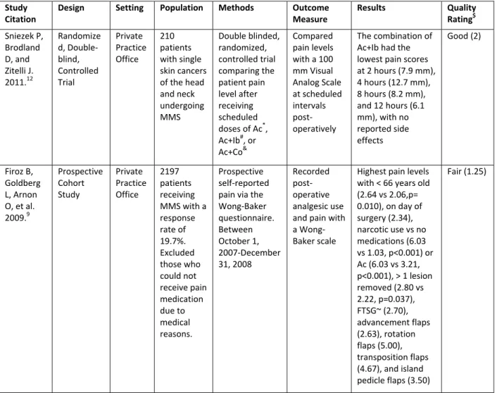
Table 1: Summary of study design characteristics
Study
Citation
Design Setting Population Methods Outcome
Measure
Results Quality
Rating$
Sniezek P,
Brodland
D, and
Zitelli J.
2011.12
Randomize d, Double‐
blind, Controlled Trial Private Practice Office 210 patients
with single
skin cancers
of the head
and neck
undergoing
MMS
Double blinded,
randomized,
controlled trial
comparing the
patient pain
level after
receiving
scheduled
doses of Ac*,
Ac+Ib#, or
Ac+Co&
Compared
pain levels
with a 100
mm Visual
Analog Scale
at scheduled
intervals
post‐
operatively
The combination of
Ac+Ib had the
lowest pain scores
at 2 hours (7.9 mm),
4 hours (12.7 mm),
8 hours (8.2 mm),
and 12 hours (6.1
mm), with no
reported side
effects
Good (2)
Firoz B,
Goldberg
L, Arnon
O, et al.
2009.9
Prospective Cohort Study Private Practice Office 2197 patients receiving
MMS with a
response
rate of
19.7%.
Excluded
those who
could not
receive pain
medication
due to
medical
reasons.
Prospective
self‐reported
pain via the
Wong‐Baker
questionnaire.
Between
October 1,
2007‐December
31, 2008
Recorded
post‐
operative
analgesic use
and pain with
a Wong‐
Baker scale
Highest pain levels
with < 66 years old
(2.64 vs 2.06,p=
0.010), on day of
surgery (2.34),
narcotic use vs no
medications (6.03
vs 1.03, p<0.001) or
Ac (6.03 vs 3.21,
p<0.001), > 1 lesion
removed (2.80 vs
2.22, p=0.037),
FTSG~ (2.70),
advancement flaps
(2.63), rotation
flaps (5.00),
transposition flaps
(4.67), and island
pedicle flaps (3.50)
Fair (1.25)
$
Quality rating was based on the average score of 4 categories, which included: 1) assembly and maintenance of
comparable groups, 2) equal, valid, and reliable measurements, 3) clear definition of the intervention, and 4) analysis
(adjusted for potential confounders in cohort studies, included intention to treat analysis in randomized controlled trials);
additional information can be found in the Methods section
* Acetaminophen
#
Acetaminophen + Ibuprofen
&
Acetaminophen + Codeine
~ Full‐thickness skin graft