O R I G I N A L
Epidural blood patch for refractory low CSF pressure headache:
a pilot study
Søren Aalbæk Madsen• Jonna Storm Fomsgaard• Rigmor Jensen
Received: 15 November 2010 / Accepted: 14 March 2011 / Published online: 2 April 2011
The Author(s) 2011. This article is published with open access at Springerlink.com
Abstract Once believed an exceedingly rare disorder, recent evidence suggests that low cerebrospinal fluid (CSF) pressure headache has to be considered an important cause of new daily persistent headaches, particularly among young and middle-aged individuals. Treatment of low CSF pressure headache consists of non-invasive/conservative measures and invasive measures with epidural blood patch providing the cornerstone of the invasive measures. In the present pilot study we therefore aimed to evaluate the treatment efficacy of epidural blood patch (EBP) in treat-ment-refractory low-pressure headache. Our primary effect parameter was total headache burden defined as area under the curve (AUC: intensity9duration) and as secondary effect parameters we identified: intensity (VAS 0-10), frequency (days per month), duration in hours (total hours/ month) and also medication days (days on medication/ month). In our primary effect parameter we found a sig-nificant reduction in AUC with more than 25% and this is considered to be clinically relevant. We found also a sig-nificant and relevant reduction at -22% in intensity. A trend towards reduction in duration was seen. We found no
statistically significant reduction in frequency. An increase in days with use of medication was found. Increased awareness of low CSF pressure headache is emphasized and a controlled larger randomized study is needed to confirm the results. However the present results, allows us to conclude that EBP in treatment-refractory low CSF pressure headache can be considered as a treatment option.
Keywords Intracranial hypotensionEpidural blood patchHeadacheLow CSF pressure headache
Purpose
To evaluate the treatment efficacy of epidural blood patch (EBP) in treatment-refractory low CSF (cerebrospinal fluid) pressure headache.
Introduction
Although first described in 1938 low CSF pressure head-ache is still an under-diagnosed headhead-ache disorder. This being partly due to unfamiliarity, and with the overlap with other forms of headache, often leading to misdiagnosis, incorrect treatment and leaving the patient with a persistent severe headache [1]. Low CSF pressure headache has significant symptom-overlap with migraine, tension type headache, post-infectious headache and most commonly post-dural puncture headache [2]. Symptoms vary greatly but the orthostatic headache, tinnitus, hypacusis, photo-phobia and/or nausea indicating low CSF pressure are the most frequently reported [4]. However, the associated symptoms may vary ranging from classic neurological symptoms to cognitive defects [3]. Diffuse pachymeningeal
S. A. Madsen (&)
Intensive Care Unit 4131, National Hospital, University of Copenhagen, Blegdamsvej 9, 2100 Copenhagen, Denmark
e-mail: [email protected]
J. S. Fomsgaard
Department of Anesthesiology, Glostrup Hospital, University of Copenhagen, Ndr. Ringvej 57, 2600 Glostrup, Denmark
R. Jensen
enhancement on brain magnetic resonance imaging (MRI) is the major finding of the classic syndrome. However, the diagnosis maybe difficult in long lasting cases as the cardinal symptom, the orthostatic headache-feature, may disappear over time and the diffuse meningeal enhance-ment may be absent. Over time various terminologies has been used to describe the syndrome:
• Spontaneous (or idiopathic) low CSF pressure headache
• Spontaneous (or primary) intracranial hypotension
• Low CSF volume headache
• Hypoliquorrhoeic headache
• Aliquorrhea
• CSF leak headache
• CSF hypovolemia
• CSF volume depletion.
The accepted etiology is leakage of cerebrospinal fluid, either spontaneously or as a result of minor/trivial trauma, instrumentation of or near the structures surrounding the spinal cord. The leak is usually located in the upper tho-racic level or at the cervico-thotho-racic junction [3]. As a result of this leakage, the brain sags providing traction on bridging veins, pain-sensitive meningeal structures and the cranial nerves, possibly explaining the associated auditory and, at times visual symptoms. In accordance with the Monroe–Kellie doctrine, secondary venodilation may develop as a compensatory measure to the low CSF pres-sure, adding a vascular component with venous dilatation to the typical MR-presentation introduced as SEEPS (subdural hygroma, enhancement, engorged veins, pituitary hyperaemia, and sagging of the brain [3]). Once believed to be an exceedingly rare disorder, recent evidence suggests that low CSF pressure headache is not that rare and has to be considered an important cause of new daily persistent headaches, particularly among young and middle-aged individuals [4]. This said few data are available regarding the demography of low CSF pressure headache. A peak incidence centered around 40 years of age has been reported, the incidence is estimated to 5 per 100,000 and women are overrepresented in a 2:1 ratio [3].
Treatment of low CSF pressure headache consists of non-invasive/conservative strategies and/or invasive mea-sures with epidural blood patch providing the cornerstone of the invasive measures. Although no randomized clinical trials are available to evaluate the effectiveness of the various treatment protocols, initial treatment for most patients consist of conservative measures. The most con-servative measure, bed rest and time, is probably effective in many patients [3]. Analgesics are often employed early as part of the conservative treatment they do though pro-vide little relief. Steroid-therapy is of anecdotal value [4]. Apart from EBP, other invasive measures are surgical repair and epidural fibrin glue, they are though, limited by
the fact that the epidural lesion has to be located which is not always possible [3]. EBP is the mainstay in the treat-ment of low CSF pressure headache, but no protocol has yet been developed. Treatment protocols vary between injection of 10–100 ml autologous blood and 1–6 EBPs, placement in the Trendelenburg position, premedication, etc. [5]. In the present pilot study we therefore aimed to evaluate the treatment efficacy of epidural blood patch (EBP) in treatment-refractory low-pressure headache.
Methods
We followed 14 patients all diagnosed at the Danish Headache Center (DHC), the national tertiary headache center, with low CSF pressure headache and treated with EBP. All the patients followed in this pilot study presented themselves with an orthostatic component as part of their symptoms although an overlap in symptoms was seen. At the time of initial evaluation, and at the time of the EBP all patients, except two, described significant worsening of the headache either when erect or when physically active.
All patients had been refractory to conservative non-invasive management and were referred to DHC for further evaluation. Before their first visit all patients receive a diagnostic headache diary and are told to record headache characteristics, including type and amount of medication used, prospectively for at least a 4-week period. They also fill out a questionnaire regarding status of health, impact on work, family and social life and prior headache treatment. A detailed headache history is obtained by means of a standardized procedure at the initial consultation. The history is supplemented by the diagnostic headache diary and the general medical questionnaire. After a complete general physical and neurological examination, all first-visit patients were classified according to ICHD-II by a headache specialist.
The headache diaries of the 14 patients were reviewed prior to treatment with EBP and a second review at least 12 months after treatment with EBP. Additional phone-interviews were performed and data from the preliminary and final examination were extracted in case of missing data. Our primary effect parameter was total headache burden defined as area under the curve (AUC: inten-sity9duration) and as secondary effect parameters we identified: intensity (VAS 0-10), frequency (days per month), duration in hours (total hours/month) and also medication days (days on medication/month).
L3/4, and L4/5). All EBP were performed with 18-G Touhy needle, using a midline approach with the patients lying on one side. Autologous blood (6–37 ml; mean 23.64 ml) was injected until patients complained of lumbar pain. After the procedure the patients were placed supine in bed for the next 24 h.
Results
In the period of 2005–2010 we intended to treat 25 patients with EBP on the indication of treatment-refractory low CSF pressure headache. During the follow-up process seven patients were excluded due to incomplete pre-treat-ment data, one patient did not fill out post-treatpre-treat-ment headache diary due to ongoing serious illness, one patient withdrew consent and two patients failed to return post-treatment headache diaries.
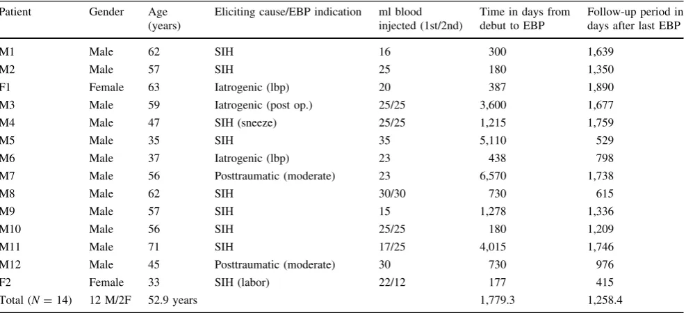
After the follow-up period our study consisted of 14 patients; 2 women and 12 men, aged 33–71 years of age (mean 52.86). All were treated and observed in the period 2005–2010. All received EBP 177–6,570 days (mean 1,779.29 days) after debut of headache. Follow-up period was 415–1,759 days (mean 1,258.4 days). Further patient characteristics are displayed in Table1.
Despite a number of eliciting causes all patients had significant overlap in symptoms and orthostatic headache as their core symptom. Nine patients presented with symptoms of spontaneous intracranial hypotension (SIH), hereof two patients with a history of tension type headache and medication overuse headache, and another two patients
reported Valsalva maneuver (sneeze, labor) as eliciting cause. Two patients reported physical trauma as the elic-iting cause. The traumas would be categorized as moderate. One patient was struck by a falling bookcase, resulting in laceration to the forehead and unconsciousness. The other trauma was caused by a falling loading ramp on a lorry also resulting in laceration to the forehead and unconsciousness. Three patients reported the eliciting cause as iatrogenic (lumbar puncture or post-operative).
All patients reported a significant orthostatic component as an initial part of their headache. Other symptoms included fatigue, tinnitus, photophobia, phonophobia, neck stiffness and concentration problems.
As our primary effect parameter total headache burden was defined as AUC (mean daily intensity9summated daily duration) and a statistically significant reduction was identified in AUC [pre-AUC 4,139.1 (±419.8 SEM) vs. post-EBP AUC 2,997.2 (±582.7 SEM); paired t test p =0.015 (CI 265.7; 2,018.1)] (see Fig.1).
As secondary effect parameters we identified intensity (mean daily VAS score) and found a statistically significant reduction [pre-EBP intensity 8.1(±0.4 SEM) vs. post-EBP intensity 6.2 (±0.6 SEM)]. This represents a reduction of 22% in intensity on VAS 0-10 scale (see Fig.2).
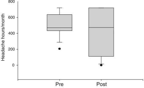
We found no statistically significant reduction in fre-quency, defined as days with headache (Wilcoxon signed rank test, p=0.313). Furthermore, a trend, but not statis-tically significant, a reduction in duration (defined as total hours of headache per month) was noted [pre-EBP duration 507.9 (±39.4 SEM) vs. post-EBP 421.6 (±71.7 SEM); paired ttest:p =0.11 (CI-194.9; 22.2)] (see Fig.3).
Table 1 Patient characteristics (N=14) Patient Gender Age
(years)
Eliciting cause/EBP indication ml blood injected (1st/2nd)
Time in days from debut to EBP
Follow-up period in days after last EBP
M1 Male 62 SIH 16 300 1,639
M2 Male 57 SIH 25 180 1,350
F1 Female 63 Iatrogenic (lbp) 20 387 1,890 M3 Male 59 Iatrogenic (post op.) 25/25 3,600 1,677 M4 Male 47 SIH (sneeze) 25/25 1,215 1,759
M5 Male 35 SIH 35 5,110 529
M6 Male 37 Iatrogenic (lbp) 23 438 798
M7 Male 56 Posttraumatic (moderate) 23 6,570 1,738
M8 Male 62 SIH 30/30 730 615
M9 Male 57 SIH 15 1,278 1,336
M10 Male 56 SIH 25/25 180 1,209
M11 Male 71 SIH 17/25 4,015 1,746
M12 Male 45 Posttraumatic (moderate) 30 730 976
F2 Female 33 SIH (labor) 22/12 177 415
An increase in days with use of medication [pre-EBP days 10.0 (Q 0;24) vs. post-EBP days 26.5 days (Q 1;30); Wilcoxon signed rank test:p=0.049] was found.
Discussion
We intended to evaluate the treatment efficacy of epidural blood patch in long-lasting treatment-refractory low CSF
pressure headache. In total, 25 patients were reviewed but due to losses in the follow-up period we ended up including 14 patients.
In our primary effect parameter, AUC, we found a sig-nificant reduction with more than 25% and this is consid-ered to be clinically relevant. Likewise a significant and relevant reduction in intensity at-22% was identified.
As expected, although statistically not significant, a trend towards reduction in duration was seen, whereas the headache frequency was unchanged. It can be theorized that lack of power is the reason for behind this. The increase in days with use of medication was expected due to a low number of patients with medication overuse and to the initiation of optimized treatment strategies in a subset of patients.
The treatment of low CSF pressure headache has never been evaluated by randomized clinical trials [3], but sev-eral series have now documented positive results for EBP in treating SIH [6–10]. These series report a success rate of 36–90% of complete recovery after one EBP increasing with the numbers of EBPs. Our series does not quite sup-port these results. Our patient population differed from other series in very long headache history, male prepon-derance and higher age. Although noticeable, neither age-specific nor gender-age-specific treatment response has ever been documented. Injected volume was very similar among studies.
We treated all our patients with lumbar EBP and placed them in a supine position for 24 h to ensure sufficient spread. Ferrante [5], Horikushi [6] and Berroir [7], etc. [8,
9] primarily performed lumbar EBPs but used different post-EBP treatment regimens. Thus, neither age, gender, site-specific EBP, nor post-EBP regimen seems to explain the differences found between this series and earlier pub-lished results.
A marked difference in this series compared to other publications is the timing of EBP treatment. In this study a mean of 1,779.29 days (177–6,570) passed from symptom onset to EBP treatment in our center.Other series report a mean of 20–62 days [5,7–9], Chung et al. [10] performed EBP treatment after 5–7 days of supportive treatment but time from symptom onset is unclear. This delay in treat-ment in our patients was due to further diagnostic workup and limitations in acute admissions availability. Also these long lasting patients may be more subtle and differently affected than the acutely and severely affected patients. Therefore, increased awareness and a detailed history of onset of headache is of major importance for the diagnosis, and some low CSF pressure headache patients may in fact be overlooked because of their atypical presentation. Sen-chakova et al. suggested that the EBP procedure was more effective the more rapidly it was performed after symptom onset, and that persisting symptoms resulted in slower and
AUC
0 1000 2000 3000 4000 5000 6000
*
Pre Post
Fig. 1 Box plot AUC pre-EBP versus post-EBP
Pre Post
Intensity
0 2 4 6 8 10
*
Fig. 2 Box plot. Intensity pre-EBP versus post-EBP
Headache hours/month
0 200 400 600 800
Pre Post
incomplete recovery. This suggests a chronification of pain or dysregulation of the intracranial blood volume [8]. Additionally, it is well known that the posture-related component becomes less prominent over time [9] and chronic disease involves a significant degree of central sensitization and stigmatization. This could be part of the explanation behind the difference between this and other series [7–10]. Worth mentioning is that Ferrante et al. and Berroir et al. only treated patients with purely orthostatic headache (40/42 and 30/30), and Chung et al. treated patients markedly younger and with a shorter delay than the patients in this study. Our patients had more mixed symptoms probably again reflecting a longer diagnostic delay contributing to the differences in results.
This study contains some apparent weaknesses. It is unblinded, retrospective and contains relatively few patients. The ever present lack of control for spontaneous recovery also applies to this study, but since the Danish Headache Center is a tertiary center, all the patients have received conservative treatment elsewhere, including a watchful wait. Patient compliance in returning post-treat-ment headache diaries proved to be a considerable chal-lenge. Also the level of detail (or lack of) lead to contact to the treated patients to clarify headache diary entries.
The headache diary, in turn, can also be considered a strength in addition to a systematic prospective approach since it allows us to monitor patients more precisely on several parameters. In turn this allows us to define more effect parameters than seen in other studies. Furthermore, as this is a ‘‘before-and-after’’ study, it contributes to considerable clinical and statistical validity.
As conclusion, in this pilot study of very long lasting low CSF pressure headache we found a significant reduc-tion in AUC, a clinically significant reducreduc-tion in intensity but also a significant increase in days with use of medi-cation after being treated with EBP. Additionally, we recorded a trend towards a reduction of headache duration but the very long lasting diagnostic delay may have com-promised the outcome. Increased awareness and early diagnosis of low CSF pressure headache is hereby emphasized and a controlled larger randomized study is
needed to confirm the results. However, the present results allow us to conclude that EBP in treatment-refractory low CSF pressure headache can be considered as a treatment option.
Acknowledgments Kim Lindelof, MD, PhD, Glostrup Hospital, is acknowledged for great help with statistics.
Conflict of interest None.
Open Access This article is distributed under the terms of the Creative Commons Attribution License which permits any use, dis-tribution and reproduction in any medium, provided the original author(s) and source are credited.
References
1. Schievink WI (2003) Misdiagnosis of spontaneous intracranial hypotension. Arch Neurol 60:1713–1718
2. (2004) The international classification of headache disorder (2nd edn). International Headache Society
3. Mokri B (2001) Spontaneous intracranial hypotension. Curr Pain Headache Rep 5:284
4. Schievink WI (2006) Spontaneous Spinal cerebrospinal fluid leaks and intracranial hypotension. JAMA 295(19):2286–2296. doi:10.1001/jama.295.19.2286
5. Ferrante E, Arpinob I, Citterioa A, Wetzlb R, Savinoc A (2010) Epidural blood patch in Trendelenburg position pre-medicated with acetazolamide to treat spontaneous intracranial hypotension. Eur J Neurol 17:715–719. doi:10.1111/j.1468-1331.2009.02913.x
6. Horikushi T et al (2006) Effectiveness of an epidural blood patch for patients with intracranial hypotension syndrome and persis-tent spinal epidural fluid collection after treatment. Eur J Neurol 13:1128–1138. doi:10.3171/2009.10.JNS09806
7. Berroir S, Loisel B, Ducros A, Boukobza M, Tzourio M, Valade D, Bousser MG (2004) Early epidural blood patch in spontaneous intracranial hypotension. Neurology 63(10):1950–1951
8. Senchakova D, Mokri B, McClelland RL (2001) The efficacy in epidural blood patch in spontaneous CSF leaks. Neurology. 57(10):1921–1923
9. Nowak DA, Takano B, Topka H (2006) Spontaneous cerebro-spinal fluid hypovolemia: a therapeutic dilemma ? Eur J Neurol 13(10):1128–1138. doi:10.1111/j.1468-1331.2006.01453.x