Beata M. Sobieszczańska
1, d, c, Urszula Kasprzykowska
1, a, B, anna duda-Madej
1, a, B,
anna Secewicz
1, B, Joanna Marciniak
2, B, Grażyna Gościniak
1, e, fRelevance of Serology for
Mycoplasma Pneumoniae
Infection Among Children with Persistent Cough
1 University of Medicine, department of Microbiology, Wroclaw Medical University, Poland 2 University of Medicine, 1st department and clinic of Paediatrics, allergology and cardiology, Wroclaw Medical University, PolandA – research concept and design; B – collection and/or assembly of data; C – data analysis and interpretation; D – writing the article; E – critical revision of the article; F – final approval of article; G – other
Abstract
Background. Mycoplasma pneumoniae is an important cause of upper and lower respiratory tract infections. cough and tracheobronchitis are the commonest features of M. pneumoniae infection but diagnosis based on clinical symptoms that may be due to other respiratory pathogens is impossible. Thus laboratory testing for M. pneumoniae is particularly important. correct and rapid diagnosis of M. pneumoniae infections is of prime importance to introduce appropriate antibiotic treatment.
Objectives. evaluation of the incidence of IgM and IgG antibodies specific to M. pneumoniae among children with pneumonia and/or chronic cough.
Material and Methods. Serum samples from 148 children with a history of chronic cough (lasting at least one month), recurrent respiratory tract infections, allergic rhinitis, and/or inflammatory changes on X-chest ray. first, all sera were screened for specific anti-M. pneumoniae antibodies using agglutination test following the detection of specific IgM and IgG anti-M. pneumoniae antibodies using immunoenzymatic assays.
Results. Out of the 148 serum samples, 57 (38.5%) gave positive screening results. However, the presence of M. pneumoniae-specific IgM and/or IgG antibodies was confirmed by immunoenzymatic assays in only 30 (52.6%) of these 57 positive samples. These results indicated that in as many as 27 (47.4%) out of the 57 serum samples screened, false-positive results occurred.
Conclusions. evaluation of acute- and convalescent-phase sera is necessary to make possible accurate interpreta-tion of the serological testing results (Adv Clin Exp Med 2014, 23, 2, 185–190).
Key words: Mycoplasma pneumoniae, serology, persistent cough.
adv clin exp Med 2014, 23, 2, 185–190 ISSN 1899-5276
ORIGINaL PaPeRS
© copyright by Wroclaw Medical University
Mycoplasma pneumoniae (M. pneumoniae) belongs to the family Mycoplasmataceae, order
Mycoplasmatales and class Mollicutes. all mem-bers of the class Mollicutes, including M. pneu-moniae, characterize the lack of a cell wall, mak-ing these microorganisms resistant to β-lactam antibiotics that inhibit bacterial cell wall synthe-sis. Moreover, the lack of a rigid cell wall confers pleomorphic natureof these organisms, inabili-ty to stain with Gram dyes and their substantial susceptibility to desiccation. Mycoplasmas be-long to the smallest microorganisms capable of a cell-free mode of living. Their small size allows mycoplasmas to pass through 0.45 µm pore size
filters and make them undetectable by light mi-croscopy [1, 2].
cell facilitates M. pneumoniae to fuse with and en-ter host cells establishing laen-ter or chronic infec-tions, although the extent to which the organism invades the host cells in vivo is not known.
M. pneumoniae is an important cause of up-per and lower respiratory tract infections as well as a wide spectrum of extrapulmonary manifestations, involving almost all organs of the human body and including neurological (e.g. M. pneumoniae is an important antecedent to Guillain-Barre syndrome, cranial nerve palsy, cerebellar ataxia, polyradic-ulopathy), hepatic, renal (e.g. Iga nephropathy, acute glomerulonephritis, renal failure) cardiac diseases, hemolytic anemia, polyarthritis, erythre-ma multiforme or Stevens-Johnson syndrome. al-though most commonly M. pneumoniae infections occur among children 5–15 years of age, they can be involved in diseases in older individuals and in children below 5 years of age [4–6]. In Poland, the epidemiological data from the period 1994–1996 showed that M. pneumoniae caused from 2 to 16% of all respiratory tract infections; however, the per-centage of infections increases every 4–5 years dur-ing epidemics and may reach even 60% [7]. accord-ing to surveillance data from european countries during the autumn of 2011 there was an increase in reporting rates of M. pneumoniae infections in northern but not in southern europe [8].
M. pneumoniae may account for as many as 25% of the clinical manifestations of the upper re-spiratory tract illness. The most common mani-festations include sore throat, hoarseness, fever, nonproductive cough, headache, chills, coryza, myalgia and general malaise [1, 2, 9]. children un-der 5 years of age are most likely to manifest cory-za and wheezing but progression to bronchopneu-monia is relatively uncommon in this age group. However, M. pneumoniae may cause up to 5% of
cases of bronchiolitis in young children. In contrast, older children are more likely to develop broncho-pneumonia. Mild and asymptomatic infections are particularly common in adults though broncho-pneumonia develops in 3 to 10% of infected indi-viduals [1]. community acquired pneumonia due to M. pneumoniae is usually mild and self-limit-ing (hence term walkself-limit-ing pneumonia) but severe pulmonary involvement requiring hospitalization can occur in otherwise healthy children and adults. Pleural effusion, pneumatocele, lung abscesses, pneumothorax, bronchiectasis, chronic interstitial fibrosis and acute respiratory-distress syndrome have been reported [2, 9]. M. pneumoniae -associ-ated respiratory tract infections may promote the exacerbation of asthmatic symptoms, which is re-lated to the immune response against these micro-organisms [10, 11]. M. pneumoniae infection was found to increase the production of interleukin-4 (IL-4) and interferon gamma (IfNγ), indicat-ing a predominant Th2-like cytokine response and creating favorable condition for Ige produc-tion [12]. Stelmach et al. [13] have shown serum Ige increase in children after acute infection with
M. pneumoniae. furthermore, lipoproteins and glycoproteins present in the cell membrane and cytoplasm of M. pneumoniae are potent inducers of cytokines that through their molecular mimicry can elicit autoimmunity.
Nonproductive cough is one of the most prominent clinical symptom of M. pneumoni-ae infection. according to Kiciński et al. [14]
M. pneumoniae infection was confirmed in 9.4% of 1710 children up to 6 years of age with cough-ing as a predominant clinical symptom of pneu-monia. Wang et al. [15] have detected M. pneu-moniae infection among 12.9% of children with a persistent cough. In the study we analyzed the incidence of IgM and IgG antibodies specific to
M. pneumoniae among children with pneumonia and/or chronic cough hospitalized in 1st depart-ment and clinic of Paediatrics, allergology and cardiology of the Wroclaw Medical University, Poland.
Material and Methods
Serum samples from 148 children were ob-tained from 1st department and clinic of Paediat-rics, allergology and cardiology, Wroclaw Med-ical University. The ages of the patients ranged from 18 months to 17 years (median age, 7 years). all patients had a history of chronic cough (last-ing at least one month), recurrent respiratory tract infections, allergic rhinitis, and/or inflammatory changes on X-chest ray.
Fig. 1. Species of Mycoplasmataceae family inhabiting mucosal surfaces of the respiratory and urogenital tract in humans [1]. Species involved in human infections are marked in bold
Site of colonization
Respiratory tract Urogenital tract
M. buccale M. faucium
M. fermentans
M. lipophilum M. orale M. salivarium M. primatum
M. pneumoniae
M. fermentans M. hominis M. genitalium
M. penetrans M. primatum M. spermatophilum
Mycoplasma Screening Assay
first, all sera were screened for specific anti- -M. pneumoniae antibodies. The Serodia Myco II gelatin particle agglutination test (fujirebio, pur-chased from Mast diagnostica, Poland) was per-formed according to the manufacturer’s instruc-tions. The test mainly measure IgM (and in a lesser extent IgG) antibody for M. pneumoniae and is based on the principle that gelatin particles sensi-tized with M. pneumoniae cell-membrane compo-nents are agglutinated by specific antibodies.Mycoplasma IgM and IgG
Antibody by Enzyme-Linked
Immunosorbent Assay (EIA)
for detection of specific IgM and IgGanti-M. pneumoniae antibodies the eIa-Platelia (Bio-Rad Laboratories, Poland) was performed. The antigen included in the test contains a high pro-portion of membrane proteins and is enriched in P1 cytoadhesin and some other cytoadherence-as-sociated proteins. The test was performed manual-ly according to the manufacturer’s recommenda-tions. The test result was validated as instructed in the kit manual. a specimen was considered pos-itive for IgM antibodies to M. pneumoniae when the absorbance value of the specimen was equal to or greater than that of the cutoff serum specimen included in the kit. The values of IgG antibodies to M. pneumoniae were validated and transformed to arbitrary units (aU) from 1 to 100 determined with the kit standards, and the results were inter-preted as recommended by the manufacturer. The specimen aU values were determined from the calibration curve and were interpreted as follows. a value of < 10 U for a single serum specimen was considered insignificant, a value of > 10 and < 40 U was considered low, and a value of ≥ 40 U was con-sidered high.
Results
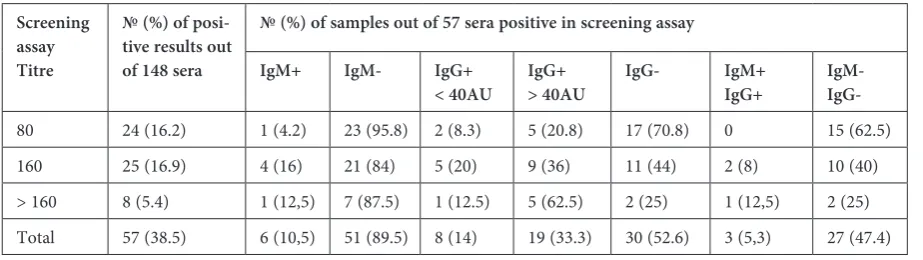
Out of the 148 serum samples, 57 (38.5%) gave positive screening results. However, the presence of M. pneumoniae-specific IgM and/or IgG an-tibodies was confirmed by eIa assays in only 30 (52.6%) of these 57 positive samples (fig. 2). These results indicated that in as many as 27 (47.4%) out of the 57 serum samples screened, false-positive re-sults occurred.
among these 30 serum samples positive in eIa-Platelia assay, M. pneumoniae-specific IgM antibodies were present in 6 (10.5%) serum
Fig. 2. The incidence of specific anti-M. pneumoniae antibodies determined by screening test and eIa- -Platelia assay. Out of the 148 serum samples examined 57 (38.5%) was positive, but in only 30 (20.3%) samples the presence of specific antibodies was confirmed by eIa-Platelia test
Screening EIA-Platelia
%
of
posi
tiv
e s
am
ples
samples. Both, IgM and IgG antibodies were pres-ent in 3 (5.3%) of serum samples examined (Ta-ble 1). Taking into consideration that all these IgM-positive samples were obtained from chil-dren presenting symptoms of the lower respiratory tract infection, i.e. persistent cough, an interstitial inflammatory changes on X-chest ray, these sero-logic assay results indicated an acute infection. as many as 19 (33.3%) serum samples showed high levels of IgG without the accompanying IgM. In all these cases the interpretation of these results is difficult as may indicate recent remote infection as well as an acute infection. Hence, to discriminate between acute and remote infection the second serum sample should be examined. In 8 (14%) of samples there were only low levels of IgG antibod-ies, indicating a remote infection. Thus, although all these children showed clinical signs of the low-er respiratory tract infection, most likely M. pneu-moniae was not the cause of these symptoms. On the other hand, taking into consideration that even among children re-infections may occur, the low levels of IgG antibody may indicate anamnes-tic response which can be confirmed by re-exami-nation of the IgG level after two weeks. These cas-es perfectly illustrate the importance of serological tests in all patients with suspected mycoplasmal pneumonia as well as the necessity to re-examine doubtful results of serologic tests e.g. low levels of IgG without accompanying IgM antibodies.
Discussion
due to other respiratory pathogens is impossible, thus laboratory testing for M. pneumoniae is par-ticularly important. correct and rapid diagnosis of M. pneumoniae infections is of prime impor-tance in order to introduce appropriate antibiot-ic treatment [16].
culture of M. pneumoniae from clinical spec-imens is highly specific but time-consuming, dif-ficult and relatively insensitive. Moreover, col-onization of the upper respiratory tract by
M. pneumoniae in asymptomatic individuals has been reported, making the clinical value of a posi-tive culture results uncertain [17]. Therefore, lab-oratory diagnosis of M. pneumoniae infection is usually established through serological or molec-ular testing. Polymerase chain reaction (PcR), similarly to culture characterize high specifici-ty, but low or variable sensitivispecifici-ty, thus PcR can-not replace serology although, may be performed in conjunction with serology [1, 16]. Several com-mercial tests utilizing passive agglutination, indi-rect immunofluorescence and immunoenzymatic assays are available and widely used for the detec-tion of antibodies specific to M. pneumoniae in human sera. The complement fixation test, being as it is unspecific and insensitive, is no longer ac-cepted [18]. However, several factors i.e. time of serum sample collection or patient’s age, influ-ence on the results of serologic tests as well as on the results interpretation. Moreover, the interpre-tation of serological tests results is complicated by the fact that low levels of antibody specific to
M. pneumoniae are found in sera of healthy indi-viduals, probably because of past M. pneumoniae
infections or repeated exposures on M. pneumoni-ae. Thus, in M. pneumoniae infection it is difficult to set up criteria to determine acute or remote in-fection [19, 20].
following an initial infection, specific IgM an-tibodies are rapidly produced, indicating acute or recent primary M. pneumoniae infection. IgM an-tibodies appear 7 to 10 days after infection, peak
after 3 to 6 weeks, followed by a gradual decline over months to years [1]. However, specific IgM antibodies do not always indicate an acute infec-tion, as they can persist for up to a year after the
M. pneumoniae infection. In addition, IgM anti-bodies may not be present if the serum sample is obtained too early in the infection. Moreover, an IgM response may be either minimal or undetect-able when adults are infected and during re-infec-tion [16, 18]. Thus, a negative result of IgM evalu-ation does not exclude current infection, especially in patients over the age of 45 years. Since children have fewer than adults opportunities for repeated exposures to M. pneumoniae, the presence of scific IgM antibodies has a high reliability in pe-diatric patients with a recent infection of at least a week’s duration, even on a single serum sam-ple [1, 20]. The overall data showed that the fre-quency of the carriage M. pneumoniae in symp-tomless children is low, ranging from 4.6% to 13.5% [2].
Specific IgG antibodies appear about 2 to 3 weeks after IgM occurrence and maximal re-sponse for IgG occur during the fifth week after onset of disease. an elevated IgG is frequently in-terpreted as evidence of acute infection, whereas low levels of IgG can indicate either an early stage of the current infection or a past illness. Howev-er, specific IgG antibodies may remain elevated for extended periods and thus do not discriminate be-tween a current or remote M. pneumoniae infec-tion. On the other hand, IgG is produced more quickly as an anamnestic response to re-infection; therefore, a second serum sample collected with an interval of 2 to 3 weeks should be examined, when a fourfold or greater increase of specific IgG titer evidences current infection [16, 21, 22]. Unfor-tunately, correct interpretation of tests results in paired sera with a rise in IgG antibody titer, delays the diagnosis.
Importantly, the serologic response may not reach detectable levels, thus producing false-Table 1. The incidence of positive and negative results of screening and immunoenzymatic tests
Screening assay Titre
№ (%) of posi-tive results out of 148 sera
№ (%) of samples out of 57 sera positive in screening assay
IgM+ IgM- IgG+
< 40AU IgG+> 40AU IgG- IgM+IgG+ IgM-
negative results, if a patient is successfully treated with antibiotics early in the course of the disease. Moreover, Uldum et al. [23] have shown that chil-dren younger than 10 years may have negative IgG whereas sera of children infected with M. pneu-moniae with high IgG value may have reduced by 20 to 40% IgM reaction, causing a risk of a false-negative IgM reactions.
Moreover, considering that many commen-sal Mycoplasma species, commonly colonizing the human oropharynx, can produce a cross-reaction with M. pneumoniae antibodies, serologic test re-sults should be interpreted with caution and in as-sociation with clinical symptoms [1]. Mycoplasmal adhesion molecules exhibit homology with human cd4 and class II major histocompatibility complex lymphocyte proteins, which could generate auto-reactive antibodies [1, 24]. In the study as many as 47.4% of serum samples showed false-positive results in a screening test, indicating that cross-reacting antibodies are common in children and may affect the actual number of infections detect-ed. although the detection of antibodies specific to
M. pneumoniae allows us to eliminate most false-positive results, this still does not help to interpret the serologic tests results correctly.
The analysis presented in this study of serolog-ic tests results perfectly illustrates the diffserolog-iculties in the interpretation of the results, arising from a sin-gle serum sample survey. In pediatric patients the
presence of IgM antibody is a good indicator of an acute M. pneumoniae infection, even in a single se-rum sample. However, the presence of IgG with-out accompanying IgM makes the interpretation of the result difficult. even high levels of IgG anti-body may result from a remote infection, but may also indicate acute infection without IgM. The presence of specific IgG without IgM, combined with clinical signs of the lower respiratory tract infection, may suggest M. pneumoniae infec-tion and incorrect treatment. after all, many oth-er infectious agents e.g. bactoth-eria, viruses and fun-gi, can cause similar to M. pneumoniae infection clinical signs and symptoms. although treatment with macrolides (e.g. erythromycin, clarithromy-cin, roxithromyclarithromy-cin, josamycin and most effec-tive azithromycin) and ketolides (telithromycin), tetracycylines (e.g. minocycline) and chinolones (e.g. levofloxacin, gatifloxacin, moxifloxacin, gemi-floxacin) that are active against mycoplasmas may be effective against other pathogens of the respira-tory tract, in many cases they can also lead to an-tibiotics abuse [4]. Taking into account the impact of mycoplasmal infections on the host immune sys-tem e.g. and their association with allergies, asth-ma and immune response disturbances, correct di-agnosis and treatment is of particular importance. Thus, evaluation of acute- and convalescent-phase sera could make accurate interpretation of the se-rological testing results possible.
References
[1] Waites KB, Talkington DF: Mycoplasma pneumoniae and its role as a human pathogen. clin Microbiol Rev 2004, 17, 697–728.
[2] Principi N, Esposito S: emerging role of Mycoplasma pneumoniae and Chlamydia pneumoniae in paediatric respi-ratory-tract infections. Lancet 2001, 1, 334–344.
[3] Rottem S: Invasion of mycoplasmas into and fusion with host cells. In Molecular biology and pathogenicity of mycoplasmas. eds.: Razin S, Herrmann R, Kluwer academic/Plenum Publishers, New York 2002, 391–402. [4] Kashyap S, Sarkar M: Mycoplasma pneumonia: clinical management. Lung India 2010, 27, 75–85.
[5] Narita M: Pathogenesis of neurologic manifestations of Mycoplasma pneumoniae infection. Pediatr Neurol 2009, 41, 159–166.
[6] Sharma MB, Chaudhry R, Tabassum I, Ahmed NH, Sahu JK, Dhawan B, Kalra V: The presence of Mycoplasma pneumoniae infection and GM1 ganglioside antibodies in Guillain-Barre syndrome. J Infect dev ctries 2011, 5, 459–464.
[7] Załęska-Ponganis J, Jackowska T: atypowe bakteryjne zapalenie płuc u dzieci. Post Nauk Med 2008, 9, 582–588. [8] european centre for disease Prevention and control, 2012/http://ecdc.europa.eu/en/press/news/Lists/News/
ecdc_dispform.aspx?List=32e43ee8-e230-4424-a783-85742124029a&Id=554
[9] Esposito S, Principi N: asthma in children: are chlamydia or mycoplasma involved? Paediatr drugs 2001, 3, 159–168.
[10] Biscardi S, Lorrot M, Marc E, Moulin F, Boutonmat-Faucher L, Heilbronner C, Iniguez JL, Chaussain M, Nicand E, Raymond J, Gendrel D: Mycoplasma pneumoniae and asthma in children. clin Infect dis 2004, 38, 1341–1346.
[11] Specjalski K: Rola zakażeń Chlamydia pneumoniae i Mycoplasma pneumoniae w przebiegu astmy. Pneumol alergol Pol 2010, 78, 284–295.
[12] Zaki MS, Raafat D, Metaal AA: Relevance of serology for Mycoplasma pneumoniae diagnosis compared with PcR and culture in acute exacerbation of bronchial asthma. am J clin Pathol 2009, 131, 74–80.
[14] Kiciński P, Wiśniewska-Ligier M, Woźniakowska-Gęsicka T: Zapalenie płuc o atypowej etiologii u dzieci do 6 roku życia. Przegl Pediatr 2011, 41, 15–21.
[15] Wang K, Chalker V, Berningham A, Harrison T, Mant D, Harnden A: Mycoplasma pneumoniae and respiratory virus infections in children with persistent cough in england: a retrospective analysis. Pediatr Infect dis J 2011, 30, 1047–1051.
[16] Zhang L, Zong ZY, Liu YB, Ye H, Lv XJ: PcR versus serology for diagnosing Mycoplasma pneumoniae infection: a systematic review and meta-analysis. Indian J Med Res 2011, 134, 270–280.
[17] Karppelin M, Hakkarainen K, Kleemola M, Miettinen A: comparison of three serological methods for diagnos-ing Mycoplasma pneumoniae infection. J clin Pathol 1993, 46, 1120–1123.
[18] Talkington DF, Shott S, Fallon MT, Schwartz SB, Thacker WL: analysis of eight commercial enzyme immuno-assay tests for detection of antibodies to Mycoplasma pneumoniae in human serum. clin diag Lab Immunol 2004, 11, 862–867.
[19] Jacobs E, Bennewitz A, Bredt W: Reaction pattern of human anti-Mycoplasma pneumoniae antibodies in enzyme-linked immunosorbent assays and immunoblotting. J clin Microbiol 1986, 23, 517–522.
[20] Petitjean J, Vabret A, Gouarin S, Freymuth F: evaluation of four commercial immunoglobulin G (IgG)- and IgM-specific enzyme immunoassays for diagnosis of Mycoplasma pneumoniae infections. J clin Microbiol 2001, 40, 165–171.
[21] Lui FC, Chen PY, Huang FL, Tsai CR, Lee CY, Lin CF: do serological testes provide adequate rapid diagnosis of Mycoplasma pneumoniae infection? Jpn J Infect dis 2008, 61, 397–399.
[22] Sillis M: The limitations of IgM assays in the serological diagnosis of Mycoplasma pneumoniae infections. J Med Microbiol 1990, 33, 253–258.
[23] Uldum SA, Jensen JS, Sondergard-Andersen J, Lind K: enzyme immunoassay for detection of immunoglobulin M (IgM) and IgG antibodies to Mycoplasma pneumoniae. J clin Microbiol 1992, 30, 1198–1204.
[24] Root-Bernstein RS, Hoobs SH: Homologies between mycoplasma adhesion peptide, cd4 and class II MHc pro-teins: a possible mechanism for HIV-mycoplasma synergism in aIdS. Res Immunol 1991, 142, 519–523.
Address for correspondence:
Beata M. Sobieszczańska department of Microbiology Wroclaw Medical University chałubińskiego 4
50-346 Wrocław Poland
Tel.: 071 784 13 08
e-mail: [email protected]
conflict of interest: None declared
![Fig. 1. Species of Mycoplasmataceae family inhabiting mucosal surfaces of the respiratory and urogenital tract in humans [1]](https://thumb-us.123doks.com/thumbv2/123dok_us/8769341.1756145/2.595.73.287.63.216/species-mycoplasmataceae-family-inhabiting-mucosal-surfaces-respiratory-urogenital.webp)