J. Serb. Chem. Soc. 85 (0) 1–14 (2020) UDC
JSCS–8813 Original scientific paper
1
Systematic profiling of ATP response to acquired drug-resistant
EGFR family kinase mutations
DINGWA ZHANG1*, DEYONG HE1, XIAOLIANG PAN2 and LIJUN LIU1**
1School of Chemistry and Chemical Engineering, Jinggangshan University, Ji’an 343009,
China and 2School of Mechanical and Electrical Engineering, Jinggangshan University, Ji’an 343009, China
(Received 24 November 2019, revised 8 April, accepted 19 May 2020)
Abstract:Kinase-targeted cancer therapy (KTCT) with ATP-competitive inhi-bitors has been widely applied in clinics. However, a number of kinase mis-sense mutations were observed to confer acquired drug resistance during ther-apy, largely limiting the clinical application of kinase inhibitors in KTCT. Ins-tead of directly influencing inhibitor binding, kinase mutations can also cause generic resistance to ATP-competitive inhibitors by increasing ATP affinity. Herein, the intermolecular interaction of the ATP molecule with clinically observed drug-resistant EGFR family kinase mutations involved in human can-cer are systematically characterize. Rigorous quantum mechanics/molecular mechanics (QM/MM) calculation and empirical Poisson–Boltzmann/surface area (PB/SA) analysis as well as in vitro kinase assay and surface plasmon resonance analysis were integrated to explore the binding capability of ATP to mutant residues in the structural context of the kinase domain, which resulted in a comprehensive profile of ATP response to acquired drug-resistant mutat-ions of four EGFR family kinases (EGFR/ErbB1, ErbB2, ErbB3 and ErbB4). From the profile, it was possible to identify those potent mutations that may influence ATP binding significantly; such mutations are potential candidates to cause generic resistance for ATP-competitive inhibitors. Consequently, the well documented generic drug-resistant mutation EGFR T790M and its count-erpart ErbB2 T798M are found to increase ATP affinity by establishing an additional S–π interaction between the side-chain thioether group of the mutant Met residue and the aromatic adenine moiety of the ATP molecule, while EGFR D761Y is identified as a new generic drug-resistant mutation that can increase ATP affinity by eliminating unfavorable electrostatic repulsion. In contrast, ErbB2 K753E and T768I are considered to be two generic drug-sen-sitive mutations that can decrease ATP affinity by unfavorable charge reversal and by impairing favorable polar interaction, respectively. In addition, the EGFR L858R mutation is located at the kinase activation loop and nearby the kinase active site, thus largely complicating the multiply dependent
relation-*,** Corresponding authors. E-mail: (*)[email protected]; (**)[email protected]
ship of kinase, ATP and inhibitor, which therefore exhibits divergent effects on different tested inhibitors.
Keywords:molecular modeling; inhibitor; biomolecular interaction; missense mutation; human cancer.
INTRODUCTION
Protein kinases play a predominant regulatory role in nearly every aspect of cell biology; they regulate apoptosis, differentiation, development, transcription, cycle and cytoskeletal rearrangement. The human epidermal growth factor rec-eptor (EGFR) family consists of four protein-tyrosine kinase members (ErbB1/ /EGFR/Her1, ErbB2/Her2, ErbB3/Her3 and ErbB4/Her4) that belong to the ErbB lineage of proteins, which are among the most documented cell signaling families in biology.1 These receptors consist of a glycosylated extracellular domain, a
single hydrophobic transmembrane segment, a juxtamembrane segment, a protein kinase domain, and a carboxyterminal tail. They are activated by ligand-induced dimerization as well as by transphosphorylation of their intracellular kinase dom-ains.2 Aberrant activation of ErbB kinases is implicated in many human cancers,
such as glioma, breast cancer, ovarian cancer, and non-small cell lung cancer.3 Given their central role in regulating cell proliferation, survival and migration in normal and cancerous cells, much effort has been devoted to the development of cancer therapeutics targeting ErbB kinases. Molecular targeting therapy against this family with small-molecule tyrosine kinase inhibitors (TKIs) has been estab-lished as a promising strategy against diverse cancers. Nowadays, several TKI drugs, such as Erlotinib, Gefitinib and Lapatinib, have been approved by the US FDA, and also, a number of second- and third-generation TKIs, such as Afatinib, Neratinib and WZ4002, are in clinical or preclinical development.4
Clinical observations revealed that many patients treated with TKIs have dramatic response initially, but would eventually develop resistance to these inhi-bitors.5 Subsequent studies found that missense mutations in ErbB kinase
T790M gatekeeper mutation causes drug resistance through a new mechanism that increases substrate ATP affinity to EGFR instead of impairing inhibitor binding to the kinase.7 It was suggested that this mutation could confer a generic
drug resistance to all ATP-competitive TKIs. However, since then only very few efforts have been made at exploring the association between kinase mutation-induced ATP affinity change and acquired drug resistance, primarily focusing on EGFR kinase and a few kinase gatekeeper mutations.9,10
In order to systematically investigate the effect of ErbB family missense mutations on ATP affinity, an attempt was made to create a comprehensive pro-file of ATP response to such mutations (not only limited to gatekeeper mutat-ions). However, it is known that experimental analysis of all kinase mutation– –ATP pairs is too time-consuming and expensive. To combat this problem, herein an integrative strategy was employed to combine in silico modeling and in vitro assay, which could be regarded as a good compromise between efficiency and accuracy. The quantum mechanics/molecular mechanics (QM/MM) method has been widely used to study biomolecular binding phenomenon.11–13 Pre-viously, we have systematically optimized the theoretical combination in QM/ /MM calculations to reproduce biomolecular recognition and interaction.14 The optimized method was also successfully employed to investigate strain energy in enzyme–substrate binding15 and to rationally design orthogonal stacking systems at the protein–peptide complex interface.16,17 In this study, a gene oncology
search was performed against the medicinal literature databases to enrich a large number of clinically observed acquired drug-resistant ErbB mutations involved in human cancer. Next, the QM/MM method was employed to determine the change in ATP binding free energy upon enriched drug-resistant ErbB mutations. A number of potential mutation–ATP pairs associated with strong sensitization or passivation were identified; their structural basis and energetic property were examined in detail, from which, a few promising pairs were tested using exp-erimental kinase assay and surface plasmon resonance analysis to substantiate the computational findings.
EXPERIMENTAL
Gene ontology enrichment of clinical drug-resistant ErbB mutations involved in human cancers
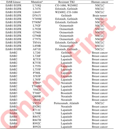
Although many mutations/variants/alternations have been observed in ErbB kinases, herein only those of clinical significance were considered, i.e., the mutations should be rel-evant to human cancers and acquired during kinase-targeted TKI therapy. In addition, only single-point somatic mutations occurring in the ErbB kinase domains were considered in this study. By gene ontology (GO) enrichment against the medicinal literature databases,18 a total
of 37 acquired drug-resistant mutations harbored on the kinase domains of four ErbB kinases that are known therapeutic targets of diverse human cancers, such as non-small-cell lung car-cinoma (NSCLC), breast cancer and brain tumor, were manually identified. These mutations have been documented to exhibit resistance to a variety of marketed or investigated kinase-targeted drugs, such as Gefitinib, Lapatinib, Osimertinib and Erlotinib. Here, the 37 GO-enriched
On
Lin
e F
irs
kinase gatekeeper mutations involved in human cancers are listed in Table S-I of the Sup-plementary material to this paper.
Computational analysis of ATP binding energy change upon ErbB mutations
The complex structures of the ATP molecule with wild-type and mutant ErbB kinase domains were computationally modeled using a protocol described in the Supplementary material, Fig. S-1. The modeled complexes of wild-type and mutant ErbB kinase domains with ATP molecule were structurally minimized using an ONIOM-based quantum mech-anics/molecular mechanics (QM/MM) method described in previous works.16,17 This method
enables different levels of theory to be applied to different parts of a large biomolecular sys-tem. The complex system was divided into a high-level QM layer and a low-level MM layer; the QM layer contained the ATP molecule and the mutated kinase residue, and was treated using the semi-empirical AM1 method, while the MM layer contained the rest of the system that was modeled with all-atom UFF force field. The combination of AM1/UFF was proved in a previous work as a good compromise between computational efficiency and accuracy.14 The
intermolecular interaction energy change (∆U) between kinase and ATP was estimated with a supermolecule approach.19,20 The Poisson–Boltzmann/surface area (PB/SA) method was used
to describe the solvent effect (∆Gslv) associated with the kinase–ATP binding.21 Considering
that ATP is a relatively rigid small molecule and its binding entropy to different kinase mut-ants could be regarded as a constant.22 Consequently, the total binding free energy of a kinase–
–ATP complex could be calculated as: ΔGttl = ∆Uint + ∆Gplr + ∆Gnplr.23,24 Therefore, the total
binding free energy change (ΔΔGttl) of ATP upon a drug-resistant kinase mutation could be
expressed as: ΔΔGttl = ΔGttlmt – ΔGttlwt, where the ΔGttlmt and ΔGttlwt are the total binding free
energy changes of ATP to wild-type and mutant kinases, respectively.25,26
Measurements
The inhibitory activities of Gefitinib and Lapatinib against wild-type ErbB1 and ErbB2 kinase domains as well as their respective mutants were determined using in vitro kinase assays, which can be found in the Supplementary material. The binding affinities of ATP molecule to wild-type ErbB2 kinase domain and its mutants were measured using surface plasmon resonance (SPR) analyses, which can be found in the Supplementary material.
RESULTS AND DISCUSSION
Creation of systematic ATP response profile to drug-resistant ErbB mutations
The complex structures of ATP with four wild-type ErbB kinase domains as well as their 37 drug-resistant mutations were computationally modeled using ligand grafting, virtual mutagenesis and energy minimization, which were then calculated using QM/MM and PB/SA to derive the binding free energy changes of ATP upon the 37 mutations. The obtained ΔΔGttl values reflect the effect of
different drug-resistant ErbB mutations on the strength of the intermolecular interaction of ATP with ErbB kinase domains, which may reveal information about the acquired drug resistance arising from mutation-induced ATP affinity change. As can be seen in Fig. 1, the profile is highly uneven and most mutations have only a moderate or modest effect on ATP binding with –1 <ΔΔGttl < 1
kcal* mol–1. In addition, most mutations are predicted to increase ATP affinity
* 1 kcal = 4184 J
On
Lin
e F
irs
(ΔΔGttl < 0), which lead to a favorable response for ATP binding, suggesting that
acquired drug-resistant ErbB mutations can generally cause drug resistance by, at least partially, improving ATP binding capability to combat ATP-competitive inhibitors for the kinase active sites. This is expected because such mutations addressing unfavorable ATP response would tend to sensitize (rather than cause resistance for) kinase inhibitors.
Fig. 1. The total binding free energy change (ΔΔGttl) profile used to characterize the
systematic response of ATP to 37 drug-resistant ErbB mutations. The asterisk (*) indicates gatekeeper mutation.
As might be expected, the well-documented EGFR gatekeeper mutation T790M can largely improve ATP affinity (ΔΔGttl = –1.97 kcal/mol), thus
signi-ficantly reducing the competitive capability of inhibitor ligands with the ATP for kinase active site, conferring generic resistance to TKIs. Interestingly, the ErbB2 gatekeeper mutation T798M exhibits a consistent behavior (ΔΔGttl = –1.78 kcal
mol–1) with the ErbB1 gatekeeper mutation T790M, both of which can largely promote ATP binding. However, another ErbB2 gatekeeper mutation T798I would moderately reduce the ATP binding affinity (ΔΔGttl = 0.23 kcal mol–1),
suggesting a considerable difference between the Met and Ile substitutions of ErbB2 gatekeeper residue Thr798. A similarly phenomenon can be observed on the ErbB3 gatekeeper mutation T768I, which also impairs ATP binding (ΔΔGttl =
= 0.58 kcal mol–1).
In addition, there are also a few of mutations that were predicted to enhance ATP affinity substantially (ΔΔGttl < –1 kcal mol–1), including ErbB1 D761Y,
G796S, G796R, L858R/ErbB2, T798M/ErbB3, Q809R/ErbB4 and L798R. It is interesting to see that these favorable mutations can be divided into three groups,
i.e., negative charge-eliminated mutations (such as ErbB1 D761Y), positive charge-added mutations (such as ErbB3 Q809R) and others (such as ErbB1 T790M). This is not unexpected because the ATP molecule is highly negatively
On
Lin
e F
irs
charged in its triphosphate moiety, which would be electrostatically attractive to positively charged residue but be electrostatically repulsive to negatively charged residues. Therefore, the mutations that introduce positive charge or eliminate negative charge to the complex system would effectively improve ATP affinity to kinase. In addition, there are two mutations, ErbB2 K753I and K753E, that can significantly impair ATP binding (ΔΔGttl = 0.58 and 1.98 kcal mol–1,
respect-ively). These mutations are expected to sensitize (but not retard) inhibitors. However, considering that the effect of a kinase mutation on TKI inhibitory pot-ency is determined by multiple factors; the mutation can influence the activity not only by indirectly changing ATP binding, but also by directly altering inhi-bitor binding. In addition, the mutation can also influence the kinase catalytic activity directly (such as eliciting constitutive activation of the kinase), which generally reduces inhibitor potency. Therefore, the mutation can cause drug res-istance to TKIs in multiple ways, but not only through the ATP approach. This would largely complicate the problem.
Case analysis of drug-resistant ErbB mutation effects on inhibitor binding
Several representative favorable and unfavorable drug-resistant ErbB mutat-ions were selected from the created systematic response profile and examined at the structural level using dynamics and machine approaches to understand their molecular mechanism and biological implication underlying the resistance.27–29
The ErbB2 gatekeeper mutations T798M and T798I were predicted to have opposite effects on ATP binding; the former can considerably promote ATP affi-nity (ΔΔGttl = –1.78 kcal mol–1 < 0), whereas the latter can moderately impair
ATP affinity (ΔΔGttl = 0.58 kcal mol–1 > 0). Here, the binding modes of ATP to
wild-type ErbB2 kinase domain and its T798M and T798I mutants were modeled and minimized computationally and compared at the structural level.30 As can be
seen in Fig. 2A, the purine ring of the ATP molecule is close enough to the polar side chain of the wild-type gatekeeper residue Thr798 to form a polar interaction between them. Substitution of the polar Thr with nonpolar Ile at residue 798 would fully cancel the polar interaction but establish a weak van der Waals inter-action between them, thus resulting in a moderate unfavorable effect on ATP binding (Fig. 2B). In contrast, substitution of the polar Thr with Met at residue 798 would introduce a sulfur atom at the residue–ATP interaction interface, which can form a strong S–π interaction to effectively promote the ATP binding (Fig. 2C). Consequently, ErbB2 gatekeeper mutations T798M and T798I display distinct effects on ATP binding. It is worth noting that the ErbB1 gatekeeper mutation T790M, a counterpart of ErbB2 gatekeeper mutations T798M, was also predicted to largely increase the ATP affinity by ΔΔGttl = –1.97 kcal mol–1,
which is suggested to share a similar mechanism with the ErbB2 T798M, result-ing in an ATP affinity increase, i.e.,by forming an additional strong S–π
inter-On
Lin
e F
irs
action between the side chain of the Met residue and the aromatic purine ring of the ATP molecule.
Fig. 2. Comparison between the effects of drug-resistant ErbB2 gatekeeper mutations T798I and T798M on the binding mode of ATP to the kinase.
The ErbB3 K753E mutation is located at the kinase HRD loop where it is spatially close to the kinase active site and thus the mutation can cause a dramatic local charge change from positively charged Lys to negatively charged Glu at residue 753. This would significantly reshape the electrostatic profile of the active site and thus largely influence the binding behavior of ATP as it is a negatively charged molecule. Evidently, the wild-type Lys753 residue is electro-statically favorable for ATP, whereas the mutant Glu753 residue is unfavorable. Consequently, the K753E mutation would largely combat the ATP binding. As calculated, the mutation causes a large binding energy loss for ATP with ΔΔGttl
On
Lin
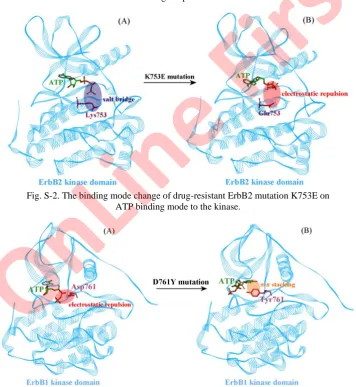
= 1.98 kcal/mol > 0, which would be significantly unfavorable for ATP binding. As can be seen in Fig. 3, the 3rd phosphate group of ATP molecule points to the Lys753 residue in the wild-type kinase–ATP complex system, forming a strong electrostatic attraction of a salt-bridging interaction (see Fig. S-2A of the Sup-plementary material). Upon substitution of Lys with Glu at residue 753, the charge is completely reversed to negative, leading to a strong electrostatic repul-sion on the negatively charged ATP phosphate, thus significantly changing the binding mode of ATP in the kinase active site and largely impairing the kinase– –ATP interaction (see Fig. S-2B). Similarly, the neutral mutation K753I only eliminates the favorable positive charge (Lys) but does not create an unfavorable negative charge at the same kinase residue, which therefore results in a moderate unfavorable effect on ATP binding (ΔΔGttl = 0.58 kcal mol–1). Generally
speak-ing, both the ErbB3 K753E and ErbB3 K753I mutations are predicted to con-siderably impair ATP binding that would sensitize the inhibitor potency. How-ever, these two mutations have been clinically observed to be associated with drug resistance. This can be explained by the fact that other factors, such as directly blocking inhibitor binding and altering the kinase activity by these mutations, may contribute to the clinically observed drug resistance.
The drug-resistant mutation D761Y is located at the beginning region of the ErbB1 activation loop where it is involved in the kinase active site and can directly contact the ATP molecule. The mutation eliminates the negative charge of wild-type Asp and introduces an aromatic Tyr at residue 761, which would largely alter the physicochemical property of the ErbB1 active site. According to computational analysis, the mutation would be highly favorable for ATP binding with ΔΔGttl = –2.14 kcal mol–1. The mutation significantly alters the binding
mode of ATP to the ErbB1 kinase domain with a large motion on the ATP triphosphate. Consequently, the unfavorable electrostatic repulsion between the wild-type Asp761 residue and the ATP ligand is eliminated (see Fig. S-3A of the Supplementary material). Instead, a favorable π–π stacking is observed to be est-ablished between the ATP purine ring and the side chain of the mutant Tyr761 residue (see Fig. S-3B). Considering that the D761Y mutation is located at the region of the kinase activation loop where it is responsible for kinase activity. Therefore, it is expected that this mutation can influence inhibitor potency through both indirectly by promoting ATP binding and directly by influencing kinase activity.
Kinase assay of drug-resistant ErbB mutations on inhibitor activity
In order to substantiate the computational findings, herein both indirect kin-ase assays and direct SPR assays were employed to determine mutation-induced ATP affinity change and its contribution to inhibitor resistance (or sensitization). In the kinase assays, the inhibitory activities (IC50 values) of gefitinib and
lapa-On
Lin
tinib against wild-type ErbB1 kinase domain and its mutants D761Y, T790M and L858R as well as wild-type ErbB2 kinase domain and its mutants K753E, T768I and T768M were measured using kinase assays and the values are listed in Table I. The two inhibitors were approved by US FDA to treat non-small cell lung can-cer (NSCLC) and breast cancan-cer, respectively,31 which have been observed to
become resistant upon many ErbB gatekeeper mutations, such as ErbB1 T790M and ErbB2 T798M.32 Yun et al. also found that the ErbB1 T790M mutation can
cause a generic resistance to Gefitinib and Lapatinib by increasing ATP affi-nity.8,33 Consistently, a considerable activity decrease of the two inhibitors upon
the mutations were observed in the present assays, with IC50 change from 760
nM to n.d. for gefitinib and from 1530 to 4730 nM for lapatinib. In addition, the ErbB2 T798M mutation also exhibits a similar effect on the two inhibitors, with
IC50 changes from 580 nM to n.d. for gefitinib and from 260 to 750 nM for
lapa-tinib, suggesting that such gatekeeper mutations can confer consensus (or gen-eric) unfavorable contribution to the inhibitor potency. Another ErbB2 gate-keeper mutation T768I was measured to address the moderate affinity decrease on the two inhibitors, with IC50 change from 580 to 470 nM for gefitinib and
from 260 to 210 nM for lapatinib. This is consistent with the computational mod-eling that the mutation can moderately impair ATP affinity (due to the removal of the polar Thr768–ATP interaction) so that it can improve the competitive cap-ability of inhibitors with ATP for the kinase active site. In addition, ErbB1 D761Y was predicted to eliminate unfavorable negative charge and contribute a favorable aromatic ring to form an additional π–π stacking with ATP, thus largely increasing ATP affinity (ΔΔGttl = –2.14 kcal mol–1).
TABLE I.The inhibitory activities (IC50 values, nM) of Gefitinib and Lapatinib against
wild-type ErbB1 and ErbB2 kinase domains and their respective mutants; n.d., not detectable
ErbB kinase Mutation Gefitinib Lapatinib
ErbB1 Wild type 760 1530
ErbB1 D761Y n.d. n.d.
ErbB1 T790M n.d. 4730
ErbB1 L858R 930 1290
ErbB2 wild type 580 260
ErbB2 K753E 106 27
ErbB2 T768I 470 210
ErbB2 T768M n.d. 750
Consistently, the inhibitory activities of gefitinib and lapatinib were mea-sured to be totally canceled upon the mutation (IC50 = n.d.), indicating that the
negative charge-eliminated mutation can confer generic resistance to kinase inhi-bitors by largely enhancing ATP binding. Similarly, the ErbB1 positive charge-added mutation L858R can contribute favorable electrostatic interaction with ATP. However, the mutation seems not to confer generic resistance to all
inhi-On
Lin
bitors, with an IC50 increase from 760 to 930 nM for gefitinib (resistance) and
decrease from 1530 to 1290 nM for lapatinib (sensitization). This is because the L858R is located at the activation loop of ErbB1 kinase and hence can influence the inhibitor potency in multiple ways, such as indirectly shifting kinase activity and/or directly altering inhibitor binding.34 In addition, the protein content may
contribute significantly to the flexible loop.35 In contrast, the ErbB2 charge-reversing mutation K753E was predicted to dramatically reduce ATP affinity (ΔΔGttl = 1.98 kcal mol–1), and therefore, it is supposed that the mutation can
generically sensitize (but not confer resistance to) kinase inhibitors. Further kinase assays confirmed that the mutation can significantly enhance the inhibit-ory activities of both gefitinib and lapatinib, with IC50 change from 580 to 106
nM and from 260 to 27 nM, respectively. Overall, the kinase assays well sup-ported computational findings, although the tested samples were limited of only two inhibitors, two ErbB kinases and six mutations.
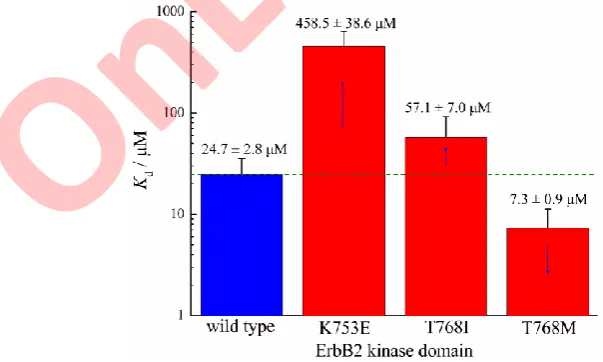
Since the kinase assays can only partially and indirectly reflect the effect of kinase mutation-induced ATP affinity change on inhibitor resistance, herein bio-physical SPR assays were employed to directly determine the binding affinities of ATP to wild-type ErbB2 kinase domain and its K753E, T768I and T768M mutants., The ATP molecule can bind to wild-type kinase with a moderate affi-nity (Kd = 24.7 μM) as shown in Fig. 3, this value falls into the normal range of
kinase–ATP binding affinities (Kd = 1100 μM). As might be expected, the ErbB2
charge-reversing mutation K753E and gatekeeper mutation T768I can largely and moderately reduce ATP affinity by 18.6-fold (Kd = 458.5 μM) and 2.3-fold
(Kd = 57.1 μM), respectively. These two mutations were also calculated to cause
ATP binding free energy loss significantly and modestly (ΔΔGttl = 1.98 and 0.23
Fig. 3. The binding affinity (Kd values) of ATP molecule to wild-type ErbB2 kinase
domain and its three mutants.
On
Lin
kcal mol–1), respectively. In contrast, the ATP affinity can be improved by 3.4- -fold upon another ErbB1 gatekeeper mutation T768M, which is in line with the large binding energy increase (ΔΔGttl = –1.78 kcal mol–1) predicted by the QM/
/MM-PB/SA method. The direct measure of ATP binding provides a molecular insight into the effect of mutation-induced ATP affinity change on inhibitor resis-tance or sensitization, which further solidified the computational findings and kinase assays.
CONCLUSIONS
Since Yun and co-workers first revealed that kinase mutation can cause acquired drug resistance by increasing ATP affinity instead of influencing inhi-bitor binding,8,38 the drug resistance conferred from mutation-induced ATP
affi-nity change has still remained largely unexplored and no systematic investigation has addressed the effect of clinically observed drug-resistant mutations on ATP binding. Therefore, it is still unclear whether the ATP approach is or not a com-mon phenomenon involved in acquired drug resistance. This is probably because experimental analysis of all kinase mutation–ATP pairs is too time-consuming and expensive. Instead, herein in silico analysis and in vitro assay were combined to investigate the effect of clinical drug-resistant kinase mutations on ATP bind-ing, which created a systematic profile of ATP response to these mutations at an acceptable cost. This would be valuable for both theoretical study and clinical practice. For example, the obtained profile can be used to explain the new mole-cular mechanism of drug resistance established by mutation-induced ATP affi-nity increase and, more significantly, to guide rational design and optimization of new kinase inhibitors to combat generic drug resistance by overcoming the increased ATP affinity.
SUPPLEMENTARY MATERIAL
Additional data are available electronically at the pages of journal website: https:// //www.shd-pub.org.rs/index.php/JSCS/index, or from the corresponding author on request.
Acknowledgements.This work was financially supported by the National Natural Sci-ence Foundation of China (No. 51762022), the Natural SciSci-ence Foundation of Jiangxi Pro-vince (Nos. 20192ACBL20029 and 20171BAB206026), the Science & Technology Research Program of Jiangxi Provincial Education Department (Nos. GJJ170636, GJJ190539 and GJJ11526), and the Educational Reform Research Program of Jiangxi Provincial Education Department (No. JXJG-10-15-3).
On
Lin
e F
И З В О Д
СИСТЕМАТИЧАН ПРЕГЛЕД ATP ОДГОВОРА НА СТЕЧЕНЕ МУТАЦИЈЕ EGFR ПОРОДИЦЕ КИНАЗА КОЈЕ СУ РЕЗИСТЕНТНЕ НА ЛЕКОВЕ
DINGWA ZHANG1, DEYONG HE1, XIAOLIANG PAN2, and LIJUN LIU1
1School of Chemistry and Chemical Engineering, Jinggangshan University и 2School of Mechanical and Electrical Engineering, Jinggangshan University, Ji’an 343009, China
Терапија тумора у којој се циљају киназе (KTCT) ATP компетитивним инхибиторима се широко примењује у клиничкој пракси. Међутим, уочено је да мутације киназа могу довести до појаве резистенције на лекове, што умањује могућност примене инхибитора киназа у овом типу клиничке терапије. Уместо да директно утичу на везивање инхибитора, мутације киназа могу изазивати резистенцију на ATP компетитивне инхибиторе тиме што повећавају афинитет за ATP. У овом раду су систематично окарактерисане међумолекулске интерак-ције молекула ATP и EGFR породице киназа које учествују у развоју тумора, а код којих је услед мутације дошло до резистенције на лекове. Везивање ATP за мутиране остатке у киназ-ном домену EGFR породице киназа (EGFR/ErbB1, ErbB2, ErbB3 и ErbB4) праћено је при-меном квантно-механичких/молекуларно-механичких (QM/MM) израчунавања, емпириј-ском анализом применом Poisson–Boltzmann једначине, као и in vitro киназним тестом и површинском плазмнонском резонанцом. Из ових података су идентификоване мутације које могу значајно утицати на везивање ATP; ове мутације су потенцијални кандидати који доводе до резистенције на ATP компетитивне инхибиторе. Већ позната мутација EGFR T790M и њен парњак ErbB2 T798M повећавају афинитет за ATP остваривањем додатних S–π
интеракција између тиоетарске групе мутираног Met остатка и ароматичног аденина ATP молекула, док је EGFR D761Y идентификована као нова генеричка мутација која изазива резистенцију на лекове и може да повећа афинитет за ATP елиминишући нежељено електро-статичко одбијање. Насупрот њима, ErbB2 K753E и T768I мутације могу смањити афинитет за ATP променом наелектрисања, односно ометањем пожељних поларних интеракција. На крају, мутација EGFR L858R је у активационој петљи киназе и близу активног места, што додатно компликује вишеструке релације између киназе, ATP и инхибитора, изазивајући различите ефекте у присуству различитих инхибитора.
(Примљено 24. новембра 2019, ревидирано 8. априла,прихваћено 19. маја 2020)
REFERENCES
1. R. Roskoski, Pharmacol. Res.79 (2014) 34 (https://doi.org/10.1016/j.phrs.2013.11.002) 2. C. L. Arteaga, Breast Cancer Res.13 (2011) 304 (https://doi.org/10.1186/bcr2829) 3. H. Zhang, A. Berezov, Q. Wang, G. Zhang, J. Drebin, R. Murali, M. I. Greene, J. Clin.
Invest.117 (2007) 2051 (https://doi.org/10.1172/JCI32278)
4. K. Ohashi, Y. E. Maruvka, F. Michor, W. Pao, J. Clin. Oncol. 31 (2013) 1070 (https://doi.org/10.1200/JCO.2012.43.3912)
5. C. Ma, S. Wei, Y. Song, J. Thorac. Dis.3 (2011) 10 ( https://doi.org/10.3978/j.issn.2072-1439.2010.12.02)
6. D. J. Riese, R. M. Gallo, J. Settleman, Bioessays29 (2007) 558 (https://doi.org/10.1002/bies.20582)
7. D. L. Gibbons, S. Pricl, J. Cortes, A. Quintás-Cardama, Cancer 118 (2012) 293 (https://doi.org/10.1002/cncr.26225)
8. C. H. Yun, K. E. Mengwasser, A. V. Toms, M. S. Woo, H. Greulich, K. K. Wong, M. Meyerson, M. J. Eck, Proc. Natl. Acad. Sci. USA105 (2008) 2070
(https://doi.org/10.1073/pnas.0709662105)
On
Lin
e F
irs
9. K. Hauser, C. Negron, S. K. Albanese, S. Ray, T. Steinbrecher, R. Abel, J. D. Chodera, L. Wang, Commun. Biol.1 (2018) 70 (https://doi.org/10.1038/s42003-018-0075-x) 10. H. Wang, Z. Yang, Y. Liu, J. Chin. Chem. Soc.67 (2020) 1
(https://doi.org/10.1002/jccs.201900435)
11. P. Zhou, C. Yang, Y. Ren, C. Wang, F. Tian, Food Chem.141 (2013) 2967 (https://doi.org/ 10.1016/j.foodchem.2013.05.140)
12. F. Tian, Y. Lv, P. Zhou, L. Yang, J. Comput. Aided Mol. Des. 25 (2011) 947 (https://doi.org/10.1007/s10822-011-9474-5)
13. P. Zhou, S. Zhang, Y. Wang, C. Yang, J. Huang, J. Biomol. Struct. Dyn.34 (2016) 1806 14. X. Guo, D. He, L. Liu, R. Kuang, L. Liu, Comput. Theor. Chem. 991 (2012) 134
(https://doi.org/10.1016/j.comptc.2012.04.010)
15. X. Guo, D. He, L. Huang, L. Liu, L. Liu, H. Yang, Comput. Theor. Chem. 995 (2012) 17 (https://doi.org/10.1016/j.comptc.2012.06.017)
16. D. Zhang, D. He, X. Pan, Y. Xu, L. Liu, Chem. Pap.73 (2019) 2469 (https://doi.org/10.1007/s11696-019-00797-8)
17. D. Zhang, D. He, X. Pan, Y. Xu, L. Liu, J. Serb. Chem. Soc.84 (2019) 1367 (https://doi.org/10.2298/JSC181221029Z)
18. M. Ashburner, C. A. Ball, J. A. Blake, D. Botstein, H. Butler, Nat. Genetics25 (2000) 25 (https://doi.org/10.1038/75556)
19. C. Yang, S. Zhang, P. He, C. Wang, J. Huang, P. Zhou, J. Chem. Inf. Model.55 (2015) 329 (https://doi.org/10.1021/ci500522v)
20. C. Yang, S. Zhang, Z. Bai, S. Hou, D. Wu, J. Huang, P. Zhou, Mol. Biosyst.12 (2016) 1201 (https://doi.org/10.1039/c5mb00800j)
21. Z. Bai, S. Hou, S. Zhang, Z. Li, P. Zhou, J. Chem. Inf. Model. 57 (2017) 835 (https://doi.org/10.1021/acs.jcim.6b00673)
22. H. Yu, P. Zhou, M. Deng, Z. Shang, J. Chem. Inf. Model.54 (2014) 2022 (https://doi.org/10.1021/ci5000246)
23. C. Yang, C. Wang, S. Zhang, J. Huang, P. Zhou, Mol. Simul. 41 (2015) 741 (https://doi.org/10.1080/08927022.2014.929127)
24. C. Yang, S. Zhang, P. He, C. Wang, J. Huang, P. Zhou, J. Chem. Inf. Model.55 (2015) 329 (https://doi.org/10.1021/ci500522v)
25. P. Zhou, C. Wang, F. Tian, Y. Ren, C. Yang, J. Huang, J. Comput. Aided Mol. Des. 27 (2013) 67 (https://doi.org/10.1007/s10822-012-9625-3)
26. P. Zhou, S. Hou, Z. Bai, Z. Li, H. Wang, Z. Chen, Y. Meng, Artif. Cells Nanomed. Biotechnol.46 (2018) 1122 (https://doi.org/10.1080/21691401.2017.1360327) 27. Z. Li, Q. Miao, F. Yan, Y. Meng, P. Zhou, Curr. Drug Metab.20 (2019) 170
(https://doi.org/10.2174/1389200219666181012151944)
28. F. Tian, R. Tan, T. Guo, P. Zhou, L. Yang, Biosystems11 (2013) 40 (https://doi.org/10.2174/10.1016/j.biosystems.2013.04.004)
29. Z. Li, F. Yan, Q. Miao, Y. Meng, L. Wen, Q. Jiang, P. Zhou P, J. Theor. Biol. 469 (2019) 25 (https://doi.org/10.1016/j.jtbi.2019.02.014)
30. H. Luo, T. Du, P. Zhou, L. Yang, H. Mei, H. Ng, W. Zhang, M. Shu, W. Tong, L. Shi, D. L. Mendrick, H. Hong, Comb. Chem. High Throughput. Screen. 18 (2015) 296
(https://doi.org/10.2174/1386207318666150305144015)
31. M. Thomas, Cancer Nurs.26 (2003) S21-S25 ( https://doi.org/10.1097/00002820-200312001-00006)
32. W. Pao, V. A. Miller, K. A. Politi, G. J. Riely, R. Somwar, M. F. Zakowski, M. G. Kris, H. Varmus, PLoS Med. 2 (2005) e73 (https://doi.org/10.1371/journal.pmed.0020073)
On
Lin
e F
irs
33. M. J. Eck, C. H. Yun, Biochim. Biophys. Acta1804 (2010) (https://doi.org/10.1016/j.bbapap.2009.12.010)
34. F. Tian, C. Yang, C. Wang, T. Guo, P. Zhou, J. Mol. Model.20 (2014) 2257 (https://doi.org/10.1007/s00894-014-2257-x)
35. P. Zhou, Q. Miao, F. Yan, Z. Li, Q. Jiang, L. Wen, Y. Meng, Mol. Omics15 (2019) 280 (https://doi.org/10.1039/c9mo00041k).
On
Lin
e F
irs
J. Serb. Chem. Soc. 85 (0) S1–S5 (2020) Supplementary material
S1
SUPPLEMENTARY MATERIAL TO
Systematic profiling of ATP response to acquired drug-resistant
EGFR family kinase mutations
DINGWA ZHANG1*, DEYONG HE1, XIAOLIANG PAN2 and LIJUN LIU1**
1School of Chemistry and Chemical Engineering, Jinggangshan University, Ji’an 343009,
China and 2School of Mechanical and Electrical Engineering, Jinggangshan University, Ji’an 343009, China
J. Serb. Chem. Soc. 85 (0) (2020) 000–000
Structural modeling of ATP complex structures with wild-type and mutant ErbB kinase domains
The complex structure of the active EGFR kinase domain with an ATP– –peptide conjugate was retrieved from the PDB database1 with id 2GS6. The
structure is a reaction intermediate of kinase-catalyzed transfer of the ATP phosphate group to the peptide substrate. Therefore, it would represent the real active binding mode of ATP to the active site of EGFR. As could be seen in Fig. S-1, the conjugate consists of an ATP molecule and a peptide substrate, in which the ATP phosphate moiety was just covalently bonded to the hydroxyl group of peptide tyrosine residue. The peptide can be readily removed from the structure leaving an ATP analog using a manual modification approach, which was manually modified to ATP molecule and then subjected to QM/MM energy minimization, finally resulting in the modeled complex structure of the EGFR kinase domain with ATP, in which, evidently, the ATP molecule adopts a “correct” binding mode to interact with the kinase active site, in which the purine ring of ATP tightly packs against the active site, while exposing its triphosphate moiety to the solvent.
The modeled complex structure of the EGFR kinase domain with ATP was used as a structural template to further model the complex structures of other three ErbB kinase domains as well as their drug-resistant mutants with ATP. In the procedure, the apo crystal structure of ErbB2, ErbB3 and ErbB4 kinase domains were retrieved from the PDB database with ids 3PP0, 3LMG and 3BCE, respectively. A ligand grafting method was employed to model the complex structures of ErbB2, ErbB3 and ErbB4 kinase domains with ATP, i.e., the apo
*,** Corresponding authors. E-mail: (*)[email protected]; (**)[email protected]
On
Lin
e F
irs
structures of Her2, Her3 and Her4 kinase domains were superposed onto the template of EGFR kinase domain–ATP complex, and then the EGFR kinase domain was manually removed from the superposed system to obtain the modeled complexes of ErbB2, ErbB3 and ErbB4 kinase domains with ATP. Next, the wild-type EGFR, ErbB2, ErbB3 and ErbB4 kinase domains in these complex structures were automatically mutated to their drug-resistant mutants in the PyMol program,2 which were then subjected to QM/MM energy
minimiz-ation. Consequently, a total 41 ATP complex structures with the 4 wild-type ErbB kinase domains and their 37 drug-resistant mutants were systematically obtained.
Fig. S-1. A) Crystal structure of EGFR kinase domain in a complex with an ATP-peptide conjugate (PDB: 2GS6). B) The peptide substrate was manually removed from the structure
to generate the complex of EGFR kinase domain with ATP, which was then minimized by QM/MM.
IN VITRO KINASE ASSAY
A standard protocol modified from previous reports3,4 was used to perform the kinase
assays. The kinase proteins were diluted in a final volume of 50 μL assay buffer containing 50 mM Tris-HCl (pH 7.8), 20 mM MgCl2, 1 mM MnCl2 and 1 mM DTT containing 10 μM
substrate peptides, 5 μM ATP and 1 μCi [γ-32P]ATP. The reaction was incubated with
increasing concentrations of inhibitor compound at room temperature for 30 min and then stopped by the addition of sample loading buffer. The samples were loaded in an SDS-PAGE gel and then exposed to X-ray beam for radioactive detection. The phosphorylated substrates were plotted against the concentration of inhibitor to determine the IC50 value for
kinase inhibition. The recombinant proteins of ErbB1 and ErbB2 kinase domains were obtained commercially. The inhibitor compounds Gefitinib and Lapatinib were suspended in DMSO and stored until use in small aliquots at –20 °C.
On
Lin
e F
irs
Surface plasmon resonance analysis
All surface plasmon resonance (SPR) experiments were performed on a Biacore T100 with active temperature control at 25 °C following the manufacturer’s protocols and previous reports.5,6 ErbB2 kinase domain proteins were immobilized onto activated a CM5 sensor chip
by standard amine coupling. All interaction experiments were performed in a buffer containing 50 mM Tris-HCl, 100 mM NaCl, 15 mM MgCl2 and 0.05 % Tween-20. The ATP
was injected at different concentrations varying between 1 and 500 nM at a flow rate of 50 μL min-1. Changes in surface concentration are proportional to changes in the refractive index on
the surface, resulting in changes in the SPR signal. For all samples, blank injection with buffer alone was subtracted from the resulting reaction surface data. The data were analyzed using Biacore evaluation software and fitted using GraphPad Prism software.
Fig. S-2. The binding mode change of drug-resistant ErbB2 mutation K753E on ATP binding mode to the kinase.
Fig. S-3. The binding mode change of drug-resistant ErbB1 mutation D761Y on ATP binding mode to the kinase.
On
Lin
e F
irs
TABLE S-I. The 37 acquired drug-resistant ErbB kinase mutations involved in human cancers
ErbB kinase Mutationa Resistant TKI Human cancer
ErbB1/EGFR L718Q CO-1686, WZ4002 NSCLC
ErbB1/EGFR L747S Erlotinib, Gefitinib NSCLC
ErbB1/EGFR L844V WZ4002, CO-1686 NSCLC
ErbB1/EGFR D761Y Gefitinib Brain tumor
ErbB1/EGFR V769M Erlotinib, Gefitinib NSCLC
ErbB1/EGFR T790M* Erlotinib, Gefitinib NSCLC
ErbB1/EGFR L792F Osimertinib NSCLC
ErbB1/EGFR L792H Osimertinib NSCLC
ErbB1/EGFR G796S Osimertinib NSCLC
ErbB1/EGFR G796R Osimertinib NSCLC
ErbB1/EGFR C797S Osimertinib NSCLC
ErbB1/EGFR T854A Erlotinib, Gefitinib NSCLC
ErbB1/EGFR L858R Osimertinib NSCLC
ErbB1/EGFR A871E Erlotinib, Gefitinib NSCLC
ErbB2 L726I Gefitinib Breast cancer
ErbB2 L726F Lapatinib Breast cancer
ErbB2 K753I Lapatinib Breast cancer
ErbB2 K753E Lapatinib Breast cancer
ErbB2 L755S Lapatinib Breast cancer
ErbB2 L755P Lapatinib Breast cancer
ErbB2 P780L Lapatinib Breast cancer
ErbB2 S783P Lapatinib Breast cancer
ErbB2 L785F Lapatinib Breast cancer
ErbB2 T798I* Neratinib Breast cancer
ErbB2 T798M* Lapatinib Breast cancer
ErbB2 V842I Lapatinib Breast cancer
ErbB3 T768I* Bosutinib Breast cancer
ErbB3 Q809R Neratinib Breast cancer
ErbB3 S846I Neratinib Breast cancer
ErbB3 V855A Pertuzumab, Afatinib NSCLC
ErbB3 E928G Neratinib Breast cancer
ErbB4 R711C Lapatinib Breast cancer
ErbB4 L798R Lapatinib Breast cancer
ErbB4 R847C Lapatinib Breast cancer
ErbB4 R847H Lapatinib Breast cancer
ErbB4 R992C Lapatinib Breast cancer
ErbB4 R992S Lapatinib Breast cancer
aThe asterisk ‘*’ indicates gatekeeper mutation.
REFERENCES
1. H. M. Berman, J. Westbrook, Z. Feng, G. Gilliland, T. N. Bhat, H. Weissig, I. N. Shindyalov, P. E. Bourne, Nucleic Acids Res.28 (2000) 235
(https://doi.org/10.1093/nar/28.1.235)
2. S. Yuan, H. C. S. Chan, Z. Hu, WIREs Comput. Mol. Sci.7 (2017) e1298 (https://doi.org/10.1002/wcms.1298)
On
Lin
e F
irs
3. X. Ai, Y. Sun, H. Wang, S. Lu, Amino Acids46 (2014) 1635 (https://doi.org/10.1007/s00726-014-1716-0)
4. Z. Wang, M. Jiang, N. Feng, C. Li, Biochimie52 (2018) 188 (https://doi.org/10.1016/j.biochi.2018.07.005)
5. K. T. Kuo, W. C. Lin, K. C. Chang, J. Y. Huang, K. C. Yen, I. C. Young, Y. J. Sun, F. H. Lin, PLoS ONE 10 (2015) e0116610 (https://doi.org/ 10.1371/journal.pone.0116610)
6. Y. Zhao, Y. Jiao, F. Sun, X. Liu, Med. Chem. Res. 27 (2018) 2160 (https://doi.org/10.1007/s00044-018-2224-7).