THE EFFECT OF USING A POWERED TOOTHBRUSH ON MCP-1 AND RANTES LEVELS IN PATIENTS WITH GINGIVITIS
Linda L. Paquette
A thesis submitted to the faculty of the University of North Carolina at Chapel Hill in partial fulfillment of the requirements for the degree of Master of Science in Dental Hygiene
Education in the Department of Dental Ecology
Chapel Hill 2008
ABSTRACT
LINDA PAQUETTE: The Effect of Using a Powered Toothbrush on MCP-1 and RANTES Levels in Patients with Gingivitis
Under the direction of Heather L. Jared, Rebecca S. Wilder, and Silvana P. Barros
ACKNOWLEDGEMENTS
A special thanks to Heather Jared, my thesis advisor, for the many hours of patient counsel and commitment during this project. I would also like to thank my thesis committee, Rebecca Wilder and Dr. Silvana Barros, for their support and encouragement.
Thanks to Kevin Moss for his patience, help and timely feedback with the statistical analysis.
I would like to thank all of the GO Health staff, especially Tracy Russell, for their support in scheduling, data collection and friendship. Also a thank you to the dental research laboratory staff, Russ Levy and Alan Welborn, who gave my project priority in their
schedules to allow me to complete my research.
TABLE OF CONTENTS
LIST OF TABLES ………v
LIST OF FIGURES ……….vi
Chapter I. INTRODUCTION ……….….…….….1
II. LITERATURE REVIEW ……….…...3
A. Cytokines ……….…...3
B. Dental Plaque ……….…….…………5
C. Periodontal Diseases ………...……6
D. Toothbrushes ………....……….……..…....7
III. MATERIALS AND METHODS ………...9
IV. RESULTS ………...…14
V. DISCUSSION ………..………...…17
VI. CONCLUSION ………..20
APPENDICES ……….. 27
LIST OF TABLES Table
1. Baseline Subject Characteristics by Group ………...……21 2. Baseline Periodontal Clinical Measurements and GCF MCP-1
LIST OF FIGURES Figure
1. Flow Diagram ………..……….…..…..24 2. Descriptive Statistics for MCP-1 for the Treatment Group Illustrating
the Trend over Time ……….25 3. Descriptive Statistics for RANTES for the Treatment Group
ABBREVIATIONS
α Alpha
β Beta
ANCOVA Analysis of covariance BOP Bleeding on probing CAL Clinical attachment level CEJ Cementoenamel junction CRF Case report form
GCF Gingival crevicular fluid
GI Gingival index
IL-1β Interleukin 1β IL-6 Interleukin 6 IL-12 Interleukin 12
MCP-1 Monocyte chemoattractant protein-1
mm Millimeter
PD Probing depth
PGE2 Prostaglandin E2
RANTES Regulated on activation, normal T cells expressed and secreted SD Standard deviation
INTRODUCTION
Gingivitis is an inflammatory condition initiated by the accumulation of biofilm on tooth surfaces in persons with inadequate oral hygiene. According to Albander,
approximately 50% of the adults in the United States have gingivitis.1 Historically, the assessment of gingival disease has been made using the static clinical parameters such as dental indices, bleeding on probing and clinical attachment levels. These tests rely on the physical changes in the periodontium but do not offer insight into the multifactorial nature of the host response.2 The host response to inflammation elicits a cascade of inflammatory mediators including cytokines.
Effective plaque control, through proper brushing and flossing, is a necessity to stop the inflammatory response and prevent further damage to the periodontal structures. Since the toothbrush is the most widely used method of plaque control, a variety of innovative powered toothbrushes, including sonic and ultrasonic, have been studied to assess their effectiveness in bacterial plaque removal.3-5 This pilot study attempted to assess the effect a powered toothbrush utilizing a combination of sonic and ultrasound technology would have on the cytokines, MCP-1 and RANTES, in a gingivitis group.
leukocytes. MCP-1 expression is highly correlated with the degree of gingival inflammation present.6 RANTES is involved in T-lymphocyte recruitment that controls cell-mediated immune response.
The potential diagnostic value of cytokines gained impetus with the identification of the episodic nature of periodontal diseases in the 1970s.7 Cytokines have been investigated as predictors of risk for periodontal diseases.8-10 The assessment of cytokines in gingival
crevicular fluid offers a method of detecting susceptible individuals and may also suggest future lines of therapy.11 “Gingival crevicular fluid is easily obtainable and can be sampled repeatedly in a painless, non-invasive manner from periodontal sites of interest.”10
REVIEW OF THE LITERATURE A. Cytokines
When a microbial challenge, as in plaque accumulation, is mounted against the host, a response is initiated which involves both saliva and the gingival crevicular fluid (GCF). Both of these have multiple capacities to deal with the bacterial challenge. Within the GCF are a variety of cells including monocytes (macrophages) which serve as antigen-presenting cells. These macrophages mediate the immune response when exposed to
lipopolysaccharides by releasing chemical messengers called cytokines.13 Studies of early or experimental gingivitis show increases in inflammatory mediators (cytokines) in the gingiva and crevicular fluid.14,15
According to Seymour and Gemmell, “It is likely that the nature of the
antigen-presenting cell is fundamental in determining the nature of the cytokine profile, which may in turn open up possibilities for new therapeutic modalities.”11 These antigen-presenting cells plus peptides and proteins (cytokines) are part of the innate immune response which provides a rapid reaction to infections causing a pathological condition know as inflammation.16
Cytokines are being studied to further the understanding of their role in the pathogenesis of a number of diseases because of their potential use as targets for therapy.17
with increased levels of a variety of inflammatory mediators”. They interact in a network; first by inducing each other, second by transmodulating cell surface receptors, and third by synergistic, additive, or antagonistic interactions on cell function.18
Among these cytokines are the chemokines MCP-1 and RANTES. Chemokines are secondary pro-inflammatory mediators that recruit a well-defined subset of leukocytes; lymphocytes, monocytes, eosinophils, and basophils. “Chemokines are induced by external signals such as IL-1 or TNF, growth factors, and viral and bacterial infection or their
products.”19 MCP-1 and RANTES are in the CC group of chemokines that are chemotatic for monocytes and a small subset of lymphocytes.20 Emingil et al suggests that MCP-1 and RANTES could play a key role in both activation and recruitment of inflammatory and immune cells in the periodontal environment.21
Monocyte chemoattractant protein-1 (MCP-1) possesses a high degree of specificity for the recruitment and activation of monocytes to sites of injury and infection. The
accumulation of plaque on the teeth induces mononuclear phagocytes and endothelial cells to express MCP-1.20 In 1993, Yu et al, reported the first identification of expression of MCP-1 or other members of the chemokine family in bacterially induced gingival inflammation.6 Yu
et al also reported that MCP-1-producing cells are commonly found in inflamed gingiva.22 In 1995 Graves reported that
“The importance of MCP-1 in gingival inflammation is underscored by three recent findings: (1) the majority of mononuclear phagocytes in inflamed gingiva express MCP-1 (Yu and Graves, 1995); (2) MCP-1 expression is highly correlated with the degree of gingival inflammation present (Yu et al, 1993); and (3) MCP-1 is the principal monocyte chemoattractant in gingival crevicular fluid (Hanazawa et al, 1993).”6,20,22,23
cell mediators.24 RANTES acts as a chemoattractant for macrophages and lymphocytes. Gamonal and colleagues reported that higher levels of cytokines, including RANTES, were detected in the GCF of inflamed sites as compared to the healthy sites; however, their increase was not significant. They stated that
“These elevated GCF levels could not be correlated with any of the clinical
parameters; probing depth (PD), clinical attachment loss (CAL) and the dichotomous measurements of supragingival plaque accumulation (PI), and bleeding on probing to the base of the crevice (BOP). Their conclusions were that; 1). clinical parameters, such as PD, CAL, and BOP do not necessarily reflect current disease activity and 2). cytokine production does not necessarily reflect the presence of gingival
inflammation but could be consistent with the divergence between clinical signs of inflammation and disease activity.”25
According to Embery and Waddington, “The collection of GCF is non-invasive, site-specific about teeth, comparatively easy to perform, and offers one of the most accessible entrées of any tissue in the body as a means of assessing the disease state”.26
B. Dental plaque
The microbial etiological factor in periodontal diseases is dental plaque biofilm. Knowledge of the composition and structure of biofilm can help to explain why dental diseases occur and progress and, also, aid in effective therapeutic modalities. Biofilm is an adherent mat of bacteria, epithelial cells, and leukocytes encased in a salivary glycoprotein and extracellular polysaccharide matrix that forms on inert surfaces. Bacteria exhibit different properties when contained within a biofilm. The biofilm associated with gingivitis is characterized by several cell layers (100-300), with bacterial stratification arranged by metabolism and aerotolerance.27 Marginal plaque, composed of filamentous microorganisms along with cocci and rods, is important in the initiation and development of gingivitis.
microbial composition.”28 Changes in the microbial composition are related to an increase in plaque thickness.28 The strength of the inflammatory response appears to be related to the amount of plaque accumulation.29 Prolonged accumulation of dental plaque can induce the clinically observable signs of inflammation in the gingiva; edema, erythema and bleeding.30
In 1965, Löe and co-workers published their classic work that demonstrated that gingival health could be reliably achieved with immaculate oral hygiene and that gingival inflammation could be caused by the accumulation of plaque on the teeth.30 In fact, many oral diseases including dental caries, periodontal disease and peri-implantitis are plaque related.27
C. Periodontal Diseases
Periodontal disease, in its various forms, represents a nearly universal ailment. Approximately 50 % of the adults in the United States have gingivitis.1,31 Gingivitis, an inflammatory disease limited to the mucosal epithelium surrounding the cervical portion of the tooth and the alveolar processes, may be attributed to an accumulation of plaque biofilm in persons with inadequate oral hygiene. It is initiated by bacterial colonization on the tooth surface. If left untreated gingivitis can lead to the irreversible condition of periodontitis, an infection affecting the periodontal structures supporting the tooth; the attachment apparatus and bone. Even though gingivitis does not always result in periodontitis, its effective treatment and prevention can improve oral health and reduce the incidence of periodontal diseases.
outcomes39-42 are being linked to periodontal disease as a possible risk factor. Recently Chen
et al suggested that periodontitis may be associated with an increased risk of peripheral arterial disease.43
Periodontal disease can no longer be thought of as an ailment with relevance only to oral health. The possible association between periodontal infection and systemic health has important implications for the treatment and management of patients. As a result of the high cost of dental diseases and its impact on systemic health, some insurance companies are evaluating risk assessment as a tool to predict costs.44 Companies, such as Cigna, provide enhanced dental benefits for their subscribers during pregnancy45 to avoid possible adverse pregnancy outcomes.
D. Toothbrushes
Bacterial plaque-induced gingivitis is the most common form of gingivitis. The literature shows that gingivitis can be treated and controlled by thorough mechanical plaque removal and this is most often accomplished by using a toothbrush. Manual toothbrushes, in their many designs, are most often used to help remove bacterial plaque and disrupt the inflammation that leads to gingivitis.46 No single manual design has demonstrated consistent superiority for either plaque removal or gingival inflammation reduction.46,47 The
thoroughness and skill of the patient are the critical determinants and the ideal toothbrush design should be innovative enough to increase effectiveness in a manner independent of the patient’s skills or specific method of brushing.48
ultrasonic brush that claims to remove plaque beyond where the bristles meet the teeth. Several in vitro studies have been conducted to test cleaning 3-4 mm beyond the reach of the bristles; however, they lack clinical support.50,51 If a powered brush enabled the user to remove plaque where the bristles do not touch the teeth (approximately 50% of the tooth surface) this would improve the oral health of the user. Studies have shown that patient compliance with brushing is improved using powered toothbrushes52-54
This study investigated the effect of a powered toothbrush utilizing licensed technology developed at the University of Washington by Professor Pierre Mourad,
incorporating both sonic and ultrasound physical processes.55 Sonic and ultrasound physical processes are a result of acoustical energy; the generation, propagation, and reception of mechanical waves and vibrations. Sonic or audible vibrations occur within the range of 20 Hz and 20,000 Hz and ultrasonic vibrations at frequencies greater than 20 KHz. Sonic vibrations produce bubbles within a liquid environment. These bubbles or cavitations are then activated by ultrasound transducers forcing them to oscillate, expand, contract and burst creating shear forces. These shear forces break up and lift off contaminants on the surface being cleaned.
MATERIALS AND METHODS
This study was a parallel study design with participants randomized to one of two treatment arms with the examiner(s) blinded to treatment. The treatment group was assigned a powered toothbrush, and the control group a manual toothbrush.
Twenty participants were recruited from printed and electronic advertisements, and from the university’s recruitment database. All participants had type I gingival disease56 with a minimum of 10 percent bleeding on probing scores and probing depths not to exceed 6 mm (excluding the distal surfaces of the most posterior teeth).
eligibility was determined participants were appointed for four additional visits over the next six months.
A medical history was collected at the screening visit and reviewed at each
subsequent visit. An intraoral exam was conducted at each visit and any adverse events were reported according to the University guidelines. For study visits 2-4 participants were
instructed to not eat within one hour of the study visit and to discontinue brushing or using any other oral hygiene aids 18-24 hours prior to the appointment. Participants were to use the test brush (either the powered or manual brush) at home (2 times per day per written instructions) with the study toothpaste. (Appendix 1) Participants were instructed not to use any products other than those assigned in the study including; mouth rinses, chewing gum, or whitening products.
During the study visits participants received a complete oral examination including plaque index, gingival index, probing depths, clinical attachment level, and bleeding upon probing. Gingival crevicular fluid samples were collected for analysis to determine the concentration of MCP-1 and RANTES. (Appendix 2) The following is a list of all procedures that were conducted during each appointment.
Six weeks Six weeks Twelve weeks
Visit 1 Screening
Visit 2 Baseline
Visit 1: Screening
• Study consent form signed
• Medical history completed and reviewed
• Inclusion and exclusion criteria reviewed
• Intra oral exam
• Gingival index (full mouth) Sites >2 recorded on CRF
• Plaque and stain index recorded
• Periodontal probing depth measurements recorded (full mouth)
• Clinical Attachment levels measured and recorded
• Bleeding on probing (BOP) recorded
• GCF and plaque sampling sites identified and recorded on CRF
• Participant compensated for screening visit Visit 2: Baseline
• Medical history reviewed
• Intra oral exam
• Baseline gingival crevicular fluid samples (GCF) will be taken from previously identified sites
• Plaque and stain index recorded
• Gingival index (full mouth)
• Probing depth and clinical attachment level data transferred from screening visit data
• Baseline plaque samples taken from previously identified sites
• Study toothbrush and toothpaste dispensed via randomization schedule
• Participant brushes with study brush and toothpaste (supervised by a dental professional)
• Adverse events documented on CRF
• Compliance telephone calls will be placed to all participants 3 weeks after visit 2
Visit 3-5:
• Medical history reviewed and compliance verified
• Intra oral exam
• GCF samples taken from pre-identified test sites (same as baseline)
• Plaque and stain index recorded
• Gingival index (full mouth)
• Probing depth measured and recorded (full mouth)
• Clinical Attachment levels measured and recorded
• BOP recorded
• Adverse Events documented on CRF
• Product use reinforced and usage instructions reviewed (2 minutes twice per day home use) (Appendix 1)
• Powered toothbrush handle checked to ensure working properly
• Powered brush head replaced or manual toothbrush replaced and additional toothpaste dispensed if needed
• Used brush heads collected
• Participants assigned to powered toothbrush completed questionnaire
periodontal pocket depth and clinical attachment levels using the UNC-15 periodontal probe. The purpose of this examiner calibration session was to quantify intra- and inter-examiner reliability of measuring periodontal soft and hard tissue parameters. All examiners
participated, and examiner calibration was conducted at the University of North Carolina School of Dentistry in the GO Health Center. The Kappa statistic for inter-examiner reliability showed almost perfect agreement for probing depth measurements and full
agreement for clinical attachment level measurements (r = 0.91 and r = 0.80, respectively).57
Data Analysis
Descriptive statistics and statistical analyses were performed with the computerized statistical packages (JMP and SAS).
Participants were randomly assigned to treatment using a randomization schedule created by the study statistician. All biological samples and data were coded and all personal information was removed and would not be linked to the data.
Initial descriptive analyses summarized subject-level mean scores within treatment groups at baseline, and in particular, any imbalances between the randomized groups at baseline were identified. The baseline characteristics of race, gender and frequency of dental visits were analyzed using a chi-square model. Age was analyzed using an unpaired t-test model. Two-sample t-tests for the comparison of mean levels of the treatment and control groups were conducted at each visit for the clinical measurements and the MCP-1 and RANTES concentrations. All MCP-1 and RANTES levels were reported in log base 10. The analysis of covariance (ANCOVA) model was used to compare the clinical
RESULTS Patient Population
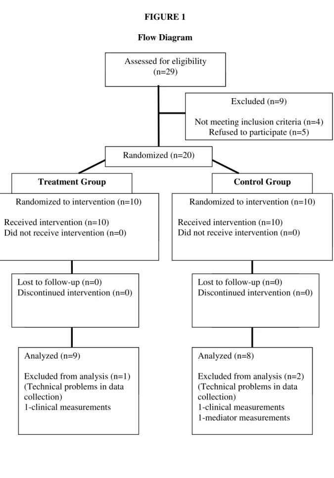
A total of 20 participants (11 males and 9 females) completed the 24 week clinical phase of this study. Of the 20 participants enrolled, 10 (50%) were randomly assigned to the powered toothbrush group and 10 (50%) to the manual toothbrush. The baseline data was recorded for all 20 participants; however, due to technical problems, the data used at 24 weeks post treatment to compute the analysis of covariance (ANCOVA) for the clinical measurements was recorded on 18 participants and the data for the GCF levels was recorded on 19 participants. (Figure 1)
No adverse events were reported during the study. There were five protocol
deviations recorded for the treatment group and seven for the control group which included doctor visits, dental visits with restorative treatment and insertion of orthodontic spacers, medication changes, out of window study visits and inability to use study brush while on vacation.
Table 1 shows, for each of the two groups (treatment and control), the baseline results for four key variables; gender, race, age, and frequency of dental visits. The non-significant, chi-square test results for baseline gender and frequency of dental visits; and the
Clinical Measurements
The baseline analysis of the dental index scores between the two groups (treatment and control) appears in Table 2. An unpaired t-test was used to determine significance between the mean dental index scores for the treatment group and the control group. There were no statistically significant differences for the clinical measurements (probing depth (PD), extent PD≥4mm., gingival index (GI), extent GI≥2mm, plaque index (PI), clinical attachment loss (CAL), extent CAL≥3mm, bleeding on probing (BOP), again indicating a balanced group at the start of the clinical trial.
Using the analysis of covariance (ANCOVA) model, Tables 3 shows that the dental index scores for PD, extent PD, GI, extent GI, PI, CAL, extent CAL, and BOP for the
treatment group and the control group were not statistically different, at baseline and 24 week post treatment, adjusting for baseline levels.
MCP-1 and RANTES
Table 2 shows the non-significant, unpaired t-test results for baseline gingival crevicular fluid (GCF) levels of MCP-1 and RANTES. These results indicate that the two groups were well balanced at baseline when comparing these mediator levels.
The analysis of covariance (ANCOVA) model was used to determine if there were differences between the mean GCF levels of MCP-1 for both groups (treatment and control) at baseline and at 24 week post therapy, adjusting for baseline levels,. Although GCF MCP-1 levels were lower at 24 weeks post therapy in the treatment group, the reduction was not statistically significant. (Table 3).
levels. Although the GCF levels of RANTES were lower at 24 weeks post therapy for both the treatment and control group, these results were not statistically significant using the analysis of covariance (ANCOVA) model.
DISCUSSION
Powered toothbrushes are being developed that utilize advancements in technology to offer the consumer products that have the potential to achieve better plaque removal in a more efficient manner. Recently, a powered toothbrush that incorporates both sonic and ultrasound physical processes was introduced. This pilot study attempted to assess the effect a powered toothbrush that utilizes this innovative technology would have on indicators of the inflammatory response, the cytokines MCP-1 and RANTES.
Most studies utilize dental indices, probing depths, clinical attachment level and bleeding on probing to quantify the amount and severity of disease and to follow any changes in gingival status over specified periods of time. The use of these clinical measurements does not necessarily reflect gingival health or the risk of disease progression. Bleeding on probing provides a much better indicator of inflammation and disease progression but is not the most sensitive or specific measure of health.
Treatment modalities which focus on the host reaction as well as the bacterial challenge offer the clinician a different therapeutic approach to treating disease.
The lack of significant findings in this pilot study could be attributed to the fact that the study did not have sufficient power to detect significant differences. In future studies having a larger patient sample size to assess GCF levels of MCP-1 and RANTES could possibly increase the significance of MCP-1 and RANTES. Even though a trend in the reduction in the biochemical mediators in patients using the powered toothbrush when compared to a manual toothbrush was observed, this reduction was not significant. The results of this study do not support the ability of either the powered toothbrush or the control brush to reduce the concentration of the inflammatory mediators MCP-1 and RANTES over a 6 month period.
The selection of the mediators, MCP-1 and RANTES, might also account for their non-significant reduction in response to therapy. MCP-1 and RANTES levels are directly related to the degree of inflammation and their expression is induced by other cytokines and cell components. It could be that gingivitis patients in this study did not have the level of disease that would illustrate a reduction in the concentration of these chemoattractant
cytokines. Further studies in populations with gingivitis using different mediators are needed to evaluate this finding.
The collection of GCF, while non-invasive, is technique sensitive. It is possible that the samples collected in this study were contaminated with plaque, blood or saliva.
This pilot study will contribute to the body of knowledge within the National Dental Hygiene Research Agenda by validating and testing mechanisms that increase health
efficacy of a powered toothbrush by utilizing cytokine measurements as indicators of a reduction in the inflammatory response. Cytokines offer an additional method of validating the efficacy of oral hygiene aids.
CONCLUSION
Innovative powered toothbrushes are being introduced to provide the consumer with products that more effectively remove plaque biofilm and consequently, improve oral health. Even though this study did not show that a powered toothbrush, combining sonic and
ultrasound technology, was more effective than a manual brush in reducing the levels of cytokines, further studies are needed to examine and evaluate this novel technology.
TABLE 1
Baseline Subject Characteristics by Group
†Statistically significant p<0.05
1
Treatment group: Subjects using the powered toothbrush
2
Control group: Subjects using the manual toothbrush Subject Characteristic Treatment
Group1
Control Group2
P value
Subjects (n {% total}) 10 (50%) 10 (50%)
Male 6 (30%) 5 (25%)
Gender
Female 4 (20%) 5 (25%)
0.65 African
American
1 (5%) 6 (30%)
Caucasian 9 (45%) 4 (20%)
Asian Native American/ Alaska Native Race Other 0.01†
6 months 8 (40%) 5 (25%)
12 months 1 (5%) 5 (25%)
Frequency of dental visits
Infrequent/ over 12 months
1 (5%) 0 (0%)
0.08
TABLE 2
Baseline Periodontal Clinical Measurements and GCF MCP-1 and RANTES Levels by Group
1
Treatment group: Subjects using the powered toothbrush
2
Control group: Subjects using the manual toothbrush Treatment Group1
Mean/SD
Control Group2 Mean/SD
P value Clinical Measurements
PD 2.03 (0.33) 2.18 (0.55) 0.49
Extent PD≥4mm 6.16 (5.56) 8.62 (7.02) 0.40
GI 0.97 (0.45) 1.28(0.22) 0.06
Extent GI ≥2 27.09 (26.37) 41.44 (19.65) 0.18
PI 0.47 (0.27) 0.79 (0.44) 0.07
CAL 1.65 (0.39) 1.58 (0.38) 0.67
Extent CAL≥3mm 13.03 (8.27) 11.50 (9.09) 0.70
BOP 47.10 (16.49) 60.33 (16.91) 0.09
GCF Levels (log)
MCP-1 0.33 (0.29) 0.36 (0.26) 0.79
TABLE 3
Baseline and 24 Weeks Post Therapy for Outcome Variables by Group
1
Treatment Group: Subjects using the powered toothbrush
2
Control Group: Subjects using the manual toothbrush Treatment Group¹ Mean/SD Control Group² Mean/SD P value Baseline 24 Weeks
Post Therapy
Baseline 24 Weeks Post Therapy Clinical
Measurements
PD 2.04 (0.33) 1.84 (0.28) 2.18 (0.55) 1.97 (0.33) 0.33 Extent
PD≥4mm 6.16 (5.56) 3.14 (4.66) 8.62 (7.02) 6.01 (6.94) 0.45 GI 0.97 (0.45) 1.11 (0.37) 1.29 (0.21) 1.24 (0.30) 0.44 Extent
GI≥2mm
27.09 (26.37) 37.78 (25.14) 41.12 (18.68) 38.15 (29.02)
0.93 PI 0.47 (0.27) 0.86 (0.51) 0.81 (0.42) 1.13 (0.42) 0.59 CAL 1.65 (0.39) 1.29 (0.32) 1.58 (0.38) 1.10 (0.20) 0.24 Extent
CAL≥3mm 13.03 (8.27) 4.59 (5.70) 11.50 (9.09) 4.40 (4.45) 0.87 BOP 47.10 (16.49) 43.72 (17.90) 60.33 (16.91) 51.96 (23.11)
0.64 GCF Levels
(log)
FIGURE 1 Flow Diagram
AssessedAssaaaa for Assessed for eligibility
(n=29)
Excluded (n=9)
Not meeting inclusion criteria (n=4) Refused to participate (n=5) Randomized (n=20)
Randomized to intervention (n=10) Received intervention (n=10)
Did not receive intervention (n=0)
Randomized to intervention (n=10) Received intervention (n=10)
Did not receive intervention (n=0)
Lost to follow-up (n=0)
Discontinued intervention (n=0)
Lost to follow-up (n=0)
Discontinued intervention (n=0)
Analyzed (n=9)
Excluded from analysis (n=1) (Technical problems in data collection)
1-clinical measurements
Treatment Group Control Group
Analyzed (n=8)
Excluded from analysis (n=2) (Technical problems in data collection)
FIGURE 2
Descriptive Statistics for MCP-1 for the Treatment Group Illustrating the Trend over Time
-0.1 0 0.1 0.2 0.3 0.4 0.5 0.6 0.7
Visit 1 Visit 2 Visit 3 Visit 4 Visit 5
p
g
/m
L Mean +1SD
Mean -1SD Mean
FIGURE 3
Descriptive Statistics for RANTES for the Treatment Group Illustrating the Trend over Time
-0.2 -0.1 0 0.1 0.2 0.3 0.4 0.5 0.6
Visit 1 Visit 2 Visit 3 Visit 4 Visit 5
p
g
/m
L Mean +1SD
Mean -1SD Mean
Appendix 1
Powered Brushing Instructions
The Ultreo has a 2-minute brushing feature which automatically turns off after 2 minutes. Follow these simple steps to maximize your brushing experience:
1. Place a small amount (approximately pea-sized) of toothpaste on the center of the brush head and wet the brush under running water. Before turning the Prototype on, place the bristles of the brush head in the back section of your mouth pointing toward the gum line.
2. Hold the handle with a light ‘finger-tip’ grip and turn on the powered toothbrush.
3. Gently move the brush in a slightly circular motion so the bristles penetrate between your teeth and into your gum line. After a few seconds, continue brushing to the next section
while you gently circulate the bristles. Do not scrub. Continue brushing throughout your mouth evenly to effectively remove plaque in those hard-to-reach places.
4. The angled brush head and slender neck help you reach the back teeth and the tooth surfaces on the inside of the mouth. Be sure to adjust your brushing to use the angle to your advantage and keep as many bristles in contact with the teeth as possible.
5. To make sure you brush evenly throughout the mouth, divide your mouth into 4 sections: 1) outside lower teeth, 2) inside lower teeth, 3) outside upper teeth and 4) inside upper teeth. Begin brushing in section 1 (outside lower teeth) and brush for 30 seconds before moving to section 2 (inside lower teeth). Continue similarly to upper teeth. You will hear a tone after 30 seconds signaling you to move on to each section until you’ve brushed for 2 minutes. 6. In order to take fully advantage of the ultrasonic feature, keep as much fluid in your mouth as comfortable during brushing.
Participant Reminders:
1. Brush two minutes twice per day with the study toothbrush and toothpaste. 2. Do not use other oral hygiene aids during the test period.
3. Do not brush or use dental floss 18 – 24 hours prior to your next study visit. 4. Do not eat 1 hour prior to your next study visit.
3
4
Manual Brushing Instructions
• Place your toothbrush at a 45-degree angle against the gums.
• Move the brush back and forth gently in short (tooth-wide) strokes.
• Brush the outer tooth surfaces, the inner tooth surfaces, and the chewing surfaces of the teeth.
• Use the "toe" of the brush to clean the inside surfaces of the front teeth, using a gentle up-and-down stroke.
• Brush your tongue to remove bacteria and freshen your breath.
Participant Reminders:
1. Brush two per day with the study toothbrush and toothpaste. 2. Do not use other oral hygiene aids during the test period.
Appendix 2 Clinical Measurements
Full Mouth Probing Depths – A manual UNC -15 periodontal probe will be used to measure probing depths at 6 sites per tooth, on all teeth present in the mouth except third molars and the distal surfaces the second molars. The distance between the gingival margin and the apical depth of the probe tip penetration will be measured. When interproximal measurements are taken, the probe will be placed with a slight angulation from the buccal toward the lingual, or visa versa, touching the contact area, but still keeping the shaft of the probe as parallel to the long axis of the root as possible. A probing force of between 20 to approximately 30 grams, which is described in the periodontal literature as “light hand pressure,” will be used. All measurements will be rounded up to the nearest millimeter.
Clinical Attachment Levels (CAL): A manual periodontal probe will be used to record recession at 6 sites per tooth, excluding 3rd molars. Recession will be measured from the cemento enamel junction (CEJ), to the free gingival margin. If the free gingival margin is below the CEJ recession will be measured and recorded on the appropriate CRF.
Bleeding on Probing (BOP): BOP will be assessed and recorded after probing measurements and CAL are taken for each quadrant. BOP will be recorded for six sites for all teeth present in the mouth.
Silness and Löe Plaque and Stain Index- Plaque and stain indices will be scored for three facial surfaces (distofacial, facial, mesiofacial) and the direct lingual surfaces of each tooth using the Silness and Löe index. The scores are determined based on a 0 - 3 scale. The tooth surface to be scored should be air dried and not disclosed. The plaque and stain index is recorded in the appropriate boxes for each tooth on the computer screen.
0 = absence of plaque or stain of the clinical crown 1 = deposits covering less than one-third of the surface
2 = deposits covering less than 2/3 of the surface 3 = deposits covering more than 2/3 of the surface
Löe and Silness Gingival Index - The degree of inflammation and/or bleeding of the gingival tissues will be evaluated using the Löe and Silness Gingival Index to evaluate improvement in gingival health over time. The Löe and Silness Gingival Index assesses gingival health (inflammation and bleeding) on a scale of 0 to 3, with 0 = no
Gingival Crevicular Fluid: Gingival Crevicular Fluid (GCF) samples will be collected from the two deepest pocket depths in each quadrant. If there are identical measurements in a quadrant then the most posterior sites will be chosen for collection. Prior to collection the dental examiner and research assistant will be gloved and care will be taken to maintain the sterility of the periopaper (Perio-strips Ora Flow Inc, Plainview NY) used to collect GCF samples.
Prior to collection, the site will be dried and isolated with a cotton roll. After
isolating the site, a periopaper strip will be inserted into the sulcus and gingival crevicular fluid will be absorbed on the filter end of the strip. Once the periopaper has absorbed the GCF it will be read using a periotron. Acceptable readings will measure between 30 -180. If a strip reads outside of these measurements an additional strip will be placed and read until an acceptable reading is obtained. Once an acceptable reading is collected, the measurement will be entered into the database and the strip will be removed from the periotron and immediately rolled into aluminum foil, taking care to avoid touching the filter end of the strip. This will continue until one strip from each quadrant is collected and rolled in the aluminum foil. Once four strips are collected and rolled together the foil will be placed in a bar-coded cryovial and dropped into liquid nitrogen chair side for snap freezing.
Plaque Samples: Plaque samples will be collected at the completion of the
periodontal examination from the deepest subgingival area in each quadrant with a sterile periodontal curette. Each sample taken will be placed in a pre-labeled vial containing 150 µl of TE buffer. Once each sample is collected, 150 µl of freshly made 0.5 M NaOH will be added and the sample will be vortexed.
Sites designated for sampling are identified to the Examiner in a report from the Dental Data Entry System (DDES), and run at the completion of the examination entry. Sites must be sampled in quadrant order and tube order, such that the sample from each quadrant is placed in the tube with the corresponding bar-coded label. Sterile TE (Tris-EDTA) buffer will be available and remains stable at room temperature. Using a Repipet device add 150µL of TE buffer to each of these vials prior to plaque sampling. TE should be added between 0-72 hours prior to plaque sampling. Using care to make sure the plaque sample from each quadrant goes into vial with the corresponding bar-coded label, place the tip of the curette into the TE and twirl the curette so that the plaque is visibly transferred into the buffer for each of the tubes. If a particularly heavy deposit adheres to the curette, it may be removed by scraping the tip of the curette onto the upper lip of the tube, and then pushing the sample down into the buffer.
However, in general, sufficient bacteria will be transferred to the buffer for DNA analysis, even if not visible by eye. Then, using a different Repipet device, clearly labeled for NaOH (Sodium Hydroxide) add 150 µL of the NaOH to each collection vial, cap tightly, and vortex to achieve good mixing of the buffer and the NaOH. Personnel should use caution whenever working with the NaOH as this is a strong base and can cause burns on skin and clothing. This procedure is repeated for the remaining 3
REFERENCES
1. Albandar JM, Brunelle JA, Kingman A. Destructive periodontal disease in adults 30 years of age and older in the united states, 1988-1994. J Periodontol. 1999 Jan;70(1):13-29. 2. Lamster IB, Celenti RS, Jans HH, Fine JB, Grbic JT. Current status of tests for periodontal
disease. Adv Dent Res. 1993 Aug;7(2):182-90.
3. Haffajee AD, Thompson M, Torresyap G, Guerrero D, Socransky SS. Efficacy of manual and powered toothbrushes (I). effect on clinical parameters. J Clin Periodontol. 2001 Oct;28(10):937-46.
4. Dentino AR, Derderian G, Wolf M, Cugini M, Johnson R, Van Swol RL, King D, Marks P, Warren P. Six-month comparison of powered versus manual toothbrushing for safety and efficacy in the absence of professional instruction in mechanical plaque control. J Periodontol. 2002 Jul;73(7):770-8.
5. Forgas-Brockmann LB, Carter-Hanson C, Killoy WJ. The effects of an ultrasonic toothbrush on plaque accumulation and gingival inflammation. J Clin Periodontol. 1998 May;25(5):375-9.
6. Yu X, Antoniades HN, Graves DT. Expression of monocyte chemoattractant protein 1 in human inflamed gingival tissues. Infect Immun. 1993 Nov;61(11):4622-8.
7. Lamster IB, Ahlo JK. Analysis of gingival crevicular fluid as applied to the diagnosis of oral and systemic diseases. Ann N Y Acad Sci. 2007 Mar;1098:216-29.
8. Champagne CM, Buchanan W, Reddy MS, Preisser JS, Beck JD, Offenbacher S. Potential for gingival crevice fluid measures as predictors of risk for periodontal diseases. Periodontol 2000. 2003;31:167-80.
9. Lin SJ, Chen YL, Kuo MY, Li CL, Lu HK. Measurement of gp130 cytokines oncostatin M and IL-6 in gingival crevicular fluid of patients with chronic periodontitis. Cytokine. 2005 May 21;30(4):160-7.
10. Offenbacher S, Collins JG, Heasman PA. Diagnostic potential of host response mediators. Adv Dent Res. 1993 Aug;7(2):175-81.
11. Seymour GJ, Gemmell E. Cytokines in periodontal disease: Where to from here? Acta Odontol Scand. 2001 Jun;59(3):167-73.
12. Zhang J, Kashket S, Lingstrom P. Evidence for the early onset of gingival inflammation following short-term plaque accumulation. J Clin Periodontol. 2002
Dec;29(12):1082-5.
14. Heasman PA, Collins JG, Offenbacher S. Changes in crevicular fluid levels of
interleukin-1 beta, leukotriene B4, prostaglandin E2, thromboxane B2 and tumour necrosis factor alpha in experimental gingivitis in humans. J Periodontal Res. 1993 Jul;28(4):241-7.
15. Kinane DF, Winstanley FP, Adonogianaki E, Moughal NA. Bioassay of interleukin 1 (IL-1) in human gingival crevicular fluid during experimental gingivitis. Arch Oral Biol. 1992 Feb;37(2):153-6.
16. Young B, Lowe J, Stevens A, Heath J. Wheater's Functional Histology, Fifth Edition In; 2006; p. 207.
17. Zhong Y, Slade GD, Beck JD, Offenbacher S. Gingival crevicular fluid interleukin-1beta, prostaglandin E2 and periodontal status in a community population. J Clin
Periodontol. 2007 Apr;34(4):285-93.
18. Niederman R, Zhang J, Kashket S. Short-chain carboxylic-acid-stimulated, PMN-mediated gingival inflammation. Crit Rev Oral Biol Med. 1997;8(3):269-90.
19. Graves DT. The potential role of chemokines and inflammatory cytokines in periodontal disease progression. Clin Infect Dis. 1999 Mar;28(3):482-90.
20. Graves DT, Jiang Y. Chemokines, a family of chemotactic cytokines. Crit Rev Oral Biol Med. 1995;6(2):109-18.
21. Emingil G, Atilla G, Huseyinov A. Gingival crevicular fluid monocyte chemoattractant protein-1 and RANTES levels in patients with generalized aggressive periodontitis. J Clin Periodontol. 2004 Oct;31(10):829-34.
22. Yu X, Graves DT. Fibroblasts, mononuclear phagocytes, and endothelial cells express monocyte chemoattractant protein-1 (MCP-1) in inflamed human gingiva. J Periodontol. 1995 Jan;66(1):80-8.
23. Hanazawa S, Kawata Y, Takeshita A, Kumada H, Okithu M, Tanaka S, Yamamoto Y, Masuda T, Umemoto T, Kitano S. Expression of monocyte chemoattractant protein 1 (MCP-1) in adult periodontal disease: Increased monocyte chemotactic activity in crevicular fluids and induction of MCP-1 expression in gingival tissues. Infect Immun. 1993 Dec;61(12):5219-24.
24. Gamonal J, Bascones A, Jorge O, Silva A. Chemokine RANTES in gingival crevicular fluid of adult patients with periodontitis. J Clin Periodontol. 2000 Sep;27(9):675-81. 25. Gamonal J, Acevedo A, Bascones A, Jorge O, Silva A. Levels of interleukin-1 beta, -8,
26. Embery G, Waddington R. Gingival crevicular fluid: Biomarkers of periodontal tissue activity. Adv Dent Res. 1994 Jul;8(2):329-36.
27. Sbordone L, Bortolaia C. Oral microbial biofilms and plaque-related diseases: Microbial communities and their role in the shift from oral health to disease. Clin Oral Investig. 2003 Dec;7(4):181-8.
28. Dalwai F, Spratt DA, Pratten J. Modeling shifts in microbial populations associated with health or disease. Appl Environ Microbiol. 2006 May;72(5):3678-84.
29. Lie MA, Danser MM, van der Weijden GA, Timmerman MF, de Graaff J, van der
Velden U. Oral microbiota in subjects with a weak or strong response in experimental gingivitis. J Clin Periodontol. 1995 Aug;22(8):642-7.
30. Loe HE, Theilade E, Jensen SB. Experimental in man. Journal of Periodontology. 1965;36:177-178.
31. Brown LJ, Loe H. Prevalence, extent, severity and progression of periodontal disease. Periodontol 2000. 1993 Jun;2:57-71.
32. Beck JD, Offenbacher S, Williams R, Gibbs P, Garcia R. Periodontitis: A risk factor for coronary heart disease? Ann Periodontol. 1998 Jul;3(1):127-41.
33. Genco R, Offenbacher S, Beck J. Periodontal disease and cardiovascular disease:
Epidemiology and possible mechanisms. J Am Dent Assoc. 2002 Jun;133 Suppl:14S-22S.
34. Mealey BL, Rose LF. Diabetes mellitus and inflammatory periodontal diseases. Curr Opin Endocrinol Diabetes Obes. 2008 Apr;15(2):135-41.
35. Nelson RG. Periodontal disease and diabetes. Oral Dis. 2008 May;14(3):204-5.
36. Taylor GW, Borgnakke WS. Periodontal disease: Associations with diabetes, glycemic control and complications. Oral Dis. 2008 May;14(3):191-203.
37. Mercado F, Marshall RI, Klestov AC, Bartold PM. Is there a relationship between
rheumatoid arthritis and periodontal disease? J Clin Periodontol. 2000 Apr;27(4):267-72.
38. Navarro-Sanchez AB, Faria-Almeida R, Bascones-Martinez A. Effect of non-surgical periodontal therapy on clinical response and glycaemic control in type 2 diabetic patients with moderate periodontitis. J.Clin.Periodontol. 2007;34:835-43.
40. Jeffcoat MK, Geurs NC, Reddy MS, Cliver SP, Goldenberg RL, Hauth JC. Periodontal infection and preterm birth: Results of a prospective study. J Am Dent Assoc. 2001 Jul;132(7):875-80.
41. Lieff S, Boggess KA, Murtha AP, Jared H, Madianos PN, Moss K, Beck J, Offenbacher S. The oral conditions and pregnancy study: Periodontal status of a cohort of pregnant women. J Periodontol. 2004 Jan;75(1):116-26.
42. Scannapieco FA, Bush RB, Paju S. Periodontal disease as a risk factor for adverse pregnancy outcomes. A systematic review. Ann Periodontol. 2003 Dec;8(1):70-8. 43. Chen YW, Umeda M, Nagasawa T, Takeuchi Y, Huang Y, Inoue Y, Iwai T, Izumi Y, Ishikawa I. Periodontitis may increase the risk of peripheral arterial disease. Eur J Vasc Endovasc Surg. 2008 Feb;35(2):153-8.
44. FitzHenry F, Shultz EK. Health-risk-assessment tools used to predict costs in defined populations. J Healthc Inf Manag. 2000 Summer;14(2):31-57.
45. Cigna. Pregnancy and oral health [Internet]. Bloomfield (CT): c2008. [cited 2008 Mar 23]. Available from: http:// www. cigna. com/ our_ plans/ dental_ health/ oral_ health_maternity_program.
46. Sauvetre E, Rozow A, de Meel H, Richebe A, Abi-Khalil M, Demeure F. Comparison of the clinical effectiveness of a single and a triple-headed toothbrushes in a population of mentally retarded patients. Bull Group Int Rech Sci Stomatol Odontol. 1995 Sep-Oct;38(3-4):115-9.
47. McDaniel TF, Miller DL, Jones RM, Davis MS, Russell CM. Effects of toothbrush design and brushing proficiency on plaque removal. Compend Contin Educ Dent. 1997 Jun;18(6):572-7.
48. Terezhalmy GT, Iffland H, Jelepis C, Waskowski J. Clinical evaluation of the effect of an ultrasonic toothbrush on plaque, gingivitis, and gingival bleeding: A six-month study. J Prosthet Dent. 1995 Jan;73(1):97-103.
49. Sicilia A, Prague I, Gallego M, Cabezas B, Cuesta S. A systematic review of powered vs manual toothbrushes in periodontal cause-related therapy. J Clin Periodontol. 2002; 29 Suppl 3:39-54;29 Suppl 3:39,54; discussion 90-1.
50. Heersink J, Costerton WJ, Stoodley P. Influence of the sonicare toothbrush on the structure and thickness of laboratory grown streptococcus mutans biofilms. Am J Dent. 2003 Apr;16(2):79-83.
51. Adams H, Winston MT, Heersink J, Buckingham-Meyer KA, Costerton JW, Stoodley P. Development of a laboratory model to assess the removal of biofilm from
52. Stalnacke K, Soderfeldt B, Sjodin B. Compliance in use of electric toothbrushes. Acta Odontol Scand. 1995 Feb;53(1):17-9.
53. Warren PR, Ray TS, Cugini M, Chater BV. A practice-based study of a power toothbrush: Assessment of effectiveness and acceptance. J Am Dent Assoc. 2000 Mar;131(3):389-94.
54. Hellstadius PA, Asman B, Gustafsson A. Improved maintainence of plaque control by electrical toothbrushing in periodontitis patients with low compliance. Journal of Clinical Periodontology. 1993;20:235-237.
55. Severson JA. UW TechTransfer FY 2007 Annual Report [Internet]. Seattle (WA): c2008.[cited 2008 Mar 2]. Available from: http://www.depts.washington.edu/ techtran/aboutus/Annual_Report/FY2007.
56. Armitage GC. Development of a classification system for periodontal diseases and conditions. Ann Periodontol. 1999 Dec;4(1):1-6.
57. Landis JR, Koch GG. An application of hierarchical kappa-type statistics in the assessment of majority agreement among multiple observers. Biometrics. 1977 Jun;33(2):363-74.