Structural genomics and interactomics of 2019 Wuhan novel coronavirus, SARS-CoV-2, indicate
evolutionary conserved functional regions of viral proteins
Authors: Hongzhu Cui1,$, Ziyang Gao1, Ming Liu1, Senbao Lu1, Winnie Mkandawire1, Oleksandr
Narykov2, Suhas Srinivasan3, Mo Sun1, and Dmitry Korkin1,2,3,*
Affiliations
1Bioinformatics and Computational Biology Program, Worcester Polytechnic Institute, Worcester, MA
01609
2Computer Science Department, Worcester Polytechnic Institute, Worcester, MA 01609
3Data Science Program, Worcester Polytechnic Institute, Worcester, MA 01609
*Corresponding author. E-mail: [email protected]
$The authors would like to note that the first eight authors are listed alphabetically.
Abstract
During its first month, the recently emerged 2019 Wuhan novel coronavirus (SARS-CoV-2) has already
infected many thousands of people in mainland China and worldwide and took hundreds of lives.
However, the swiftly spreading virus also caused an unprecedentedly rapid response from the research
community facing the unknown health challenge of potentially enormous proportions. Unfortunately, the
experimental research to understand the molecular mechanisms behind the viral infection and to design a
vaccine or antivirals is costly and takes months to develop. To expedite the advancement of our
knowledge we leverage the data about the related coronaviruses that is readily available in public
databases, and integrate these data into a single computational pipeline. As a result, we provide a
comprehensive structural genomics and interactomics road-maps of SARS-CoV-2 and use these
information to infer the possible functional differences and similarities with the related SARS
coronavirus. All data are made publicly available to the research community at
Importance
Having infected over 60,000 people and killed more than 1,300, the new Wuhan coronavirus,
SARS-CoV-2, has a major health and economic impact worldwide. With a genome of the new virus sequenced,
scientists learned that this virus is closely related to other coronaviruses, such as SARS and MERS. This
discovery, however, leads to other questions. When looking at the proteins of SARS-CoV-2, virus' main
functional building blocks, how different those proteins are from those ones of SARS? Does the new
virus target the same proteins in human cells as the known coronaviruses? Most importantly, can the
currently developed SAR or MERS drug candidates or other promising compounds be used to treat the
new infection? This study lays out the first 3D roadmap of SARS-CoV-2 viral proteins and their
functional complexes, enabling scientists to answer the above questions. The roadmap can be used to
streamline the search for new antivirals and vaccines.
Introduction
Within month and a half of its initial discovery in Wuhan, China, the novel deadly Coronavirus
(SARS-CoV-2) has infected more than 37,000 people, with the death toll already surpassing that of the 2003
SARS epidemic [1-3]. In spite of the instantaneous reaction by the scientific community and extensive
world wide efforts to address this health crisis, vaccines may be months and even years away [4, 5]. For
instance, a Phase I trial of a vaccine for a closely related to SARS-CoV-2, severe acute respiratory
syndrome (SARS) virus, was announced in December 2004, two years after the disease outbreak [6].
Additionally, a vaccine for Middle East respiratory syndrome (MERS), another coronavirus that emerged
in 2012, was patented in 2019, the same year Phase I trials were introduced [7, 8]. Nevertheless, in the
past two decades, a massive amount of work has been done to understand the molecular basis of the
coronavirus evolution and infection, develop effective treatment in forms of both vaccines and antiviral
individual proteins of SARS, MERS, and related coronaviruses, as well as their biological interactions
with other viral and host proteins have been explored along with the experimental testing of the anti-viral
properties of small-molecule ihibitors [16, 19-31].
However, experimental investigation of the same scale for SARS-CoV-2 may take the research
community years to obtain. Can it be facilitated? The answer lies in the use of the modern bioinformatics
methods that can drastically streamline knowledge discovery by quickly providing the important insights
about possible molecular mechanisms behind infection, pinpointing likely protein targets for the anti-viral
treatments, and predicting the efficacy of the existing antivirals developed for other coronaviruses. By
leveraging previously known information on genome sequences as well as protein structure and function,
bioinformaticians have been successfully helping virologists by structurally characterizing proteins of
novel viruses, determining the evolutionary trajectories, identifying interactions with host proteins, and
providing other important biological insights. In particular, a plethora of results has been achieved
through comparative, or homology, modeling principles [32, 33]. In addition to the global structural
genomics, an initiative that is focusing on determining the 3D structures of individual proteins on a
genome scale [34], as well as to the specific efforts aimed at rapid structural characterization of proteins
in emerging viruses [35-38], multiple works have used comparative modeling to predict the structures of
protein-protein interaction complexes [39-41], facilitate structure-based drug discovery [33, 42, 43], infer
protein functions [44], determine the macromolecular interaction network [45-47], and provide molecular
insights into viral evolution [48-50].
Here, using an integrated bioinformatics approach, we provide a comprehensive structural genomics and
interactomics analysis of the Wuhan CoV-2. The structural information on the individual
SARS-CoV-2 proteins and their interactions with each other and human proteins allows us to accurately
provide us with a structure-based perspective of the evolutionary diversity of SARS-CoV-2, allowing the
estimation of how similar the function of SARS-CoV-2 virus is when compared with SARS.
Consequently, we can forecast how likely the antibodies and candidate ligands that are efficient in
inhibiting the SARS functions will be efficient in doing the same for SARS-CoV-2.
Results
The recently sequenced genomes of SARS-CoV-2 strains combined with the comparative analysis of the
SARS genome organization and transcription allowed us to construct a tentative list of gene products
[51]. It has been suggested that SARS-CoV-2 has 16 predicted non-structural proteins (referred to as
wNsp1-wNsp16 here) constituting a polyprotein (wORF1ab), followed by (at least) 13 downstream open
reading frames (ORFs): Surface, ORF3a, ORF3b, Envelope, Membrane, ORF6, ORF7a, ORF7b, ORF8,
Nucleocapsid, ORF9a, ORF9b, and ORF10 that we refer in this work as wS, wORF3a, wORF3b, wE,
wM, wORF6, wORF7a, wORF7b, wORF8, wN, wORF9a, wORF9b, and wORF10, respectively. The
three viral species whose proteins shared the highest similarity were consistently the same: human SARS
(SARS-CoV), bat coronavirus (BtCoV), as well as another bat betacoronavirus (BtRf-BetaCoV).
Comparative analysis of SARS-CoV-2 proteins with the evolutionary related coronavirus proteins reveals
unevenly distributed large genomic insertions
Searching against UniProt database [52] resulted in matches for the polyprotein (wORF1ab), all four
structural proteins (wS, wE, wM, and wN), and six ORFs (wORF3a, wORF6, wORF7a, wORF7b,
wORF8, and wORF10) also referred to as accessory proteins. The closest protein matches from UniProt
shared sequence identity with the related SARS-CoV-2 proteins as high as 91% (with wORF1ab and wN)
and as low as 57% (with wORF8) (Supl. Table S1). The majority of differences were single-residue
substitutions spread across the protein sequence (see multiple sequence alignment files for all proteins in
Perhaps the most profound differences lie in the sequences of the multi-domain protein wNsp3 and
surface protein wS: our analysis revealed that, compared to related coronavirus proteins, the two proteins
had large sequence inserts (multiple sequence alignments for both proteins can be found in Suppl.
Materials). In particular, wNsp3 had a novel large (25–41 res., depending on the alignment method) insert
between its two putative functional domains, which are homologous to N-terminal domain and adenosine
diphosphate ribose 1″ phosphatase (ADRP) of SARS [53, 54] (Suppl. Materials). (Suppl. Materials).
Interestingly, the closest matching peptide, apart from the virus itself, was found in C-Jun-amino-terminal
kinase-interacting protein 4 (Seq. identity is 46%) of Labrus bergylta, a species of marine ray finned fish.
Being significantly more diverse than the other three structural proteins, wS was found to have 4 inserts
(4–6 res.) that seemed unique to SARS-CoV-2 and two additional inserts shared with human SARS
proteins (Suppl. Materials).
Three recent strains of bat SARS-like coronavirus from 2013, 2015, and 2017 share extremely high
proteome similarity with SARS-CoV-2
The unusually low conservation of ORF6, ORF8, and surface proteins between SARS-CoV-2 and human
SARS, bat coronavirus, as well as another bat betacoronavirus, BtRf-BetaCoV, prompted us to perform
an expanded search for ORF8 homologs using NCBI BLAST’s blatp tool against a large non-redundant
protein sequence repository (nr) [55]. Our search resulted in three new homologs of ORF8 from three
different isolates of bat SARS-like coronavirus: bat-SL-CoVZC45 (GenBank ID: MG772933, collected in
2017), bat-SL-CoVZXC21 (GenBank ID: MG772934, collected in 2015), and RaTG13 (GenBank ID:
MN996532, collected in 2013) that shared a striking similarity with wORF8, unseen in other strains
before: the sequence identities between each of these three homologs and wORF8 ranged between the 94
and 95%. Further analysis showed that the proteomes of these isolates shared even higher sequence
In spite of the significant similarity of the three isolates to SARS-CoV-2, important differences were
observed. First, similar to other viruses, the 2017 and 2015 isolates did not have the four sequence inserts
that were found in wS. Second, neither of the two isolates had the large insert between the two domains of
wNsp3 from wORF1ab described above. On the contrary, the 2013 isolate had both, the four sequence
inserts in its surface protein, matching those in wS, and the large insert in Nsp3, although the sequence of
the large insert is different from that one in wNsp3. The main difference between SARS-CoV-2 and the
2013 isolate is the lack of Orf10 in the latter: While the genomic sequences of both the 2015 and 2017
isolates can be translated into a protein product that shares 97.4% sequence identity (Supl. Table S1),
because of a single nucleotide deletion in that region one cannot translate a full-length ORF10 in RaTG13
because the resulting frameshift causes a premature stop-codon when translating the sequence.
Structural genomics and interactomics analysis of SARS-CoV-2
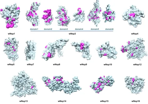
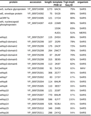
Next, as a result of a comprehensive comparative modeling effort, we were able to structurally
characterize 17 individual proteins, including 13 non-structural proteins of wORF1ab (wNsp1, wNsp3,
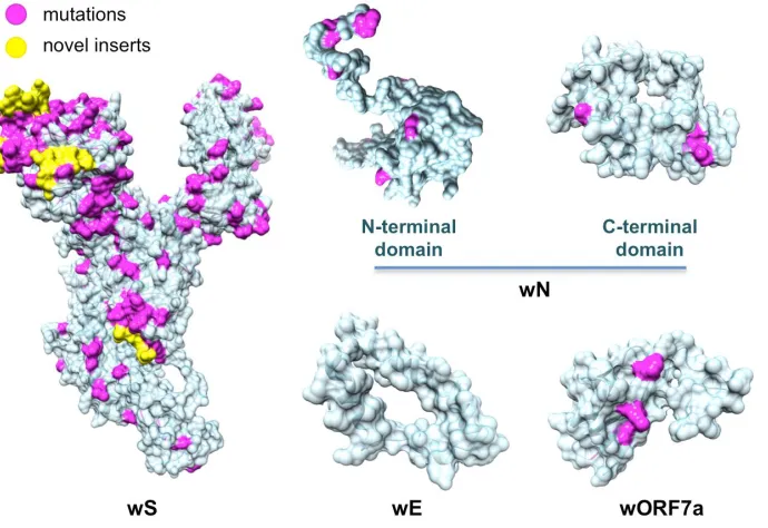
wNsp4, wNsp5, wNsp7, wNsp8, wNsp9, wNsp10, wNsp12, wNsp14, wNsp15, wNsp16), three structural
proteins (wE, wN, and wS), as well as one ORF (wORF7a). For two proteins, wNsp3 and wN, multiple
individual domains were modeled (Fig.1, Fig.2). The templates for the majority of the models were
homologous protein structures from other coronaviruses, with a high target-to-template sequence
similarity (seq. ids: 75–96%, except for ORF8). For two proteins, wN and wNsp3, multiple domains were
modeled. N-terminal and C-terminal domains of wN correspond to N-terminal RNA-binding domain and
C-terminal dimerization domain of SARS, respectively. The modeled domains 1–6 of wNsp1 correspond
to (1) N-terminal domain; (2) adenosine diphosphate ribose 1″ phosphatase domain; (3) SUD domain
(SARS-specific unique domain) containing two macrodomains; (4) SUD domain C; (5) the papain-like
domain of SARS Nsp3 was mapped to the sequence of wNsp3, but could not be modeled due to the lack
of a template structure.
The structural analysis of the modeled proteins combined with the sequence conservation analysis
revealed several findings. First, we found that the mutated residues tend to locate on the protein’s surface,
supporting previous observations in other families of RNA viruses that the core residues of viral proteins
are more conserved than the surface residues [48, 56, 57]. Furthermore, in a substantial number of
proteins, distributions of mutated positions exhibited spatial patterns, with groups of mutations found to
form clusters on the protein surfaces (Fig.2). These obtained models were then used as reference
structures to map and analyze the protein-binding and ligand-binding sites.
Next, using comparative modeling we structurally characterized protein interaction complexes, for both
intra-viral (homo- and hetero-oligomers) and host-viral interactions where host proteins were exclusively
human. In total, we obtained structural models for 16 homo-oligomeric complexes, three
oligomeric complexes, and eight human-virus interaction complexes (Fig. 3). The intra-viral
hetero-oligomeric complexes included exclusively the interactions between the non-structural proteins (wNsp7,
wNsp8, wNsp10, wNsp12, wNsp14, and wNsp16). The modeled host-viral interaction complexes
included three types of interactions: non-structural protein wNsp3 (papain-like protease, PLpro, domain)
interacting with human ubiquitin-aldehyde, surface protein wS (in its trimeric form) interacting with
human receptor angiotensin-converting enzyme 2 (ACE2) in different conformations, as well as the same
protein wS interacting with several neutralizing antibodies. Based on the obtained models, the protein
interaction binding sites were extracted and analyzed with respect to their evolutionary conservation.
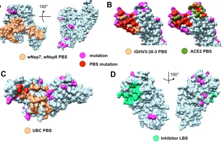
The analysis of the evolutionary conservation of protein binding sites revealed several patterns. First, we
found that all protein binding sites of non-structural proteins involving in the intra-viral heteromeric
complexes, wNSP7-wNsp8-wNsp12, wNsp10-wNsp14, and wNsp10-wNsp16, are either fully conserved
or allow at most one mutation on the periphery of the binding region, in spite of the fact that each proteins
had multiple mutations on their surfaces (Fig. 4A, see Suppl. Materials for the alignments of individual
proteins annotated with the protein binding sites). Furthermore, we observed the same behavior when
analyzing the interaction between the papain-like protease PLpro domain of wNsp3 and human
ubiquitin-aldehyde (Fig. 4B, see Suppl. Materials): the only two mutated residues were located on a border of the
binding region and thus were unlikely to disrupt the protein-protein interaction.
The surface of wS presents a striking contrast to the majority of SARS-CoV-2 proteins due to its heavily
mutated surface (Fig. 3; see Suppl. Materials for the alignment of wS with related proteins). First, the four
novel sequence inserts and two inserts shared with the closest strains were expected to affect the protein’s
function. Interestingly, three of the novel inserts are located in the first NTD domain, while the fourth one
is located immediately before the S2 cleavage site and inside the homo-trimerization interaction interface.
While the RBD domain of wS was not affected by those inserts it was the most heavily mutated region of
wS with likely disruptive functional effects on the interactions with the human ACE2 receptor and
monoclonal antibodies mAb 396 [58] and mAb 80R [59] (Fig. 4B). The NTD domain has been
considered a target of another antibody Ab G2 previously shown to work in MERS [60]. However, based
on the structural superposition of the NTD domain, it is likely that the expected interaction will be
disrupted by the novel sequence inserts to wS.
Finally, the analysis of seven ligand binding sites (LBS) for multiple candidate inhibitors previously
identified for four proteins in SARS and MERS showed that many of the LBS were intact in the
proteins annotated with the ligand binding sites) such as wN, wNsp3, wNsp5, and wNsp16. For wNsp14,
the LBS for several inhibitors was mutated while a co-localized binding site for another ligand was intact.
Joint intra-viral and human-virus protein-protein interaction network for SARS CoV indicates potential
system-wide roles of SARS-CoV-2 proteins
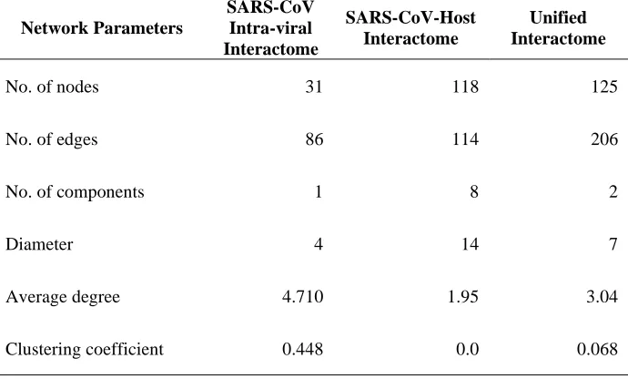
After constructing the individual networks of intra-viral interactions and virus-host interactions, they were
merged to form a unified network of SARS-CoV interactions, with the hypothesis that most interactions
would be conserved in SARS-CoV-2 (Fig. 6). The network analysis of all three networks showed that
unifying the intra-viral and virus-host networks reduces the number of components (islands) in the
SARS-CoV-Host interactome (Table 2). This suggested that the virus-host interactome map missed some viral
interactions that increased the number of components. The clustering coefficient and average node degree
were both higher in the unified interactome compared to the virus-host interactome.
The network topology of the unified interactome indicated the presence of several viral hubs for the
structural, non-structural, and accessory proteins (Fig. 6). The viral proteins form all the hubs and there
are a few of specific interest. ORF9b is one of the largest hubs in the network thought to play a secondary
role only in intra-viral interactions and not necessary for replication [61], but a recent study has shown
that ORF9b hinders immunity by targeting the mitochondria and limits host cell responses [62]. NSP8 is
the other major hub and interacts with other replicase proteins, including two other hubs, i.e. NSP7 and
NSP 12, which together play a crucial role in replication [63].
Another crucial non-structural protein is NSP1, also present in the SARS-CoV-2, which is known to be a
primary disruptor of innate immunity. The host interaction partners of NSP1 modulate the
Calcineurin/NFAT pathway that plays an important role in immune cell activation [64]. Overexpression
severe SARS cases. Additionally, inhibition of cyclophilins/immunopilins (host interaction partners) by
cyclosporine A (CspA) blocks the replication of CoVs of all genera.
There is an important interaction between the SARS spike protein (S) and the host angiotensin-converting
enzyme 2 (ACE2), as it is associated with cross-species and human-to-human transmissions. The same
interaction is also inferred from structural modeling between SARS-CoV-2 wS and ACE2, but might be
disrupted due to a substantial number of mutations in the receptor binding site of wS though another
recent study proposed that the interaction will be preserved [65]. Similar to SARS-CoV [66], wS protein
in SARS-CoV-2 is expected to interact with type II transmembrane protease (TMPRSS2) [67] and is
likely to be involved in inhibition of antibody-mediated neutralization. Thus, wS remains an important
target for vaccines and drugs previously evaluated in SARS and MERS while a neutralizing antibody
targeting the wS protein could provide passive immunity [68]. In addition, there are 7 interactions from
SARS-CoV determined by structural characterization of the protein complexes that are predicted to be
either conserved or potentially disrupted in SARS-CoV-2 (green edges in Fig. 1). An important target for
vaccines and drugs is the surface (S) protein which has been evaluated in SARS-CoV and MERS-CoV
with the idea that a neutralizing antibody targeting wS protein could provide passive immunity for
SARS-CoV-2 [68]. We also structurally modeled interactions between the SARS-SARS-CoV-2 wS protein and three
human monoclonal antibodies that were previously studied in SARS-CoV for immunotherapy and
mapped the information about the evolutionary conserved and diverse surface residues. We find that two
interactions, with mAb 396 [58] and mAb 80R [59], are likely to be disrupted due to the heavily mutated
binding sites while another, Ab G2 [60], is likely disrupted due to a novel insert into the sequence of wS.
These findings may provide guidelines in the search for potential antibody candidates for treatment.
Discussion
This work provides an initial large-scale structural genomics and interactomics effort towards
computational work is two-fold. First, by making the structural road map and the related findings fully
available to the research community, we aim to facilitate the process of structure-guided research where
accurate structural models of proteins and their interaction complexes already exist. Second, by providing
a comparative analysis between the new virus and its closest relatives from the perspective of protein- and
ligand-binding, we hope to help experimental scientists in their deciphering of the molecular mechanisms
implicated in infection by the new coronavirus as well as in vaccine development and antiviral drug
discovery.
Through integrating the information on structure, function, and evolution in a comparative study of
SARS-CoV-2 and the closely related coronaviruses, one can make several preliminary conclusions. First,
the extended peptide sequence newly introduced to wNsp3 between two structurally and possibly
functionally independent domains of this protein, might act as a long inter-domain linker, thus extending
the conformational flexibility of this multi-domain protein. Second, the presence of the four novel inserts
and one highly variable region of the surface protein wS and the analysis of this large-scale sequence
change with respect to intra-viral and viral-host interactions leads us to conclude that these inserts might
have structural impact on the homo-trimeric form of the protein as well as impact the functions carried
out by NTD domain. Third, the structurally modellable repertoire of SARS-CoV-2 proteome also
pinpoints to the interesting targets for structural biology and hybrid methods. For instance, the whole
structures of the multi-domain proteins wN and wNsp3 could be resolved by integrating individual
models with the lower-resolution but whole-protein covering techniques such as cryogenic electron
microscopy (CryoEM). The structure of wNsp3 is especially interesting because of the presence of the
novel peptide introduced between the two structural domains of the protein.
SARS-CoV-evolutionary conservation of the protein binding sites on the surfaces of non-structural proteins wNsp7,
wNsp8, wNsp10, wNsp12, wNsp14, and wNsp16 that correspond to the intra-viral interactions for three
complexes is consistent with our previous observations that the intra-viral interactions are significantly
more conserved than viral-host interactions [48, 49, 69]. However, the near-perfect conservation of the
human ubiquitin-aldehyde protein binding site on the surface of wNsp3 is rather intriguing, suggesting the
critical role of this interaction in the functioning of SARS-like coronaviruses. Lastly, the conservation of
the ligand-binding sites for many putative inhibitors previously developed for SARS and MERS suggests
the development of antiviral drugs as a promising direction for addressing this new global health threat.
Materials & Methods
The goal of this work is to identify the evolutionary differences between SARS-CoV-2 and the closest
coronavirus species, human SARS coronavirus and bat SARS-like coronavirus, and to predict their
possible functional implications. To do so, we structurally characterize individual proteins as well as
intra-viral and human-virus protein complexes, extract the information on their interaction interfaces and
ligand binding, and superpose the evolutionary difference and conservation information with the binding
information. Specifically, our integrative computational pipeline will include the following five steps.
First, for each of the candidate SARS-CoV-2 proteins a set of sequentially similar coronavirus proteins is
determined and aligned. Second, structural models of SARS-CoV-2 proteins are obtained using
template-based, or homology, modeling. Third, the protein-protein interaction complex structures of SARS-CoV-2
proteins interacting with each other and/or with the human proteins are determined using a multi-chain
comparative modeling protocol. Fourth, the protein-binding sites will be extracted from the obtained
models of protein-protein interaction complexes, and protein-ligand binding sites will be extracted from
the evolutionary close coronavirus protein-ligand templates and mapped to the relevant structural models
of SARS-CoV-2 through structural alignment. Fifth, the information on the evolutionary differences and
extracted from the above protein sequence alignments will mapped onto the structural models of the
SARS-CoV-2 proteins to determine if the protein- and ligand-binding sites are functionally conserved.
Lastly, a joint human-virus and virus-virus interactome is predicted through homology and analyzed. All
the obtained models and sequence alignments have been made publicly available to the research
community.
Protein sequence data collection and analysis
Available sequences for protein candidates wS, wORF3a, wE, wM, wORF6, wORF7a, wORF7b,
wORF8, wN, and wORF10 are extracted from NCBI Virus repository [70] (collected on January 29,
2019) and then used in the sequence analysis and structural modeling (Suppl. Materials). The Uniprot
BLAST-based search is performed for each of the proteins using default parameters. From the results of
each search, the final selection is done based on the pairwise sequence identity (>60%) as well as the
relationship (Coronaviridae family). Each of SARS-CoV-2 proteins is then aligned with the
corresponding coronavirus proteins using the multiple sequence alignment method Clustal Omega [71].
Structural characterization of protein and protein complexes
The structure of each protein is determined using a single-template comparative modeling protocols with
the MODELLER software package [72]. First, the template for each protein sequence is identified using a
PSI-BLAST search in Protein Data Bank (PDB) [73]. In general, a structural template with the highest
sequence identity is selected out of those that cover at least 50 residues of the target sequence with at least
30% sequence identity. The polyprotein wORF1ab was first split into 16 putative proteins based on its
alignment with the human SARS polyprotein, with each protein independently searched against PDB. In
total, structural templates for 17 proteins were chosen (Table 1, Figs. 1-3). In some cases, several
independent templates, each covering an individual protein domain, of a target SARS-CoV-2 protein are
Each model is assessed using the DOPE statistical potential [74]; the best-scoring model is selected as a
final prediction.
To model a protein-protein interaction complex, a multi-chain modeling protocol is used [39].
Specifically, we align the corresponding pairs of homologous proteins and combine them into a single
alignment where the individual chains are separated by “/” symbol. The alignment is used as an input
together with the multi-chain structural template of a homologous complex. In case of viral-human
interaction (these are the only virus-host structural templates found), the human protein remains the same
in the alignment. In total, 18 structural templates of protein complexes involving homologs of 14
SARS-CoV-2 proteins were retrieved (Fig. 3). Similar to the single-protein protocol, five candidate models are
generated for each complex conformation, and each model is assessed using the DOPE statistical
potential following selection of the best-scoring model as a final prediction.
Mapping of functional regions and evolutionary conservation
We next extract protein- and ligand-binding sites and map them onto the models of SARS-CoV-2
proteins. For protein binding sites, the obtained modeled structures of protein complexes that involve
SARS-CoV-2 proteins are considered. For each SARS-CoV-2 protein in a protein-protein interaction
complex, we identify all binding residues that constitute its protein-binding site. Given an interaction
between two proteins, a residue on one protein is defined as a protein binding site residue if there is at
least one pair of atoms, one from this residue and another from a residue in the second protein, with Van
Der Waals (VDW) surfaces not farther than 1.0 Å. Using this definition, the binding sites are identified
with UCSF Chimera [75]. The ligand binding sites are identified and mapped using a different protocol—
we rely on the default definition of protein-ligand binding site residues from PDB 3D Ligand View, since
this standard is widely accepted. Once all ligand binding site residues are identified for a related
structural alignment between the two proteins, which puts residues from both proteins into a one-to-one
correspondence.
Inferring intra-viral and virus-host protein-protein interaction networks
Next, we leverage the obtained protein homology information to predict and map all possible intra-viral
and virus-host protein interactions at a systems level. Since the SARS-CoV-2 genome exhibits substantial
similarity to the 2002 SARS-CoV genome [76] and proteome [77], we hypothesize that many of the
interactions observed in the SARS proteome will be preserved in the SARS-CoV-2 proteome as well,
unless the corresponding binding sites are affected. The information in the interactome will help us
understand the global mechanistic processes of the viral molecular machinery during viral infection,
survival within the host, and replication. With this knowledge, we can discern the protein interactions that
are crucial for transmission and replication—such interactions are potential candidates for inhibitory
drugs [78]. Furthermore, using the systems approach, we can identify hubs and bottlenecks new to
SARS-CoV-2 that could again be targeted by the antiviral drugs.
For this purpose, we create a comprehensive integrated SARS-CoV interactome that consists of both
intra-viral and virus-host interactions. The SARS intra-viral interactome was created using published data,
while the SARS-CoV ORFeome was cloned and a genome-wide analysis of viral protein interactions
through yeast-two-hybrid (Y2H) matrix screens was performed [61]. The Y2H matrix screen was
summarized by combining interactions in one direction, both directions and self-interaction. We also
included intra-viral interactions gathered from the literature review [61]. The aggregated intra-viral
We then construct the SARS-CoV-Host interactome through published interaction data, where first the
SARS-CoV ORFeome was cloned for Y2H screens and used as a bait [64]. Specifically, a cDNA library
encoding 5,000 different human genes were used that acted as prey molecules. We also include virus-host
interactions mined from literature survey of 5,000 abstracts [64]. The curated virus-host interaction
network consists of 118 proteins, including 93 host proteins, and 114 unique virus-host interactions. Next,
to create an integrated network, the two individual networks were imported in Cytoscape [79] and merged
to form a unified interactome representing both intra-viral and virus-host interactions. Finally, we include
our predictions from structural modeling of SARS-CoV-2 intra-viral and virus-host interactions, surveyed
recent literature on predicted SARS-CoV-2 interactions and annotated them accordingly in the unified
interactome. The unified interactome consists of 125 proteins (94 host proteins) and 200 unique
interactions. In addition, network analysis is performed to compute a set of topological statistics (degree
distribution, clustering coefficient, and other important characteristics) to characterize parameters of the
three networks, before which the networks are pruned for duplicate edges. Finally, the 200 putative
interactions were annotated based on the extent to which the protein binding sites of the SARS-CoV-2
proteins were altered, compared to their SARS homologs. Based on the evolutionary conservation
analysis of the putative protein binding sites extracted from the modeled complexes, some interactions
were annotated as potentially disrupted.
Availability: All data generated in this work is freely available to the research community via Korkin Lab
web-server (korkinlab.org/wuhan).
Funding
Figures
Tables
Table 1. The list of SARS-CoV-2 proteins analyzed and structurally characterized in this work.
protein accession length template PDB id
Trgt-tmplt Seq ID
organism
wS, surface glycoprotein YP_009724390 1273 6ACK 75% SARS
wE, envelope protein YP_009724392 75 5X29 89% SARS
wORF7a YP_009724395 121 1YO4 90% SARS
wN, nucleocapsid
phosphoprotein YP_009724397 419 2JW8 96% SARS
1SSK 83% SARS
4UD1 51% MERS
wNsp1 YP_009725297 115 2HSX 86% SARS
wNsp3-domain1 YP_009725299 107 2GRI 79% SARS
wNsp3-domain2 YP_009725299 175 2ACF 72% SARS
wNsp3-domain3 YP_009725299 264 2WCT 76% SARS
wNsp3-domain4 YP_009725299 67 2KAF 70% SARS
wNsp3-domain5 YP_009725299 315 3E9S 82% SARS
wNsp3-domain6 YP_009725299 113 2K87 82% SARS
wNsp4 YP_009725300 91 3VC8 60% MHV
wNsp5 YP_009725301 306 2GT7 96% SARS
wNsp7 YP_009725302 83 1YSY 67% SARS
wNsp8 YP_009725304 114 6NUR 85% SARS
wNsp9 YP_009725305 110 3EE7 99% SARS
wNsp10 YP_009725306 121 2G9T 98% SARS
wNsp12 YP_009725307 770 6NUR 97% SARS
wNsp13 YP_009725308 596 6JYT 100% SARS
wNsp14 YP_009725309 526 5C8U 95% SARS
wNsp15 YP_009725310 346 2H85 86% SARS
Table 2. Network Parameters of the SARS-CoV intra-viral, virus-host and unified networks. The
table shows topological statistics for the three networks. Among the many computed statistics, the shown
parameters include the number of nodes and edges in the networks, the average degree, number of
components (independent networks), diameter (maximum shortest path), and clustering coefficient.
Network Parameters
SARS-CoV Intra-viral Interactome
SARS-CoV-Host Interactome
Unified Interactome
No. of nodes 31 118 125
No. of edges 86 114 206
No. of components 1 8 2
Diameter 4 14 7
Average degree 4.710 1.95 3.04
References
1. Huang, C., et al., Clinical features of patients infected with 2019 novel coronavirus in Wuhan,
China. The Lancet, 2020.
2. Hui, D.S., et al., The continuing SARS-CoV-2 epidemic threat of novel coronaviruses to global health—The latest 2019 novel coronavirus outbreak in Wuhan, China. International Journal of Infectious Diseases, 2020. 91: p. 264-266.
3. Zhu, N., et al., A novel coronavirus from patients with pneumonia in China, 2019. New England Journal of Medicine, 2020.
4. Nabel, G.J., Designing tomorrow's vaccines. New England Journal of Medicine, 2013. 368(6): p. 551-560.
5. Plotkin, S.A. and S.L. Plotkin, The development of vaccines: how the past led to the future. Nature Reviews Microbiology, 2011. 9(12): p. 889-893.
6. Martin, J.E., et al., A SARS DNA vaccine induces neutralizing antibody and cellular immune responses in healthy adults in a Phase I clinical trial. Vaccine, 2008. 26(50): p. 6338-6343.
7. Zumla, A., et al., Coronaviruses—drug discovery and therapeutic options. Nature reviews Drug discovery, 2016. 15(5): p. 327.
8. Zumla, A., et al., Vaccine against Middle East respiratory syndrome coronavirus. The Lancet Infectious Diseases, 2019. 19(10): p. 1054-1055.
9. Cotten, M., et al., Transmission and evolution of the Middle East respiratory syndrome
coronavirus in Saudi Arabia: a descriptive genomic study. The Lancet, 2013. 382(9909): p.
1993-2002.
10. de Wit, E., et al., SARS and MERS: recent insights into emerging coronaviruses. Nature Reviews Microbiology, 2016. 14(8): p. 523.
11. Dyall, J., et al., Repurposing of clinically developed drugs for treatment of Middle East respiratory
syndrome coronavirus infection. Antimicrobial agents and chemotherapy, 2014. 58(8): p.
4885-4893.
12. Li, F., Structure, function, and evolution of coronavirus spike proteins. Annual review of virology, 2016. 3: p. 237-261.
13. Lu, G., et al., Molecular basis of binding between novel human coronavirus MERS-CoV and its
receptor CD26. Nature, 2013. 500(7461): p. 227-231.
14. Luk, H.K., et al., Molecular epidemiology, evolution and phylogeny of SARS coronavirus. Infection, Genetics and Evolution, 2019.
15. Millet, J.K. and G.R. Whittaker, Physiological and molecular triggers for SARS-CoV membrane fusion and entry into host cells. Virology, 2018. 517: p. 3-8.
16. Song, W., et al., Cryo-EM structure of the SARS coronavirus spike glycoprotein in complex with its
host cell receptor ACE2. PLoS pathogens, 2018. 14(8): p. e1007236.
17. Walls, A.C., et al., Cryo-electron microscopy structure of a coronavirus spike glycoprotein trimer. Nature, 2016. 531(7592): p. 114-117.
18. Yuan, Y., et al., Cryo-EM structures of MERS-CoV and SARS-CoV spike glycoproteins reveal the
dynamic receptor binding domains. Nature communications, 2017. 8: p. 15092.
19. Cho, C.-C., et al., Macro Domain from Middle East Respiratory Syndrome Coronavirus (MERS-CoV) Is an Efficient ADP-ribose Binding Module CRYSTAL STRUCTURE AND BIOCHEMICAL
STUDIES. Journal of Biological Chemistry, 2016. 291(10): p. 4894-4902.
20. Gui, M., et al., Cryo-electron microscopy structures of the SARS-CoV spike glycoprotein reveal a
21. Jacobs, J., et al., Discovery, synthesis, and structure-based optimization of a series of N-(tert-butyl)-2-(N-arylamido)-2-(pyridin-3-yl) acetamides (ML188) as potent noncovalent small molecule inhibitors of the severe acute respiratory syndrome coronavirus (SARS-CoV) 3CL
protease. Journal of medicinal chemistry, 2013. 56(2): p. 534-546.
22. Jia, Z., et al., Delicate structural coordination of the Severe Acute Respiratory Syndrome
coronavirus Nsp13 upon ATP hydrolysis. Nucleic acids research, 2019. 47(12): p. 6538-6550.
23. Kankanamalage, A.C.G., et al., Structure-guided design of potent and permeable inhibitors of MERS coronavirus 3CL protease that utilize a piperidine moiety as a novel design element. European journal of medicinal chemistry, 2018. 150: p. 334-346.
24. Kirchdoerfer, R.N., et al., Stabilized coronavirus spikes are resistant to conformational changes induced by receptor recognition or proteolysis. Scientific reports, 2018. 8(1): p. 1-11.
25. Kirchdoerfer, R.N. and A.B. Ward, Structure of the SARS-CoV nsp12 polymerase bound to nsp7
and nsp8 co-factors. Nature communications, 2019. 10(1): p. 1-9.
26. Li, Y., et al., A humanized neutralizing antibody against MERS-CoV targeting the receptor-binding
domain of the spike protein. Cell research, 2015. 25(11): p. 1237-1249.
27. Ma, Y., et al., Structural basis and functional analysis of the SARS coronavirus nsp14–nsp10
complex. Proceedings of the National Academy of Sciences, 2015. 112(30): p. 9436-9441.
28. Ratia, K., et al., Structural basis for the ubiquitin-linkage specificity and deISGylating activity of
SARS-CoV papain-like protease. PLoS pathogens, 2014. 10(5).
29. Shimamoto, Y., et al., Fused-ring structure of decahydroisoquinolin as a novel scaffold for SARS 3CL protease inhibitors. Bioorganic & Medicinal Chemistry, 2015. 23(4): p. 876-890.
30. Su, D., et al., Dodecamer structure of severe acute respiratory syndrome coronavirus
non-structural protein nsp10. Journal of virology, 2006. 80(16): p. 7902-7908.
31. Wang, N., et al., Structural Definition of a Neutralization-sensitive Epitope on the MERS-CoV S1-NTD. Cell reports, 2019. 28(13): p. 3395-3405. e6.
32. Martí-Renom, M.A., et al., Comparative protein structure modeling of genes and genomes. Annual review of biophysics and biomolecular structure, 2000. 29(1): p. 291-325.
33. Cavasotto, C.N. and S.S. Phatak, Homology modeling in drug discovery: current trends and
applications. Drug discovery today, 2009. 14(13-14): p. 676-683.
34. Burley, S.K., et al., Structural genomics: beyond the human genome project. Nature genetics, 1999. 23(2): p. 151-157.
35. Yan, L., et al., Assessment of putative protein targets derived from the SARS genome. FEBS letters, 2003. 554(3): p. 257-263.
36. Wichapong, K., et al., Homology modeling and molecular dynamics simulations of Dengue virus
NS2B/NS3 protease: insight into molecular interaction. Journal of Molecular Recognition: An
Interdisciplinary Journal, 2010. 23(3): p. 283-300.
37. Ekins, S., et al., Illustrating and homology modeling the proteins of the Zika virus. F1000Research, 2016. 5.
38. Prabakaran, P., X. Xiao, and D.S. Dimitrov, A model of the ACE2 structure and function as a
SARS-CoV receptor. Biochemical and biophysical research communications, 2004. 314(1): p. 235-241.
39. Davis, F.P., et al., Host–pathogen protein interactions predicted by comparative modeling. Protein Science, 2007. 16(12): p. 2585-2596.
40. Russell, R.B., et al., A structural perspective on protein–protein interactions. Current opinion in structural biology, 2004. 14(3): p. 313-324.
41. Zhang, Q.C., et al., Structure-based prediction of protein–protein interactions on a genome-wide
42. Cavasotto, C.N., et al., Discovery of novel chemotypes to a G-protein-coupled receptor through
ligand-steered homology modeling and structure-based virtual screening. Journal of medicinal
chemistry, 2008. 51(3): p. 581-588.
43. Wang, S.-Q., Q.-S. Du, and K.-C. Chou, Study of drug resistance of chicken influenza A virus
(H5N1) from homology-modeled 3D structures of neuraminidases. Biochemical and biophysical
research communications, 2007. 354(3): p. 634-640.
44. Loewenstein, Y., et al., Protein function annotation by homology-based inference. Genome biology, 2009. 10(2): p. 207.
45. Durmuş, S. and K.Ö. Ülgen, Comparative interactomics for virus–human protein–protein
interactions: DNA viruses versus RNA viruses. FEBS open bio, 2017. 7(1): p. 96-107.
46. Zhang, A., L. He, and Y. Wang, Prediction of GCRV virus-host protein interactome based on
structural motif-domain interactions. BMC bioinformatics, 2017. 18(1): p. 145.
47. Vidalain, P.-O. and F. Tangy, Virus-host protein interactions in RNA viruses. Microbes and infection, 2010. 12(14-15): p. 1134-1143.
48. Warren, S., et al., Extreme evolutionary conservation of functionally important regions in H1N1
influenza proteome. PloS one, 2013. 8(11).
49. Wang, L., et al., From mosquitos to humans: genetic evolution of Zika virus. Cell host & microbe, 2016. 19(5): p. 561-565.
50. Patel, H. and A. Kukol, Prediction of ligands to universally conserved binding sites of the influenza a virus nuclear export protein. Virology, 2019. 537: p. 97-103.
51. Sawicki, S.G., D.L. Sawicki, and S.G. Siddell, A contemporary view of coronavirus transcription. Journal of virology, 2007. 81(1): p. 20-29.
52. UniProt: the universal protein knowledgebase. Nucleic acids research, 2017. 45(D1): p.
D158-D169.
53. Saikatendu, K.S., et al., Structural basis of severe acute respiratory syndrome coronavirus
ADP-ribose-1 ″-phosphate dephosphorylation by a conserved domain of nsP3. Structure, 2005. 13(11):
p. 1665-1675.
54. Serrano, P., et al., Nuclear magnetic resonance structure of the N-terminal domain of
non-structural protein 3 from the severe acute respiratory syndrome coronavirus. Journal of virology,
2007. 81(21): p. 12049-12060.
55. Johnson, M., et al., NCBI BLAST: a better web interface. Nucleic acids research, 2008.
36(suppl_2): p. W5-W9.
56. Cheng, S. and C.L. Brooks III, Viral capsid proteins are segregated in structural fold space. PLoS computational biology, 2013. 9(2).
57. Patel, H. and A. Kukol, Evolutionary conservation of influenza A PB2 sequences reveals potential target sites for small molecule inhibitors. Virology, 2017. 509: p. 112-120.
58. Zhu, Z., et al., Potent cross-reactive neutralization of SARS coronavirus isolates by human
monoclonal antibodies. Proceedings of the National Academy of Sciences, 2007. 104(29): p.
12123-12128.
59. Sui, J., et al., Effects of human anti-spike protein receptor binding domain antibodies on severe acute respiratory syndrome coronavirus neutralization escape and fitness. Journal of virology, 2014. 88(23): p. 13769-13780.
60. Coughlin, M.M., J. Babcook, and B.S. Prabhakar, Human monoclonal antibodies to
SARS-coronavirus inhibit infection by different mechanisms. Virology, 2009. 394(1): p. 39-46.
61. von Brunn, A., et al., Analysis of intraviral protein-protein interactions of the SARS coronavirus
62. Shi, C.-S., et al., SARS-coronavirus open reading frame-9b suppresses innate immunity by
targeting mitochondria and the MAVS/TRAF3/TRAF6 signalosome. The Journal of Immunology,
2014. 193(6): p. 3080-3089.
63. Subissi, L., et al., One severe acute respiratory syndrome coronavirus protein complex integrates
processive RNA polymerase and exonuclease activities. Proceedings of the National Academy of
Sciences, 2014. 111(37): p. E3900-E3909.
64. Pfefferle, S., et al., The SARS-coronavirus-host interactome: identification of cyclophilins as
target for pan-coronavirus inhibitors. PLoS pathogens, 2011. 7(10).
65. Wan, Y., et al., Receptor recognition by novel coronavirus from Wuhan: An analysis based on
decade-long structural studies of SARS. Journal of Virology, 2020.
66. Glowacka, I., et al., Evidence that TMPRSS2 activates the severe acute respiratory syndrome coronavirus spike protein for membrane fusion and reduces viral control by the humoral immune
response. Journal of virology, 2011. 85(9): p. 4122-4134.
67. Hoffmann, M., et al., The novel coronavirus 2019 (SARS-CoV-2) uses the SARS-coronavirus
receptor ACE2 and the cellular protease TMPRSS2 for entry into target cells. bioRxiv, 2020.
68. Kruse, R.L., Therapeutic strategies in an outbreak scenario to treat the novel coronavirus
originating in Wuhan, China. F1000Research, 2020. 9(72): p. 72.
69. Voitenko, O.S., et al., Patterns of amino acid conservation in human and animal
immunodeficiency viruses. Bioinformatics, 2016. 32(17): p. i685-i692.
70. Brister, J.R., et al., NCBI viral genomes resource. Nucleic acids research, 2015. 43(D1): p. D571-D577.
71. Sievers, F. and D.G. Higgins, Clustal Omega for making accurate alignments of many protein
sequences. Protein Science, 2018. 27(1): p. 135-145.
72. Eswar, N., et al., Comparative protein structure modeling using Modeller. Current protocols in bioinformatics, 2006. 15(1): p. 5.6. 1-5.6. 30.
73. Berman, H.M., et al., The protein data bank, in Protein Structure. 2003, CRC Press. p. 394-410. 74. Shen, M.y. and A. Sali, Statistical potential for assessment and prediction of protein structures.
Protein science, 2006. 15(11): p. 2507-2524.
75. Pettersen, E.F., et al., UCSF Chimera—a visualization system for exploratory research and
analysis. Journal of computational chemistry, 2004. 25(13): p. 1605-1612.
76. Chan, J.F.-W., et al., Genomic characterization of the 2019 novel human-pathogenic coronavirus isolated from a patient with atypical pneumonia after visiting Wuhan. Emerging Microbes & Infections, 2020. 9(1): p. 221-236.
77. Dong, N., et al., Genomic and protein structure modelling analysis depicts the origin and infectivity of SARS-CoV-2, a new coronavirus which caused a pneumonia outbreak in Wuhan,
China. bioRxiv, 2020.
78. De Chassey, B., et al., Virus-host interactomics: new insights and opportunities for antiviral drug
discovery. Genome medicine, 2014. 6(11): p. 115.
79. Shannon, P., et al., Cytoscape: a software environment for integrated models of biomolecular