www.impactjournals.com/Genes&Cancer
Genes & Cancer, Vol. 6 (5-6), May 2015
Histone acetyltransferases and histone deacetylases in B- and
T-cell development, physiology and malignancy
Leila Haery
1, Ryan C. Thompson
1and Thomas D. Gilmore
1 1 Department of Biology, Boston University, Boston, MA, USACorrespondence to: Thomas D. Gilmore, email: [email protected]
Keywords: HAT, HDAC, acetylation, B cells, T cells
Received: May 1, 2015 Accepted: May 12, 2015 Published: May 15, 2015
This is an open-access article distributed under the terms of the Creative Commons Attribution License, which permits unrestricted use, distribution, and reproduction in any medium, provided the original author and source are credited.
ABSTRACT
The development of B and T cells from hematopoietic precursors and the regulation of the functions of these immune cells are complex processes that involve highly regulated signaling pathways and transcriptional control. The signaling pathways and gene expression patterns that give rise to these developmental processes are coordinated, in part, by two opposing classes of broad-based enzymatic regulators: histone acetyltransferases (HATs) and histone deacetylases (HDACs). HATs and HDACs can modulate gene transcription by altering histone acetylation to modify chromatin structure, and by regulating the activity of non-histone substrates, including an array of immune-cell transcription factors. In addition to their role in normal B and T cells, dysregulation of HAT and HDAC activity is associated with a variety of B- and T-cell malignancies. In this review, we describe the roles of HATs and HDACs in normal B- and T-cell physiology, describe mutations and dysregulation of HATs and HDACs that are implicated lymphoma and leukemia, and discuss HAT and HDAC inhibitors that have been explored as treatment options for leukemias and lymphomas.
INTRODUCTION
B and T cells have a variety of cellular subtypes that
arise through a complex series of developmental events.
The function of these various immune cell subtypes can
be altered by numerous extracellular factors, including
antigens, cytokines, and growth factors. Many of these
developmental and functional processes are controlled
by large-scale changes in gene expression, either due
to epigenetic changes in chromatin structure or to the
activity of specific transcription factors (TFs). Histone
acetyltransferases (HATs) and histone deacetylases
(HDACs) are two opposing classes of enzymes that play
widespread roles in regulating transcription either by
altering chromatin structure or by modulating the activity
of specific TFs. Thus, it is perhaps not surprising that
HATs and HDACs play roles in maintaining hematopoietic
precursors and in coordinating their maturation into
various subtypes of B and T cells.
As with many proteins that have important roles in
normal developmental and cell-specific proliferation and
survival processes, HAT and HDAC activity is altered
in many B- and T-cell malignancies. Moreover, several
HDAC inhibitors (HDACi) have been found to reduce
the proliferation of B and T cancer cells
in vitro
and
in
vivo
. As an outcome of such basic research, there are four
FDA-approved HDACi being used clinically to treat T-cell
lymphoma and multiple myeloma, and there are several
clinical trials using HDACi for the treatment of B- and
T-cell cancers.
In this review, we describe the roles of HATs and
HDACs in normal B- and T-cell development and function,
and also discuss alterations in HAT/HDAC activity in
B- and T-cell malignancies. Finally, we summarize the
current status of HAT and HDAC inhibitors as potential
therapies for cancers affecting B and T cells.
Overview of the Regulation of Transcription by
HATs and HDACs
an acetyl group prevents the formation of positive charges
on the lysine amino group, and thus, can affect protein
activity. Through this reversible catalytic event, HATs
and HDACs can regulate transcription in two general
ways: 1) by altering histone acetylation patterns, thereby
modulating chromatin structure and its accessibility
to transcriptional regulatory proteins [1, 2], and 2) by
acetylating and affecting the activity of non-histone
substrates that directly regulate transcription, including a
diverse array of TFs [3].
HATs are a subtype of transcriptional coactivators,
in that they possess intrinsic acetyltransferase activity and
can enhance the ability of a TF to activate transcription.
In general, HAT-mediated acetylation of nucleosomal
histones increases the accessibility of DNA to TFs
and leads to increased transcription at a given DNA
locus. Acetylation of specific TFs by HATs can also
increase their ability to bind DNA, resist proteasomal
degradation, or interact with other TFs or coactivators,
and consequently, direct acetylation of TFs can also be a
transcriptional activating event [3]. In addition, by serving
as protein scaffolds, HATs can promote the formation of
transcriptional activating complexes near a gene promoter.
This scaffolding function does not necessarily require HAT
enzymatic activity, but rather is defined by the
protein-interaction domains of these relatively large molecules.
HDACs, on the other hand, generally act as
transcriptional corepressors by deacetylating nucleosomal
histones, which can lead to chromosomal condensation
and the exclusion of transcriptional activating complexes.
Additionally, large HDAC-containing repressor complexes
can localize to specific gene loci and exclude activating
molecules, including HATs, from interacting with TFs.
HDACs can also deacetylate specific TFs, decreasing
their DNA-binding activity, inducing their degradation, or
changing their subcellular localization or protein-protein
interactions [4].
Families of Human HATs and HDACs
HAT families
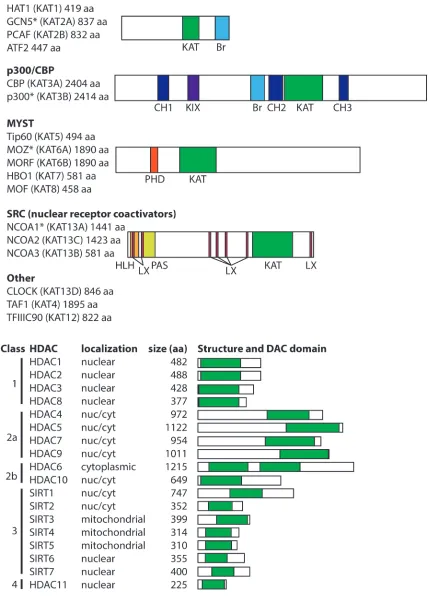
There are 17 human HATs, which are divided into
five families based primarily on the extent of sequence
similarity [5] (Figure 1). Although HATs can act on
a broad range of substrates
in vitro
, HATs are usually
directed to specific targets
in vivo
, and thus, HAT families
generally have distinct biological functions. The
non-catalytic domains of HATs are responsible for dictating
this substrate specificity, and HAT families generally have
conserved protein-protein interaction and reader domains
(e.g., bromodomains, PHD fingers), which enable them to
localize to particular genomic sites and recognize specific
chemical or epigenetic modifications. The size of the
catalytic HAT domain and the mechanism of catalysis also
differ between HAT families. For example, CBP and p300
utilize a “hit and run” kinetic model defined by an initial
binding of acetyl-CoA followed by transient binding to the
target lysine [6, 7], whereas the GNAT family HATs adopt
a ternary complex during catalysis [8].
GNAT family HATs (GCN5, HAT1, PCAF, ATF2)
are generally part of large, multi-protein complexes
that contain TBP-associated factors (TAFs) and a single
catalytic HAT subunit (reviewed in [9]). Two
well-characterized complexes found in humans are the 700 kDa
(i.e., ATAC) and 2 MDa (i.e., TFTC, STAGA, and PCAF)
complexes. These large HAT-containing complexes play
roles in global chromatin acetylation (i.e., the deposition
of acetyl marks on histones) and as coactivators of genes
when recruited to DNA by specific TFs or regulatory
proteins. Members of the GNAT family, especially PCAF,
also acetylate specific TFs and modulate their activity
(e.g., p53, BRCA2, PTEN). GNAT family members have
a conserved C-terminal bromodomain, which has been
shown to be an acetyl-lysine targeting motif. GNAT family
member ATF2 is the only sequence-specific DNA-binding
transcriptional activator with intrinsic HAT activity.
The CBP/p300 HATs are large (~300 kDa),
highly related proteins with a single HAT domain, a
bromodomain, and several cysteine–histidine-rich (CH)
domains that participate in a variety of protein-protein
interactions [5]. Indeed, CBP/p300 have been shown to
acetylate over 75 target proteins, including all histone
proteins, as well as numerous TFs. By virtue of their
multiple protein-protein interaction domains, CBP/p300
can also promote transcriptional activation by nucleating
transcriptional complexes at promoters in a non-enzymatic
manner. Although they generally act as coactivators,
in some cases, CBP/p300 appear to be involved in
transcriptional repression [10].
The MYST family of HATs (TIP60, MOZ,
MORF, HBO1, MOF) is characterized by a conserved
MYST domain that contains the catalytically active
HAT domain. The two largest family members, MOZ
and MORF, also have a PHD zinc finger domain, which
recognizes methyl-lysine-containing motifs [11], and
a C-terminal transactivation domain that interacts with
various transcription factors, including hematopoietic
cell regulators PU.1 and Runx1 [12-14]. Most MYST
family HATs act as catalytic subunits of large multiprotein
complexes, including the ING family of tumor suppressors
[15].
The steroid receptor coactivators (SRCs) include
three HATs (NCOA1, NCOA2, NCOA3) that enhance
transcription of genes responsive to liganded nuclear
with other nuclear receptors and cofactors; and 3) two
C-terminal transcriptional activation domains (AD1 and
AD2). Although SRCs have been associated with various
human cancers, currently they are not known to have a
role in hematopoiesis or B-/T-cell function.
Other HATs that are not clearly part of a family
include the following; TAF1 (TAF
II250), a subunit of the
TFIID general TF complex; CLOCK, which is primarily
involved in circadian rhythm; and the 90 kDa subunit
of TFIIIC, which is involved in the control of general
transcription in a complex with RNA polymerase III.
HDAC families
To counterbalance the impact of HATs on protein
function and genome structure, there are 18 human
HDACs, which are commonly divided into four major
classes based on homology to yeast orthologs (Figure
1): class 1 (HDAC1, 2, 3 and 8), class 2 (HDAC4, 5, 6,
7, 9, and 10), class 3 (aka sirtuins; SIRT1, 2, 3, 4, 5, 6,
and 7), and class 4 (HDAC11) [17-20]. HDAC classes
differ in their structure, substrate specificity, enzymatic
mechanism, subcellular localization, and tissue-specific
expression. Even though most HDACs contain a nuclear
localization signal (NLS) and, in some cases, a nuclear
export signal (NES), HDACs often localize to specific
subcellular regions due to protein-protein interactions with
proteins that direct their cellular localization.
The “classical” HDACs are those in classes 1, 2a,
2b, and 4, and they have a conserved ~390 aa catalytic
domain and Zn
2+-dependent deacetylase activity. The
conserved, ~275 aa catalytic domain of the class 3 sirtuins
is NAD
+-dependent and unrelated to the catalytic domain
of the classical HDACs [21, 22]. These differences in their
catalytic mechanisms have implications for inhibition of
HDAC activity, and thus, many of the HDAC inhibitors
(HDACi) used in cancer therapeutics target the classical
HDACs (discussed below).
Class 1 HDACs are ubiquitously expressed and
localize almost exclusively to the nucleus. The class 2
HDACs are generally much larger than class 1 HDACs,
show tissue-specific expression patterns, and often
shuttle in and out of the nucleus. In general, HDACs in
both classes 1 and 2 are found in large transcriptional
repressing complexes, and are recruited to DNA either
by other proteins in those complexes or by other
DNA-binding proteins. These large protein complexes play roles
in HDAC localization and substrate specificity, can act as
scaffolds to recruit DNA-binding proteins, and provide
the cofactors required for HDAC function. Indeed, lack
of these cofactors limits the activity of some recombinant
HDACs [23]. HDAC 11 is the only class 4 HDAC, and
although it shows sequence similarity to class 1 and 2
HDACs, it does not exist within any of the known HDAC
complexes.
Class 3 sirtuins vary in their subcellular localization
and interact with a diverse array of TFs and other,
primarily non-histone, substrates [24]. For example,
SIRT1 can directly interact with substrates involved in
the stress response, including p53, FOXO proteins, and
NF-κB. The mitochondrially-localized sirtuins (SIRT3,
4, 5) can regulate mitochondrial function, respiration,
and energy consumption [25]. Some sirtuins (SIRT4 and
6) lack deacetylase activity, but possess
ADP-ribosyl-transferase activity and play roles in metabolism and DNA
repair.
HATs and HDACs in B- and T-cell Development
HATs and HDACs in early hematopoietic
development
B- and T-cell development involves controlled
stages of gene expression programs and genomic
instability, which ultimately give rise to the diversity of
cells that provide adaptive immunity. These developmental
stages are tightly regulated by a large variety of TFs and
are coupled with the accessibility of DNA to factors
that coordinate chromosomal rearrangements. HATs
and HDACs play major roles in normal B- and T-cell
development because they can interact with hematopoietic
regulators and TFs, as well as affect DNA accessibility by
modifying chromatin structure near relevant target genes.
In this section, some roles of HATs and HDACs in normal
hematopoiesis, lymphopoiesis, and B- and T-cell function
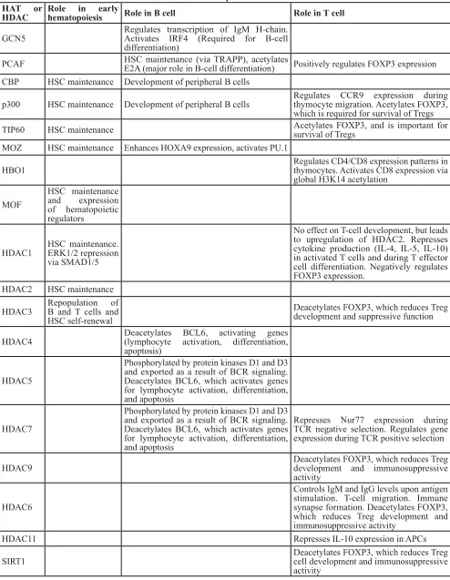
are discussed (summarized in Table 1).
Much of what is known about the role of HATs and
HDACs in the development of mammalian B and T cells
comes from the study of whole knockout (KO) mice and
of mice with tissue-specific inactivation of individual
HATs and HDACs. In most cases, whole-mouse HAT and
HDAC KOs are embryonic lethal. Therefore, to explore
the roles of these HATs/HDACs in hematopoiesis, either
hematopoietic progenitors have been isolated from KO
mice or hematopoietic lineage-specific gene KOs have
been generated.
Mice with cell-specific KOs of CBP or p300
have defects in maintenance and differentiation of
hematopoietic stems cells (HSCs) [26, 27]. The defect
in hematopoiesis in p300-null stem cell lines can be
rescued by re-expression of wild-type p300 or when an
extra copy of CBP is placed under control of the p300
locus, suggesting that the total dosage of HAT activity by
CBP/p300 is critical for hematopoietic maintenance and
differentiation rather than the specific activity of either
individual HAT [26, 27]. Likewise, in the MYST family,
whole animal KO of MOZ is embryonic or perinatal
lethal, and MOZ KO embryos show a dramatic reduction
in the number and repopulation capacity of hematopoietic
Table 1: Roles of HATs and HDACs in B- and T-cell development and function
HAT or
HDAC
Role in early
hematopoiesis
Role in B cell
Role in T cell
GCN5
Regulates transcription of IgM H-chain.
Activates IRF4 (Required for B-cell
differentiation)
PCAF
HSC maintenance (via TRAPP), acetylates
E2A (major role in B-cell differentiation)
Positively regulates FOXP3 expression
CBP
HSC maintenance Development of peripheral B cells
p300
HSC maintenance Development of peripheral B cells
Regulates CCR9 expression during
thymocyte migration. Acetylates FOXP3,
which is required for survival of Tregs
TIP60
HSC maintenance
Acetylates FOXP3, and is important for
survival of Tregs
MOZ
HSC maintenance Enhances HOXA9 expression, activates PU.1
HBO1
Regulates CD4/CD8 expression patterns in
thymocytes. Activates CD8 expression via
global H3K14 acetylation
MOF
HSC maintenance
and expression
of hematopoietic
regulators
HDAC1
HSC maintenance.
ERK1/2 repression
via SMAD1/5
No effect on T-cell development, but leads
to upregulation of HDAC2. Represses
cytokine production (IL-4, IL-5, IL-10)
in activated T cells and during T effector
cell differentiation. Negatively regulates
FOXP3 expression.
HDAC2
HSC maintenance
HDAC3
Repopulation of
B and T cells and
HSC self-renewal
Deacetylates FOXP3, which reduces Treg
development and suppressive function
HDAC4
Deacetylates BCL6, activating genes
(lymphocyte activation, differentiation,
apoptosis)
HDAC5
Phosphorylated by protein kinases D1 and D3
and exported as a result of BCR signaling.
Deacetylates BCL6, which activates genes
for lymphocyte activation, differentiation,
and apoptosis
HDAC7
Phosphorylated by protein kinases D1 and D3
and exported as a result of BCR signaling.
Deacetylates BCL6, which activates genes
for lymphocyte activation, differentiation,
and apoptosis
Represses Nur77 expression during
TCR negative selection. Regulates gene
expression during TCR positive selection
HDAC9
Deacetylates FOXP3, which reduces Treg
development and immunosuppressive
activity
HDAC6
Controls IgM and IgG levels upon antigen
stimulation. T-cell migration. Immune
synapse formation. Deacetylates FOXP3,
which reduces Treg development and
immunosuppressive activity
HDAC11
Represses IL-10 expression in APCs
with a HAT deletion of MOZ often show intermediate
phenotypes, suggesting a dose-dependent requirement for
activity of this HAT [13, 28].
To study the role of specific p300 domains in
hematopoiesis, a series of p300 deletion mutants were
re-expressed in p300-null embryonic stem cells (ESCs),
and their ability to contribute to hematopoiesis was
analyzed. These studies showed that p300 mutants
lacking the KIX or CH1 domain had reduced abilities
to induce hematopoiesis, and these defects were similar
to the parental p300-null cells [27]. This reduction in
hematopoiesis is thought to be due to an inability of
KIX and CH1 deletion mutants to interact with the TF
MYB [27, 29]. Interestingly, the presence of a functional
HAT domain in p300 appears to play a role in limiting
the proliferation of hematopoietic precursors, in that
expression of a HAT-deficient p300 mutant in
p300-null cells leads to increased numbers of hematopoietic
cell populations, as compared to re-expression of
wild-type p300 [27]. The dispensability of the CBP/p300
HAT domain for hematopoiesis may be due to their
interaction with the HAT PCAF, which provides catalytic
HAT activity in some CBP/p300 signaling contexts
(e.g., myogenic differentiation) [30]. Thus, unlike the
MYST family, which relies on HAT activity for proper
proliferation of HSCs (described below), the HAT domains
of CBP/p300 may actually reduce the proliferation of
some hematopoietic cell types.
Among the MYST family proteins, the role of
MOZ in hematopoiesis was determined by analyzing the
hematopoietic progenitors in MOZ KO mice.
Whole-mouse MOZ KO reduces the number of HSCs, and
also affects the ability of these stem cells to renew and
reconstitute the hematopoietic system [13]. HSCs from
MOZ KO mice have reduced HOXA9 expression, which
is known to reduce the differentiation potential of HSCs.
MOZ is also a transcriptional coactivator of PU.1, and
reduced PU.1 activity can explain many of the phenotypes
seen in MOZ KO mice [13, 31, 32]. In mice expressing
HAT-deficient MOZ, hematopoietic progenitors are
severely defective in competitive repopulation assays,
demonstrating a critical role of the MOZ catalytic domain
in HSC functionality [33]. These defects were linked to a
marked deficiency in the proliferative capacity of
HAT-deficient MOZ precursors [33]. Loss of
MYST family
member MOF in ESCs is associated with a reduction in the
expression of some hematopoietic genes, suggesting a role
for MOF in hematopoiesis [34]. Additionally, conditional
KO of the coactivator TRRAP, which can act as a subunit
of TIP60 and PCAF HAT-containing complexes, leads to
loss of HSCs [35].
HDAC1 and 2 have overlapping critical roles in
early hematopoiesis and HSC homeostasis, largely by
acting in a SIN3A/HDAC1/2-repressor complex. While
mice with bone marrow-specific deletions of either
HDAC1 or HDAC2 show only moderate phenotypes, the
simultaneous deletion of HDAC1 and HDAC2 (or SIN3A
alone) leads to nearly complete loss of hematopoietic
progenitors, causing severe reduction in the numbers of
spleen, thymic and bone marrow cells [36-38]. During
HSC emergence in zebrafish, HDAC1 is recruited to
the
erk
promoter by SMAD1/5, and represses
erk1/2
expression by deacetylating H3K9 and H3K27 [39].
Conditional KO studies have shown that HDAC3 is
required for DNA replication in HSCs, which is essential
for their ability to produce B- and T-cell progenitors [40].
HATs and HDACs in B-cell development and
function
Disruption of p300 or CBP at the pro-B cell stage
results in a 25-50% reduction in the number of B cells in
the peripheral blood; however, the number of pro-B, pre-B,
and immature B cells in the bone marrow is unaffected
[41]. Loss of CBP at this stage does not drastically perturb
gene expression in resting B cells, as ~99% of microarray
transcripts measured in CBP-null cells were within
1.7-fold of controls [41].
These results indicate that loss of
either p300 or CBP starting at the pro-B cell stage is not
required for B-cell function, possibly due to functional
redundancy of these two HATs. In contrast to the single
KOs, the double KO of CBP and p300 in pro-B cells
causes a dramatic reduction in the number of peripheral
B cells [41].
With the exception of mature B cells, the HAT
activity of MOZ is required for the cell proliferation
required to maintain healthy numbers of hematopoietic
precursors
.
That is,
mice expressing a HAT-deficient
MOZ protein show an approximately 50% reduction in the
numbers of pro/pre-B cells and immature B cells, whereas
the number of mature B cells and their ability to carry out
antibody responses is unaffected [33].
KO of GCN5 in the chicken immature B-cell line
DT40 showed that GCN5 regulates transcription of the
IgM H-chain gene, and GCN5 deficiency decreased
membrane-bound and secreted forms of IgM proteins [42].
GCN5 also directly activates expression of the TF IRF4,
which is required for B-cell differentiation [43]. PCAF
acetylates the TF E2A, which plays a major role in the
differentiation of B lymphocytes [44].
HDACs also appear to play a role in signaling
from the B-cell receptor (BCR). During BCR activation,
HDACs 5 and 7 are phosphorylated by protein kinases
D1 and D3 and exported from the nucleus, suggesting a
link between BCR function and epigenetic regulation of
chromatin structure [45].
A major regulator of B-cell differentiation is the TF
BCL6, which represses a set of target genes during proper
germinal center (GC) development [46]. BCL6 also serves
as an anti-apoptotic factor during an immune response,
[47, 48]. To achieve GC-specific gene expression, BCL6
is recruited to a large repressor complex that contains
HDAC4, 5, and 7, and localizes to the nucleus to regulate
its target genes [49]. Treatment of cells with an HDACi
results in hyper-acetylation of BCL6, which derepresses
expression of BCL6 target genes involved in lymphocyte
activation, differentiation, and apoptosis [50, 51].
In B cells, HDAC1 and 2 play a key, redundant role
in cell proliferation and at certain stages of development.
That is, in early B cells the combined KO of HDAC1 and 2
results in a loss of further B-cell development and the few
surviving pre-B cells undergo apoptosis due to a cell cycle
block in G1, whereas individual KOs of these HDACs has
no effect [52]. In mature B cells, the combined KO of
HDAC1 and 2 has no effect on cell survival or function
in the resting state, but these double KO cells fail to
proliferate in response to lipopolysaccharide and IL-4
[52].
HATs and HDACs in T-cell development and
function
HATs and HDACs also play roles in T-cell
development and function. For example, the HAT p300
is important for the expression of chemokine CCR9,
which is expressed in thymocytes during their migration
and development into mature T cells [53]. Early in this
developmental process, NOTCH signaling prevents p300
recruitment to, and acetylation of, core histones at two
CCR9 enhancers, thus reducing CCR9 expression [53].
This NOTCH-dependent repression of CCR9 occurs via
effects on p300 in multipotent progenitor cells and is also
observed in T-lymphoma cell lines [53].
Thymus-specific deletion of the
bromodomain-containing protein BRD1, which is a subunit of the HAT
HBO1 complex [54], alters the pattern of CD4/CD8
expression in thymocytes and decreases the abundance of
CD8
+mature T cells in the periphery [55]. Furthermore,
the HBO1-BRD1 complex is responsible for activating
CD8 expression by increasing global acetylation of
H3K14 in developing T cells [55].
T cell-specific KO of HDAC1 does not affect
late T-cell development or the number of T cells in the
periphery [56]. The lack of an effect is likely due to
compensation by HDAC2, whose expression is increased
when HDAC1 is inactivated [56]. Moreover, T
cell-specific KO of both HDAC1 and 2 results in arrested
T-cell development [57], similar to what is seen in HSCs
and early B cells (see above). Nevertheless, T cell-specific
KO of HDAC1 alone does cause an increased Th2-type
inflammatory response in a mouse model of asthma,
which is characterized by elevated expression of 4,
IL-5, and IL-10, suggesting that HDAC1 represses cytokine
production in activated T cells and during T effector
(T
eff) cell differentiation [56]. Of note, the
HDAC1-deficient increased expression of IL-4 in T cells is seen
only after several rounds of cell division, suggesting that
the effect of HDAC1 on IL-4 expression occurs via an
epigenetic mechanism, and that the removal of repressive
marks occurs during DNA replication [56]. In T cells, an
HDAC1/mSIN3A complex represses IL-2 expression [58],
and during T-cell activation, HDAC1-mediated repression
of IL-2 is relieved by phosphorylation of mSIN3A by
CDK5, which disrupts the formation of the HDAC1/
mSIN3A complex [58].
HDAC6-null mice exhibit normal B-cell
development, but have reduced IgM and IgG levels
following antigen stimulation [59]. This defect may be due
to the role of HDAC6 in immune synapse formation and
T-cell migration [60, 61].
In developing T cells, the interaction of class
2 HDACs with the TF MEF2D plays a major role in
regulating T-cell receptor (TCR)-mediated apoptosis
during negative selection of T cells with a TCR that
interacts with self-antigen. For example, HDAC7 is
recruited to the NUR77 promoter by MEF2D, where
it acetylates chromatin and represses the expression
of this apoptotic regulator [62]. In T cells undergoing
negative selection, HDAC7 or class 2 HDACs become
phosphorylated near their N-termini by protein kinase
D, which allows recognition by 14-3-3, disruption of the
interaction with MEF2D, and results in nuclear export of
the repressive HDAC [62-64]. In addition to its role in
negative selection, HDAC7 regulates the expression of
genes involved in positive thymic selection. For example,
HDAC7 KO and mutation studies in mouse thymocytes
have shown that transcription of HDAC5 is regulated by
MEF2D but not by NUR77; thus, HDAC5 may be a direct
target for transcriptional regulation by HDAC7 during
positive, but not negative T-cell selection [65, 66]
Class 4 HDAC11 represses the expression of
IL-10 in antigen-presenting cells (APCs) by interacting with
the distal region of the IL-10 promoter [67]. HDAC11
localization at the IL-10 distal promoter is coupled to
increased binding of the transcriptional repressor PU.1
at the distal promoter and decreased acetylation of
histones H3 and H4 at the proximal IL-10 promoter [67].
APCs that overexpress HDAC11 are able to restore the
responsiveness of tolerant CD4
+T cells [67].
HATs and HDACs in the development of
T-regulatory cells
T-regulatory cells (Tregs) play an important role in
limiting T-cell immune responses, and HATs and HDACs
have a variety of roles in Treg function. p300 and other
HATs maintain the stability and function of Tregs by
acetylating the TF FOXP3, whose transcriptional output
is required for Treg-mediated immunosuppression [68].
or negatively regulated by the TF KLF10 through its
association with PCAF or SIN3-HDAC1, respectively
[69]. Interestingly, p300-deficient Tregs show many of
the same defects in activity, survival and proliferation
that occur in FOXP3-deficient Tregs [68], suggesting that
the effects of p300 deficiency on Treg function are due
to a reduction in FOXP3 activity. FOXP3 has also been
found in a transcriptional regulatory complex with TIP60,
HDAC7, HDAC9, and other proteins [70].
The acetylation of FOXP3 by either p300 or TIP60
both protects FOXP3 from degradation and increases its
DNA-binding activity, and as a result, p300- or
TIP60-deficient Tregs have defects in activity, survival, and
proliferation [68, 71]. Deleting either p300 or CBP
in FOXP3
+Tregs in mice does not affect the overall
proportion of T cells under basal conditions, and thus,
these two HATs appear to have redundant roles in Treg
production under resting conditions [72, 73]. When the
Tregs are activated, however, p300- or CBP-deficient
FOXP3
+Tregs undergo apoptosis, are unable to suppress
homeostatic T
effcell proliferation, and reject transplanted
allografts [72, 73]. Mice with simultaneous Treg-specific
deletion of p300 and CBP develop severe autoimmunity,
as both p300 and CBP interact not only with FOXP3, but
also with many FOXP3-regulating TFs including NFAT,
STAT1, FOXO1, FOXO3, NF-κB, RUNX1, and STAT5
[72, 74-78]. Nevertheless, p300 and CBP also have
distinct roles in Tregs; for example, only p300 is required
for efficient GATA-3 expression, which is important for
FOXP3 expression and Treg accumulation [72].
Countering the HATs, the HDACs deacetylate
FOXP3, which reduces Treg development and
immunosuppressive function, and also provides a
therapeutic target for enhancing immunosuppressive (and
potentially anti-tumor) activity in patients [79-81]. FOXP3
can be deacetylated by certain HDACs (i.e., HDAC3, 6,
7, 9 and SIRT1), which decreases FOXP3 protein levels
and activity [80, 82, 83]. Of note, HDAC6, which is
normally cytoplasmic, translocates to the nucleus of some
Tregs where it can deacetylate FOXP3 [81]. Treg-specific
deletion of these HDACs or treatment with HDACi has
been shown to enhance immunosuppressive activity and
Treg function [79]. Taken together, these results suggest
that acetylation of FOXP3 favors Treg development.
However, while Treg development is important in limiting
host autoimmunity, it may also reduce host immune
responses and anti-tumor activity. Thus, inhibition of
FOXP3 acetylation is a promising anti-tumor strategy
[73].
Regulation of Immune Cell-Related TFs by
Acetylation
The activities of several TFs that play key roles
in immune responses are affected directly and indirectly
by HATs/HDACs. In most cases, the ability of CBP/
p300 to acetylate a given TF and affect its activity has
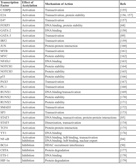
been investigated (summarized in Table 2). There
are four general ways that direct acetylation has been
shown to affect TF function: 1) lysine acetylation can
increase protein stability by blocking ubiquitination of
the same lysines that promote proteasome-mediated
degradation; 2) lysine acetylation within the
DNA-binding domain can decrease the ability of the TF to bind
DNA; 3) lysine acetylation can increase (or decrease)
protein-protein interactions with TF regulators; and 4)
acetylated lysines on TFs can serve as a docking domain
for the bromodomain of HATs, which can increase
their transactivation activity [18]. For example, NF-κB
and STAT subunits are important regulators of B- and
T-cell development and function. Both of these TFs
undergo acetylation/deacetylation at several lysines,
and acetylation at different residues can positively or
negatively impact their activity in distinct ways depending
on the lysine residue [84, 85]. Furthermore, the activity of
NF-κB and STAT can be indirectly affected by acetylation,
for example, by acetylation of their specific co-activators,
the TFs that interact with them, or histones at their target
gene promoters. Moreover, the ability of acetylation to
affect NF-κB and STAT activity can depend on the specific
target gene studied.
In a small number of cases, it is known how
acetylation alters the activity of a TF in a way that
affects B- or T-cell function. As described above, the
TF FOXP3 is a direct substrate of p300 and other HATs,
and acetylation of FOXP3 plays a key role in Treg
development and maintenance [68]. Acetylation increases
FOXP3 activity by stabilizing the protein and enhancing
its DNA-binding activity at certain promoters [68].
BCL6 is a transcriptional repressor that is
essential for GC formation and lymphocyte function
and proliferation [86, 87]. p300 can directly bind to
and acetylate BCL6, which interferes with its ability to
bind HDAC-containing complexes and consequently
inactivates its repressor activity [50]. CBP/p300 mutant
proteins found in some diffuse large B-cell lymphomas
(DLBCLs) show a reduced ability to acetylate BCL6 [88],
and therefore such lymphoma cells have increased BCL6
activity, which is related to the oncogenic state of these
cells.
GATA-3 plays a key role in T-cell differentiation
and survival, and acetylation has been shown to increase
GATA-3 transactivation activity [89]. Moreover,
overexpression of an acetylation-defective GATA3 protein
affects T-cell homing to lymph nodes and increases T-cell
survival after antigen stimulation [89].
HATs and HDACs in B- and T-cell Malignancy
Given their broad role in control of lymphoid
cell gene expression and TF activity, it is not surprising
As described in more detail below, in B- and T-cell
cancers one often finds gene deletions and mutations
that inactivate or reduce HAT activity (e.g., in CBP/
p300) or overexpression of non-mutant forms of HDACs.
As a consequence, reduction of global histone and TF
acetylation appears to be correlated with B- and T-cell
proliferation and survival, whereas increased acetylation
is associated with B- and T-cell tumor growth arrest and
cell death.
Mutations of HATs in B- and T-cell leukemia/
lymphoma
Although chromosomal translocations involving
p300, CBP and MYST are well-documented in acute
myeloid leukemia [90], they have not been found in B-
and T-cell malignancies. However, other types of HAT
gene mutations are common in certain types of B- and
T-cell cancers. Namely, the genes encoding CBP and
Table 2: B- and T-cell transcription factors that are regulated by acetylation
Transcription
Factor
Effect of
Acetylation
Mechanism of Action
Refs
C/EBPβ
Activation
Transactivation
[155]
E2A
Activation
Transactivation, protein stability
[156, 157]
E47
Activation
Transactivation
[157]
FOXP3
Activation
DNA binding, protein stability
[68]
GATA-2
Activation
DNA binding
[158]
GATA-3
Activation
Transactivation
[89]
IRF2
Activation
Transactivation
[159]
JUN
Activation
Protein-protein interaction
[160]
MYB
Activation
Transactivation
[161]
MYC
Activation
Protein stability
[162]
NFATc1
Activation
DNA binding
[163]
NOTCH1
Activation
Protein stability
[164]
NOTCH3
Activation
Protein stability
[165]
p53
Activation
Protein stability
[166]
PAX5
Activation
Transactivation
[167]
PU.1
Activation
Transactivation
[168]
RUNX1
Activation
DNA binding/transactivation
[169]
RUNX2
Activation
Protein stability
[170]
RUNX3
Activation
Protein stability
[171]
SMAD3
Activation
Transactivation
[172]
SP3
Activation
Transactivation
[173]
STAT3
Activation
DNA binding, transactivation; protein-protein interactions
[85]
STAT5
Activation
Dimerization, transactivation
[174]
TCF4
Activation
Protein-protein interaction
[175]
YY1
Activation
DNA binding
[176]
NF-κB
Activation
Inhibition
DNA binding, IκB binding, transactivation
DNA binding; IκB binding, nuclear export
[84]
BCL6
Inhibition
HDAC recruitment interference
[50]
CIITA
Inhibition
Protein degradation
[177]
ETS-1
Inhibition
DNA binding
[178]
p300 harbor point mutations or deletions in approximately
20–40% of DLBCL [88, 91, 92], about 70% of follicular
lymphomas (FL) [93], and less frequently in T-cell
leukemia, acute lymphoblastic leukemia (ALL) and
myelodysplastic syndrome [94, 95]. The TIP60 gene
frequently suffers mono-allelic loss and reduced expressed
in several types of B-cell lymphoma [95]. Moreover, our
analysis of the Cancer Cell Line Encyclopedia (CCLE)
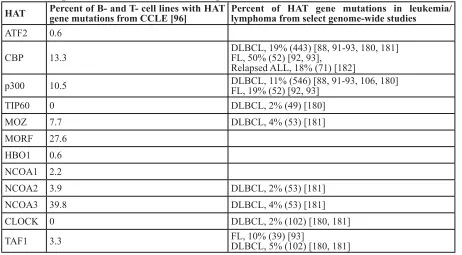
database [96] finds that mutations in CBP/p300 and other
HATs (especially MORF) are common in a variety of B-
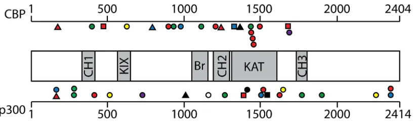
and T-cell cancer cell lines (Table 3). With CBP and p300,
the majority of these lymphoma mutations occur within
or near the HAT domain or introduce frame-shifts or stop
codons N-terminal to the HAT domain (see Figure 2).
Thus, many of the CBP/p300 mutations found in DLBCL
and FL are predicted to reduce acetyltransferase activity
[88]. Indeed, several of these point mutations have been
demonstrated to impair the affinity of CBP for
acetyl-CoA and consequently compromise the ability of CBP to
acetylate the TFs BCL6 and p53 [88]. Of note, acetylation
of BCL6 decreases its gene repressing activity, whereas
acetylation of p53 is required for its gene activation
function (Table 2) [50, 97]. Thus, DLBCL cells with HAT
gene mutations have higher levels of active BCL6 and
lower levels of active p53 [88], consistent with decreased
acetylation being associated with increased tumor cell
growth.
In contrast to the more common point mutations,
genomic alterations that completely remove the HAT
domain in CBP or p300 are present in a minority of
DLBCL and FL tumors and cell lines [88, 93]. Expression
of C-terminally truncated CBP/p300 proteins missing the
HAT domain has been demonstrated in some DLBCL
cell lines [88, 98, 99]. A variety of evidence suggests
that these HAT-deficient p300 mutants play an active
role in lymphomagenesis. First, expression of the
HAT-deficient p300 proteins is preferentially retained in
these cell lines, whereas the wild-type allele is silenced
[98, 99]. Second, knockdown of p300 mutant protein
expression reduces the growth of some DLBCL cell lines
[98, 99]. Third, HAT-deficient p300 mutants localize to
sites of active transcription in cell nuclei, interact with
the lymphomagenic TF REL, and affect transcription of
REL target genes [98-100]. Moreover, expression of a
HAT-deficient mutant of p300 increases the proliferation
of HSCs that lack wild-type p300 [27]. DLBCL cell lines
that express HAT-defective p300 mutants have generally
lower levels of histone H3 acetylation at K14 and K18
[99]. Low levels of H3K14 and H3K18 acetylation have
been associated with proliferation in other cell types [101],
and CBP/p300 catalyze nearly all H3K18 acetylation in
mice [102]. Thus, even though they are defective for
HAT activity, C-terminally truncated p300 proteins
appear to contribute to B-cell transformation, at least in
part, by acting as aberrant scaffolds that organize altered
transcription complexes at a variety of gene promoters/
enhancers to cause a broad-range of transcriptional
deregulation. For example, we have previously suggested
that dampening of global REL/NF-κB-dependent gene
transcription is one oncogenic effect of p300 mutants in
DLBCL [99].
Similar to the effects of mutations on HAT activity
in lymphoma, the HBZ protein of Human T-cell Leukemia
Virus type 1 (HTLV-1) binds to and inactivates the HAT
domains of CBP and p300, and consequently reduces
cellular levels of H3K18 acetylation [103, 104]. Thus,
inhibition of CBP/p300 HAT activity may also be
important for HTLV1-induced T-cell leukemia.
Interestingly, the TF BCL6, which is upregulated
in and required for the growth of approximately 70%
of DLBCLs [105], appears to be a direct transcriptional
repressor of the p300 gene [106]. Furthermore, induced
expression of p300 is required for the anti-proliferative
effects of BCL6 inhibitors on DLBCL cell lines [106].
Consequently, DLBCL cell lines with defective p300
proteins are resistant to the anti-growth effects of BCL6
inhibitors, and in these cell lines, HDACi synergize with
BCL6 inhibitors for inhibition of DLBCL cell growth
[106].
Overall, there are no good mouse models for HAT
gene mutations in B- and T-cell malignancy. In one
report [107], a single mouse reconstituted with CBP-null
HSCs developed a thymic lymphoma that arose from
the CBP-null cells, but that mouse has not been further
characterized. Based on the inactivation of the
wild-type
EP300
allele in DLBCLs containing certain p300
mutations [98, 99], expression of truncated or mutant
p300 proteins (from human DLBCLs) in p300
-/-B-cell
precursors may lead to B-cell malignancy in a transgenic
mouse model.
HDAC dysregulation in B- and T-cell lymphoma/
leukemia
Unlike the case with HATs, mutations in genes
encoding HDACs have not been found in any B- and
T-cell malignancies. However, HDACs have been reported
to have altered (usually increased) expression in a variety
of B- and T-cell malignancies, including DLBCL, FL,
and chronic lymphocytic leukemia (CLL) (Table 4).
For example, HDAC1 is overexpressed in some T-cell
lymphomas [108-112], while HDAC6 has been reported
to be both overexpressed [110, 113] and underexpressed
[114] in DLBCL.
At this point, two of the most relevant questions are
whether altered expression of a specific HDAC contributes
to the growth or survival of the tumor cells (and how it
does so) and whether altered HDAC expression can be
prognostic for therapy. In a smattering of cases, there are
data addressing these questions, but the overall picture
is still not clear. Inhibition of HDAC8 induces apoptosis
in T cell-derived lymphoma and leukemic cells, but
not in solid tumors [115]. High HDAC4
expression
is
associated with a poor response to prednisone in ALL, and
siRNA-mediated inhibition of HDAC4 has been shown
to sensitize a T-ALL cell line to etoposide-induced cell
death [116]. Moreover, the interaction of HDAC4 with
the leukemic PLZF-RARα fusion protein contributes to
oncogenesis because it is required for the repression of
differentiation-associated genes [117].
In childhood acute lymphoblastic leukemia (ALL),
high HDAC3 expression has been associated with a better
prognosis, whereas overexpression of HDAC7 and 9 have
been associated with a poorer prognosis [118]. A study of
over 200 adult CLL B-cell tissue samples reported that
overexpression of HDAC7 and 10 and underexpression of
HDAC6 and SIRT3 are correlated with a poor prognosis
Table 3: HAT gene mutations in B- and T-cell malignancies
HAT
Percent of B- and T- cell lines with HAT
gene mutations from CCLE [96]
Percent of HAT gene mutations in leukemia/
lymphoma from select genome-wide studies
ATF2
0.6
CBP
13.3
DLBCL, 19% (443) [88, 91-93, 180, 181]
FL, 50% (52) [92, 93],
Relapsed ALL, 18% (71) [182]
p300
10.5
DLBCL, 11% (546) [88, 91-93, 106, 180]
FL, 19% (52) [92, 93]
TIP60
0
DLBCL, 2% (49) [180]
MOZ
7.7
DLBCL, 4% (53) [181]
MORF
27.6
HBO1
0.6
NCOA1
2.2
NCOA2 3.9
DLBCL, 2% (53) [181]
NCOA3 39.8
DLBCL, 4% (53) [181]
CLOCK 0
DLBCL, 2% (102) [180, 181]
TAF1
3.3
FL, 10% (39) [93]
DLBCL, 5% (102) [180, 181]
[119]. HDAC6 overexpression correlates with a more
favorable outcome in DLBCL, but with a negative
outcome in peripheral T-cell lymphoma [110]. Although
HDAC1, 2 and 3 are all overexpressed in Hodgkin’s
Table 4: Misregulated expression of HDACs in B- and T-cell malignancies
HDAC
Disease
Expression
Refs
1
T-ALL
Increased
[118]
B-ALL
Increased
[119]
ALL
Increased
[116]
HL
Increased
[120]
DLBCL
Increased
[110]
CLL
Increased
[183]
2
ALL
Increased
[116, 118]
B-ALL
Reduced
[119]
HL
Increased
[120]
DLBCL
Increased
[110]
3
ALL
Increased
[118]
CLL
Increased
[183]
DLBCL
Increased
[184]
HL
Increased
[120]
4
T-ALL
Increased
[118]
5
B-ALL
Increased
[118]
6
ALL
Increased
[118]
B-ALL
Increased
[119]
CLL
Increased
[183]
DLBCL
Increased
[110]
7
ALL
Increased
[118]
B-ALL
Increased
[119]
CLL
Increased
[183]
8
ALL
Increased
[116, 118]
9
CLL
Increased
[183]
10
CLL
Increased
[183]
11
B-ALL
Reduced
[119]
B-ALL
Increased
[119]
SIRT1
CLL
Increased
[183]
SIRT3
B-ALL
Increased
[119]
SIRT4
B-ALL
Decreased
[119]
SIRT5
B-ALL
Decreased
[119]
SIRT6
B-ALL
Increased
[119]
CLL
Increased
[183]
SIRT11 B-ALL
Increased
[119]
lymphoma tissue samples, only high HDAC1 expression
is correlated with a worse outcome [120].
As with HATs, there are no good mouse models for
the role of HDACs in cancer. Based on most evidence,
it is unlikely that overexpression of any HDAC would,
by itself, be oncogenic. Thus, one method for evaluating
the molecular mechanisms by which increased HDAC
expression contributes to oncogenesis might be to create
transgenic mice with B and/or T cell-specific expression
of a relevant HDAC (e.g., HDAC6) and cross such mice to
other common transgenic mouse tumor models (e.g.,
Eµ-MYC mice). One could then determine whether increased
HDAC expression leads to enhanced tumor development
or if such mice develop chemo- or HDACi-resistant
tumors.
HAT and HDAC Inhibitors in the Treatment of
B- and T-cell Cancers
Given that mutations and dysregulation of HATs and
HDACs occur in many B- and T-cell cancers, as well as
their global effects on protein activity and gene expression,
these enzymes have been investigated for therapeutic
targeting. Below we discuss the types of compounds that
have been found to inhibit HAT and HDAC activity, and
examples of such molecules being used in the treatment of
lymphoid cancer cells. Overall, HDACi have been more
useful in such settings than HATi, and HDACi are being
used in the clinic to treat lymphoid cell cancers.
HAT inhibitors (HATi)
Several types of compounds have been characterized
as HAT inhibitors, including a variety of synthetic
compounds and natural products and their derivatives.
In general, such compounds are pan-HAT inhibitors or
inhibitors of GCN5 or CBP/p300 [121, 122].
There are few reports of HATi as inhibitors of B- or
T-cell cancers, and no HATi are currently FDA approved.
Anacardic acid, found in cashew nuts, is a potent inhibitor
of p300, PCAF, and TIP60 [123, 124]. Anacardic acid
and derivatives have been shown to inhibit Jurkat T-cell
leukemia cells at micromolar concentrations [125]. Of
note, Jurkat cells have been shown to express two TIP60
variants, including one with a deleted HAT domain [126].
The natural products gallic acid and curcumin have both
been shown to act as HAT inhibitors [127, 128], and can
induce proliferation arrest and apoptosis in lymphoma
cells [129, 130]. However, gallic acid and curcumin are
not especially potent inhibitors of lymphoma cell growth
and both have many protein targets [131, 132]; thus, it is
not clear that their effects on lymphoma cell growth are
due to their anti-HAT activity. The synthetic compound
C646 is a specific p300 inhibitor, however, it was not
especially effective against leukemia cell lines in a screen
of the National Cancer Institute (NCI) 60-cell line panel
[133].
HDAC inhibitors (HDACi)
Given that increased HDAC expression and activity
is found is many lymphoid malignancies (see above),
it is perhaps not surprising that HDACs should emerge
as targets for therapy. In contrast to HATi, HDACi have
been extensively studied for anti-cancer activity. Indeed,
since 2001, a number of HDACi have been used in the
clinic for the treatment of various cancers, including B-
and T-cell cancers [134]. Alone or in combination with
other anti-cancer agents, a variety of HDACi have been
shown to induce apoptosis in many different types of B-
and T-cell lymphoma and leukemia cell lines (Table 5).
Following from those studies, several HDACi have been
tested clinically for the treatment of such human cancers,
including cutaneous T-cell lymphoma (CTCL), DLBCL,
multiple myeloma (MM), FL, Hodgkin’s lymphoma (HL),
and several others (Table 6), and as of April 2015, there
are at least 12 ongoing clinical trials testing HDACi alone
or in combination with other cancer therapeutics for the
treatment of several B- and T-cell malignancies (Table 7).
HDACi fall into five main classes, based in part on
their chemical structures and in part on their specificity.
These include the following: 1) hydroxyamic acids, 2)
cyclic tetrapeptides, 3) benzamides, 4) ketones, and 5)
aliphatic acids. In addition, HDACi can have broad-based
pan-HDAC inhibitory activity, have class specificity, or
even isozyme specificity. Currently, four HDACi have
received FDA approval for clinical use. The first two
FDA-approved inhibitors are the pan-HDACi vorinostat
(aka suberoylanilide hydroxamic acid [SAHA]), which
is available as an oral medication, and the class I
HDACi romidepsin (a bacterial cyclic peptide), which is
administered intravenously. HDACi treatment is especially
effective in the treatment of CTCL, with favorable
response rates (from a number of trials) of approximately
70% when using romidepsin (Table 6). Although it is not
known why CTLC responds well to HDACi treatment,
increased expression of HDAC2 and histone H4
acetylation have been correlated with aggressive CTCL
[110]. The HDACi belinostat was approved in 2014 for
relapsed and refractory peripheral T-cell lymphomas [135].
Finally, the HDACi panobinostat, another hydroxamate,
has been approved by the FDA for refractory multiple
myeloma [136]. Because of the role of HDACs in normal
immune cell function, one problem with using HDACi
treatment for anti-cancer therapy is the concomitant
suppression of host immune responses required for
anti-tumor therapy (see also
HATs and HDACs in development
of T-regulatory cells
section above) [137].
In the simplest scenario, HDACi treatment increases
the level of acetylation of histones on chromatin, thereby
Table 5: HDACi compounds that induce apoptosis in B- and T-cell cancer cells
HDACi
Class
Target of
HDACi
Clinical
trial
stage
Hematopoietic
malignancy (Patient and
Cell Line)
Combination treatments
that induce apoptosis
Refs
Butyrate (NaB) Short-chain
fatty acid
Class 1, 2a Phase I,
II
B-lymphoma, BL
Cisplatin, Etoposide
[142,
185,
186]
Valproic Acid
(VPA)
Short-chain
fatty acid
HDAC1-5,
7, 8, 10
Phase I,
II, III
AML, CML, CLL, DLBCL,
HL, MM, NHL, NK cell
lymphoma, SLL, T-cell
lymphoma
5-Azacytidine, ATRA,
Bortezomib, Cambinol,
Cyclophosphamide,
Decitabine, Enzastaurin,
Etoposide,
EX527,
Imatinib, Pioglitazone,
Prednisone, Rituximab,
Sirtinol, Temozolomide,
Vincristine
[142,
187-193]
Tricostatin A
(TSA)
Hydroxamate Class 1, 2, 4
AML,
DLBCL, EBV+ BL, NHL Decitabine
B-lymphoma,
[142,
146,
186,
194-197]
Vorinostat,
Suberoylanilide
hydroxamate
(SAHA)
Hydroxamate Class 1, 2, 4 Phase I,
II, III
ALL, AML, CLL, CML,
CTCL, DLBCL, HL, MCL,
MM, NHL
17-AAG,
ABT-737,
Azacitidine, Bexarotene,
Bortezomib, Carboplatin,
Carfilzomib, Cladribine,
Cyclophosphamide,
Decitadine, Eltrombopag,
Enzastaurin, Etoposide,
Ifosfamide, Lenalidomide,
Melphalan, NPI-0052,
Prednisone, Rituximab
[67,
134,
142,
146,
198-209]
Belinostat
(PDX101)
Hydroxamate Class 1, 2, 4 Phase I,
II, III
ALL, AML, CLL, MM,
MCL, NHL, PTCL, T-cell
lymphoma
17-AAG,
Azacitidine,
Bortezomib
[142,
188,
206,
210-214]
Dacinostat
(LAQ824)
Hydroxamate Class 1, 2, 4 Phase I
Acute eukemia, AML
progenitor cells, CLL,
meyloid leukemias
Decitabine
[142,
215-219]
Panobinostat
(LBH589)
Hydroxamate Class 1, 2, 4 Phase I,
II, III
AML, CLL, CML, CTCL,
DLBCL, HL, MCL, MM,
NHL, NK/T-cell lymphoma,
PTCL
17-AAG, Bortezomib,
Carboplatin, Cytarabine,
Etoposide, Everolimus,
Idarubicin, Imatinib,
Ifosfamide, Lenalidomide,
Pemetrexed, Rituximab
[134,
142,
203,
220-224]
Suberic
bishydroxamate
(SBHA)
Hydroxamate Class 1, 2, 4
ALL, Leukemia, MM
ABT-737
[142,
145,
225,
226]
Azelaic
bishydroxamate
(ABHA)
Hydroxamate Class 1, 2, 4
EBV+ B-cell lines
ABT-737
[142,
227]
SK-7041
Hydroxamate HDAC1, 2
Meyloid leukemia
Imatinib
[228,
229]
ITF-A
and
ITF-B
Hydroxamate Class 1, 2, 4 Phase I DLBCL, MCL, SMZL
[151]
Tubacin
Hydroxamate Class 2b
ALL, AML, CML, EBV+
BL, MM
17-AAG, Bortezomib
[142,
230-233]
JNJ 26481585
Hydroxamate Class 1, 2, 4 Phase I CTCL, Leukemia, MM
Bortezomib,
Dexamethasone
[234]
acetylation of non-histone proteins. For lymphoid cell
TFs, increased acetylation can either increase or decrease
their activity (Table 2). Because of the myriad effects of
acetylation/deacetylation on gene expression and protein
activity, it is almost certain that the effects of HDACi on
tumor cell growth and survival are not through single or
even a small number of targets. Moreover, the effects
of HDACi would be expected to vary among tumor cell
types, within a given tumor type, and, due to tumor cell
heterogeneity, even within a given tumor.
Consistent with those hypotheses, treatment of
CTCL cell lines with vorinostat showed that HDACi
treatment leads to hyperacetylation of all core histones,
which is associated with changes in the expression of
genes involved in regulation of the G1/S and G2/M
transitions, apoptosis, anti-proliferation, and MAPK
signaling [138]. Overall, gene expression profiling showed
that up to 22% of genes are altered by HDACi as early
as four hours post treatment in several cell types
[139-141]. Nevertheless, there does appear to be a common set
of genes that change expression in response to HDACi
treatment, and these genes include several cyclins,
the cell-cycle inhibitor p21, p53, BAX, BCL2, MYC,
PKCδ, ICAM-1, IL-6 receptor, IL-2, IL-8, IL-10, VEGF,
NOTCH, GADD45 and GADD45, TGF receptor, CTP
synthase, and TYMS (reviewed by [17, 134, 142]. At
least in part, HDACi induce cell-cycle arrest by causing
accumulation of hyperacetylated p53, which then induces
expression of p21, leading to inhibition of cyclins D and
A, which are required for cell-cycle progression (reviewed
by [134, 142]). However, because CTCL tumors typically
grow quite slowly in patients, it is unclear how the
reported effects of HDACi on cell-cycle progression in
rapidly growing cell lines
in vitro
reflect its effects on
CTCL tumors
in vivo
.
HDACi treatment of tumor cells has been frequently
linked to the modulation of BCL2 family expression
to favor a pro-apoptotic expression pattern (reviewed
by [134, 142, 143]). In many cases, HDACi-induced
apoptosis occurs via increased expression of pro-apoptotic
BCL2 family members BIM, BAX, PUMA, and NOXA
(reviewed by [142, 144, 145]). Moreover, resistance to
HDACi-induced apoptosis can be achieved in DLBCL
cells lines by artificial or induced expression of
anti-Entinostat
(MS-275)
Benzamide
HDAC1-3 Phase I,
II
ALL, AML, CML, HL,
MM
Azacitadine,
Imatinib, Isotretinoin,
Sargramostim, Sorafenib,
Rapamycin, Rituximab
[142,
237-241]
Mocetinostat
(MGCD-0103) Benzamide
HDAC1-3,
10, 11
Phase I,
II
AML, CLL HL, NHL
5-azacitadine,
Bortezomib, Docetaxel,
Gemcitabine, GX15-070
[242-250]
Romidepsin
(FK228)
Cyclic
tetrapeptide
Class 1, 2, 4 Phase I,
II, III
ALL, AML, CLL, CTCL,
DLBCL, MM, MCL, NHL,
PTCL, SLL
Bortezomib, Carboplatin,
Cyclophosphamide,
Decitadine, Etoposide,
Ifosfamide, Prednisone,
Rituximab, Vincrstine
[206,
210,
223,
251-260]
Apicidin
Cyclic
tetrapeptide
HDAC1, 3
APL, CML
Imatinib, TRAIL
[142,
261-264]
Nicotinamide
Vitamin B
member
Sirtuins
CLL
[265]
Tenovin-6
(TV-6)
Small
Molecule
SIRT1
CML
Imatinib
[266]
Amurensin G
Natural
Product
SIRT1
TRAIL-resistant leukemia
[267]
Cell types: ALL, acute lymphoblastic leukemia; AML, acute myeloid leukemia; APL, acute promyelocytic leukemia; BL, Burkitt’s lymphoma; CLL, chronic lymphocytic leukemia; CML, chronic myelogenous leukemia; CTCL, cutaneous T-cell lymphoma; DLBCL, diffuse large B-cell lymphoma; EBV+ BL, Epstein-Barr virus-positive BL; HL, Hodgkin’s lymphoma; MCL, mantle cell lymphoma; MM, multiple myeloma; NHL, non-Hodgkin’s lymphoma; PTCL, peripheral T-cell lymphoma; SLL, small lymphocytic lymphoma.