Original Research Article
Assessment of effectiveness of extracapsular cataract extraction versus
manual small incision cataract surgery: an observational study
Mohin M. Sakre
1, Sana Nizami
2, Ranjana Singh
1*, Anuhya Raghavendra
3,
Anant Arunrao Takalkar
4INTRODUCTION
Cataract blindness has been assigned a pivotal and major slant in control and eradication in the WHO initiative of Vision 2020: the right to sight.1 It has been estimated that around 50 to 80% of all the cases of double-sighted blindness can be attributed to cataract in India.2 It has been predicted that on an average, 3 million individuals develop cataract each year.3 The most extensively performed surgery that accounts for most of the in-patient burden in ophthalmic setups has been attributed to be
cataract surgeries. Although manual small incision cataract surgery has been widely employed as the go-to option for the operative treatment of cataract.4,5 Extracapsular cataract extraction with posterior chamber intraocular lens implantation (PCIOL) had been the most widely performed cataract corrective surgery before 15 Years and is still employed in some setups.6
Extra capsular cataract surgery was deemed as one of the most successful and safe procedures for correction of cataract until the wide and prevalent shift to manual small
ABSTRACT
Background: Dilemma of cost effectiveness of manual small incision cataract surgeries (MSICS) in the terms of training and equipment has been widely pondered upon in developing areas. Objective of the study was to compare the manual small incision cataract surgery and extra capsular cataract extraction.
Methods: A prospective study was conducted among the IPD patients of the Ophthalmology Department of Khaja Bandanawaz Teaching and General Hospital, Kalaburagi, from June to December 2017. Statistical Analysis was performed using Microsoft Excel 2013, SPSS 23.0 and Chi-square test was performed.
Results: Out of the 160 individuals who underwent extra capsular cataract extraction (ECCE), 06 (3.75%), 91 (56.88%) and 63 (39.37%) of the study subjects had poor (5/50), moderate (6/60-6/24) and good (6/18-6/6) visual acuity respectively. Highest incidence was that of lens prolapse (25%) and corneal complications (25%) in ECCE. Among the subjects who underwent MSICS, highest incidence of intra operative complication noticed was that of lens prolapse, iris prolapse and anterior chamber collapse, each at 20%.
Conclusions: It was concluded that the restoration of visual acuity was fairly good and uniform in both the procedures. Certain intra operative complications such as lens prolapse, iris prolapse and anterior chamber collapse were noticed in MSICS and capsular flaps and vitreous loss were noticed only in ECCE.
Keywords: ECCE, MSICS, Visual actuity, Lens prolapse, Iris prolapse
1Department of Community Medicine, Saraswathi Institute of Medical Sciences, Hapur, UP, India
2Department of Ophthalmology, Khaja Bandanawaz Institute of Medical Sciences, Kalaburagi, 3Subbiah Institute of
Medical Sciences, Shivamogga, Karnataka, India
4Department of Community Medicine, MIMSR Medical College, Latur, Maharashtra, India
Received: 13 July 2019 Accepted: 22 July 2019
*Correspondence: Dr. Ranjana Singh,
E-mail: [email protected]
Copyright: © the author(s), publisher and licensee Medip Academy. This is an open-access article distributed under the terms of the Creative Commons Attribution Non-Commercial License, which permits unrestricted non-commercial use, distribution, and reproduction in any medium, provided the original work is properly cited.
incision cataract surgery. The reason can be attributed to complications of ECCS such as oedema, rise in IOP, leaking or rupture, astigmatism and retinal detachment among others.6 Smaller incision surgeries have been employed in recent times with high prevalence because of evident advantages of lesser rehabilitation time and less chances of occurrence of astigmatism.7 Also, manual SICS, in which a 6 to 6.5 mm opening in the sclera is used to deliver the nucleus is deemed to have similar advantages to that of phacoemulsification.8
However, the dilemma of cost effectiveness of manual small incision cataract surgeries in the terms of training and equipment has been widely pondered upon in developing areas. Especially considering its economic constraints in underdeveloped areas of the country. Such a study to compare the manual small incision cataract surgery and extra capsular cataract extraction has not been conducted in the vicinities of Khaja Bandanawaz Institute of Medical Sciences, Kalaburgi. This study was conducted in the in-patient department patients of Khaja Bandanawaz Teaching and General Hospital, Kalaburgi to compare Manual SICS and ECCE.
METHODS
A prospective study was conducted among the IPD patients of the Ophthalmology Department of Khaja Bandanawaz Teaching and General Hospital, Kalaburagi, from June to December 2017. All the patients who were not excluded via the exclusion criteria and who came in with clinically significant cataract wanting to get operated were included in the study after obtaining written consent. Consent was obtained after explaining the pros and cons of both the surgical procedures and ensuring that the patient was aware of the procedure being employed on them. Each case was examined with detailed history regarding their complaints and other significant clinical findings. All the individuals between the age of 40 to 70 years who registered in the hospital for cataract surgery, not being eliminated through the exclusion criteria and who gave consent were included. Exclusion criteria included age below 40 and above 70 years, co-morbidities such as liver and kidney disorders. Apart from consent and exclusion criteria, other reasons such as preferred phacoemulsification, glaucoma and the ones who refused consent were also excluded (Table 1). Among 587 patients who enrolled for cataract surgery in the hospital during the above mentioned period, eventually, 320 participated in the study.
The two surgical procedures that were employed were extra capsular cataract extraction and manual small incision cataract surgery. PCIOL was introduced in both the procedures. One among the four ophthalmic surgeons employed in Khaja Bandanawaz Teaching and General Hospital was randomly assigned to perform the surgery. The patients were subjected to thorough general physical examination, LFT’s, RFT’s, test for visual acuity, keratometry, tests to exclude other eye diseases, A scan
and calculation of the power of the IOL aimed towards restored emmetropia. Follow up was done by different surgeons post operatively and after one, three and six weeks to look out for uncorrected vision and the extent of it. If needed, spectacles were prescribed at the 6th week. Intra operative complication were noted and included in the study. Ethical clearance regarding the procedure, its complications and consequences was obtained from the institutional ethical clearance committee before beginning with the study.
The four surgeons performing the procedures were efficient and experienced and it was ensured that the surgical protocol and the procedure followed were standardized. The outcome was measured mainly regarding the visual acuity of 6/18 or better at the 6th week follows up, intra-operative and post-operative complications. The total number of male patients were 171 and female patients were 149 (Table 2).
Helsinki’s protocol was followed for obtaining consent and the ethical committee of the Khaja Bandanawaz Institute of Medical Sciences had approved the study design and procedures.18 It was ensured that standard measures of care were followed in both the procedures.
The patients were randomly assigned to either ECCE or MSICS using the lot system where it was ensured that there was always a 50% chance of picking either of the two surgeries. The selection was done a day before the opted day of surgery and the patient was informed and explained about the procedure. Out of the 320 participating individuals, 160 were operated with ECCE and the remaining 160 with MSICS. Any doubts and queries the patients had regarding the procedure were answered. The surgical procedure was conducted in front of another non–operating ophthalmologist, anaesthetist and an operation theatre attender. Statistical analysis was performed using Microsoft Excel 2013, SPSS 23.0 and Chi-square test was performed.
RESULTS
All the individuals on whom surgeries were performed came in to the out-patient department of Department of Ophthalmology for the 1st, 3rd and the 6th week follow up. Visual acuity was checked on the 6th week, whereas the post-operative complications presented themselves immediately or on the 1st and 3rd week follow-up. The findings have been reported here under.
suggesting that neither procedure was more or less risky in regard to post-operative uncorrected visual acuity. Overall, among the patients of both the surgeries combined, out of 320 patients, 121 (37.82%) had good visual acuity whereas only 10 (3.125%) had poor visual acuity (Table 3).
Table 1:Reasons for denial from participating in the study.
S. no. Reasons for exclusion from
the study
No. of patients
1. Wanted phacoemulsification 18 2. Wanted MSICS 86 3. Refused consent 69
4. Glaucoma 18
5. Severe hypertension and
diabetes mellitus 58 6. Other ophthalmic
morbidities 18
Total 267
Table 2:Distribution of the study subjects according to gender, operating surgeon and the type of
procedure employed.
Gender and
surgeon ECCE MSICS
Male 96 75
Female 64 85
Surgeon 01 36 30 Surgeon 02 52 42 Surgeon 03 40 46 Surgeon 04 32 42
Table 3:Visual acuity among the study subjects post operatively (uncorrected).
Visual acuity
ECCE MSICS Total
N (%) N (%) N (%) 5/50 (Poor) 06 (3.75) 04 (2.5) 10 (3.125) 6/60-6/24
(Moderate)
91 (56.88) 98 (61.25) 189 (59.06)
6/18-6/6 (Good)
63 (39.37) 58 (36.25) 121 (37.82)
Total 160 (100) 160 (100) 320 (100)
Chi-square: 0.8659; P value: 0.648602. The result is not significant at p<0.05.
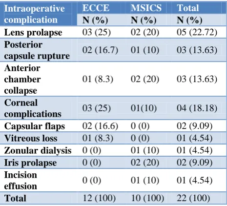
Intra operative complications that were noticed during the procedures, highest incidence was that of lens prolapse (25%) and corneal complications (25%) in ECCE. Among the subjects who underwent MSICS, highest incidence of intra operative complication noticed was that of lens prolapse, iris prolapse and anterior chamber collapse, each at 20%. No cases of zonular dialysis, iris prolapse and incision effusion were noticed in ECCE where as they were encountered in MSICS at the
incidence of 10%, 20% and 10% respectively; vice-versa, no cases of capsular flaps and vitreous loss were noticed in MSICS which were encountered in ECCE with the incidence of 16.6% and 8.3% respectively. The difference in the incidence rates wasn’t significant to be deemed clinically significant to be associated with respective procedures, however, MSICS was found to be associated exclusively with zonular dialysis, iris prolapse and incision effusion and ECCE was found to be exclusively associated with capsular flaps and vitreous loss (Table 4).
Table 4:Distribution of intraoperative complications among the study subjects.
Intraoperative complication
ECCE MSICS Total
N (%) N (%) N (%) Lens prolapse 03 (25) 02 (20) 05 (22.72) Posterior
capsule rupture 02 (16.7) 01 (10) 03 (13.63) Anterior
chamber collapse
01 (8.3) 02 (20) 03 (13.63)
Corneal
complications 03 (25) 01(10) 04 (18.18) Capsular flaps 02 (16.6) 0 (0) 02 (9.09) Vitreous loss 01 (8.3) 0 (0) 01 (4.54) Zonular dialysis 0 (0) 01 (10) 01 (4.54) Iris prolapse 0 (0) 02 (20) 02 (9.09) Incision
effusion 0 (0) 01 (10) 01 (4.54) Total 12 (100) 10 (100) 22 (100)
Chi-square: 8.757; P value: 0.36320814. The result is not significant at p<0.05.
Table 5:Distribution of postoperative complications among the study subjects.
Post-operative complications
ECCE MSICS Total
N (%) N (%) N (%) PCO 01 (25) 0 (0) 01 (11.1) Dislocated IOL 0 (0) 01 (20) 01 (11.1) Increased IOP 0 (0) 0 (0) 0 (0) Endophthalmitis 01 (25) 02 (40) 03 (33.3) Corneal oedema 0 (0) 0 (0) 0 (0) Iridocyclitis 0 (0) 01 (20) 01 (11.1) Residual cortex 02 (50) 01 (20) 03 (33.3) Total 04 (100) 05 (100) 09 (100)
PCO: Posterior capsular opacity; Chi-square: 3.6; P value: 0.730621. The result is not significant at p<0.05.
Dislocated IOL at 20% each. It was also noted that both the procedures had a common ground in endophthalmitis and residual cortex as a post-operative complication. In undivided exclusion it was seen that posterior capsular opacity was associated only with ECCE whereas dislocated IOL and iridocyclitis was seen to be associated only with MSICS (Table 5).
DISCUSSION
In this study it was evident that there was a slightly higher chance of poor post-operative uncorrected visual acuity in ECCE (3.75%) than in MSICS (2.5%). This finding was similar to the finding of a study conducted in a community eye care setting in western India and another study conducted by Ang et al, where in, in both the studies it was found that MSICS had lesser chances of poor visual acuity as compared to ECCE as an outcome of the surgery. 9,10
It was noted that in accordance to both the surgeries combined, the highest prevalence of intraoperative complications was that of lens prolapse and corneal complications. Most common lens related complications were dropped nucleus, retained lens matter and loss of posterior lens fragments. This finding of our study was coherent to a study conducted in Tehran, Iran that showed similar findings.11 In extracapsular cataract extraction, lens prolapse and corneal complications were seen to be most dominant. In our study capsular flaps and vitreous loss was noticed exclusively in ECCE. This finding was similar to the finding of a study conducted in Nigeria where 7.5% of the intraoperative complications were capsular flaps and 5% were vitreous loss.12 Among the patients who were selected for MSICS, it was exclusively noted that zonular dialysis, iris prolapse and incision effusion presented themselves as an intraoperative complication. That these findings were more likely to be found in MSCIS than ECCE was also presented in a review presented by Gogate in 2009.4
In regard to the post-operative complications noticed in the study, overall prevalence of endophthalmitis and residual cortex was highest at 33.3%. High prevalence of endophthalmitis can be attributed to poor hygiene and post-operative negligence as national standards for cleanliness and precaution of endophthalmitis were explicitly maintained during the surgery, however, intraoperative reasons also cannot be ruled out in the scope of this study. This finding was extremely higher than the prevalence rate of post-operative endophthalmitis as reported by Niyanduropola et al. They reported the prevalence to be around 1%.13
The finding of our study where residual cortex was seen to be prevalent as a post-operative complication was also higher than the prevalence reported Tiwari et al.14 Endophthalmitis had the highest prevalence as a post-operative complication of MSICS was justified by Goghate in 2009.4 However, studies conducted by
Ravindran suggested otherwise where endophthalmitis had more chances of occurring in ECCE than in MSICS.15 In this study it was noticed that posterior capsular opacification was seen only in ECCE and no cases were noticed in MSICS, similar finding was reflected in a review where it was suggested that posterior capsular opacification was more prevalent in ECCE than in MSICS.16 In our study it was seen that dislocated IOL and iridocyclitis was associated only with MSICS. Even though significant exclusive association cannot be associated with MSICS, it can be substantiated that there are higher chances of developing iridocyclitis in MSICS by a study conducetd by Khanna among resident ophthalmologists that suggests the same.17
CONCLUSION
It was concluded that the restoration of visual acuity was fairly good and uniform in both the procedures. Certain intra operative complications such as lens prolapse, iris prolapse and anterior chamber collapse were noticed in MSICS and capsular flaps and vitreous loss were noticed only in ECCE. In regard to the post-operative complications, residual cortex and endophthalmitis were seen to be most prevalent as a common ground to both the surgeries and posterior capsular opacification was seen only after ECCE and dislocated IOL and iridocyclitis was seen only after MSICS.
ACKNOWLEDGEMENTS
We thank all the participants, surgeons, staff of Khaja Bandanawaz Teaching and General Hospital, Khaja Bandanawaz Institute of Medical Sciences and everyone else who was directly or indirectly involved in the study.
Funding: No funding sources Conflict of interest: None declared
Ethical approval: The study was approved by the Institutional Ethics Committee of Khaja Bandanawaz, Institute of Medical Sciences
REFERENCES
1. Foster A. Cataract and Vision 2020: The Right to Sight Initiative. Br J Ophthalmol. 2001;85(6):635-9. 2. Murthy G, Gupta SK, John N, Vashist P. Current status of cataract blindness and Vision 2020: The Right to Sight Initiative in India. Indian J Ophthalmol. 2008;56(6):489-94.
3. Minnassian DC, Mehra V. 3.8 million blinded by cataract each year: projections from the first epidemiological study of incidence of cataract blindness in India. Br J Ophthalmol. 1990;74(6):341-3.
4. Gogate PM. Small incision cataract surgery: complications and mini review. Indian J Ophthalmol. 2009;57(1):45-9.
Ophthalmologists. Indian J Ophthalmol. 1998;46(1):47-50.
6. Ezegwui IR, Aghaji A, Okpala N, Onwasigwe E. Evaluation of complications of Extracapsular Cataract Extraction Performed by Trainees. Ann Med Health Sci Res. 2014;4(1):115-7.
7. Maghraby A, Anwar M. Effect of Incision size on early post-operative visual rehabilitation after cataract surgery and intraocular lens implantation. J Cataract Refract Surg. 1993;19(4):494-8.
8. Ehlrich J. Manual Small Incision Cataract Surgery. American Academy of Ophthalmology. Accessed 25 October 2016. Available at http://eyewiki.aao.org/Manual_Small_Incision_ Cataract_Surgery. Accessed 10 May 2019.
9. Gogate PM, Deshpande M, Wormald RP, Deshpande R, Kulkarni SR. Extra Capsular Cataract Surgery compared with Manual Small Incision Cataract Surgery in Community eye care setting in western India: a randomised controlled trial. Br J Ophthalmol. 2003;87(6):667-72.
10. Ang M, Mehta JS, Evans JR. Manual small incision cataract surgery (MSICS) with posterior chamber intraocular lens versus extracapsular cataract extraction (ECCE) with posterior chamber intraocular lens for age-related cataract. Cochrane Database Syst Rev. 2012;18:(4):CD008811. 11. Hashemi H, Rezvan F, Etemad K, Gilasi H, Asgari
S, Mahdavi A, et al. Intraoperative complications of cataract surgery in Tehran Province, Iran. Optom Vis Sci. 2016;93(3):266-71.
12. Ezegwui IR, Aghaji A, Okpala N, Onwasigwe E. Evaluation of complications of Extracapsular Cataract Extraction Performed by Trainees. Ann Med Health Sci Res. 2014;4(1):115-7.
13. Niyandurupola N, Astbury N. Endophthalmitis: Controlling infection before and after cataract surgery. Community Eye Health. 2008;21(65):9-10. 14. Tiwari A, Shah GK. Cataract Complication: The
Retinal Perspective. Review of Ophthalmology, 2006. Available at https://www.reviewofophthal mology.com/ article/ cataract-complications-the-retinal-perspective. Accessed 10 May 2019.
15. Ravindran RD, Venkatesh R, Chang DF, Sengupta S, Gyatsho G, Talwar B. Incidence of post-cataract endophthalmitis at Aravind Eye Hospital. J Cataract Refract Surg. 2009;35(4):629-36.
16. Sinha R, Shekhar H, Sharma N, Titiyal JS, Vajpayee RB. Posterior capsular opacification: a review. Indian J Ophthalmol. 2013;61(7):371-6.
17. Khanna RC, Kaza S, Shantha GPS, Sangwan VS. Comparative out comes of manual small incision cataract surgery and phacoemulsification performed by ophthalmology trainees in a tertiary eye care hospital in India: a retrospective cohort design. BMJ Open. 2012;2(5):1-2.
18. Lurie P, Wolfe SM. Proposed revisions to the declaration of Helsinki. Paving the way for globalisation in research. Western J Med. 2012;171(1):6.