T cell development within the human tonsil
Susan McClory, … , Gerard Nuovo, Michael A. Caligiuri
J Clin Invest. 2012;
122(4)
:1403-1415.
https://doi.org/10.1172/JCI46125
.
The development of a broad repertoire of T cells, which is essential for effective immune
function, occurs in the thymus. Although some data suggest that T cell development can
occur extrathymically, many researchers remain skeptical that extrathymic T cell
development has an important role in generating the T cell repertoire in healthy individuals.
However, it may be important in the setting of poor thymic function or congenital deficit and
in the context of autoimmunity, cancer, or regenerative medicine. Here, we report evidence
that a stepwise program of T cell development occurs within the human tonsil. We identified
5 tonsillar T cell developmental intermediates: (a) CD34
+CD38
dimLin
–cells, which
resemble multipotent progenitors in the bone marrow and thymus; (b) more mature
CD34
+CD38
brightLin
–cells; (c) CD34
+CD1a
+CD11c
–cells, which resemble committed T
cell lineage precursors in the thymus; (d) CD34
–CD1a
+CD3
–CD11c
–cells, which resemble
CD4
+CD8
+double-positive T cells in the thymus; and (e) CD34
–CD1a
+CD3
+CD11c
–cells.
The phenotype of each subset closely resembled that of its thymic counterpart. The last 4
populations expressed RAG1 and PTCRA, genes required for TCR rearrangement, and all
5 subsets were capable of ex vivo T cell differentiation. TdT
+cells found within the tonsillar
fibrous scaffold expressed CD34 and/or CD1a, indicating that this distinct anatomic region
contributes to pre–T cell development, as does the subcapsular region of the thymus. Thus,
we […]
Research Article
Immunology
Find the latest version:
Evidence for a stepwise program
of extrathymic T cell development
within the human tonsil
Susan McClory,1,2 Tiffany Hughes,2 Aharon G. Freud,3 Edward L. Briercheck,1,2 Chelsea Martin,4 Anthony J. Trimboli,4 Jianhua Yu,5,6 Xiaoli Zhang,6,7 Gustavo Leone,1,2,4,6,8 Gerard Nuovo,9 and Michael A. Caligiuri1,2,5,6,8
1Medical Scientist Training Program and 2Integrated Biomedical Sciences Graduate Program, The Ohio State University, Columbus, Ohio, USA. 3Department of Pathology, Stanford University School of Medicine, Palo Alto, California, USA. 4Department of Molecular Genetics, 5Division of Hematology,
Department of Internal Medicine, 6Comprehensive Cancer Center and Arthur G. James Cancer Hospital and Richard J. Solove Research Institute, 7Center for Biostatistics, 8Department of Molecular Virology, Immunology, and Medical Genetics, and 9Department of Pathology,
The Ohio State University, Columbus, Ohio, USA.
The development of a broad repertoire of T cells, which is essential for effective immune function, occurs in
the thymus. Although some data suggest that T cell development can occur extrathymically, many researchers
remain skeptical that extrathymic T cell development has an important role in generating the T cell repertoire
in healthy individuals. However, it may be important in the setting of poor thymic function or congenital deficit
and in the context of autoimmunity, cancer, or regenerative medicine. Here, we report evidence that a stepwise
program of T cell development occurs within the human tonsil. We identified 5 tonsillar T cell developmental
intermediates: (a) CD34
+CD38
dimLin
–cells, which resemble multipotent progenitors in the bone marrow and
thymus; (b) more mature CD34
+CD38
brightLin
–cells; (c) CD34
+CD1a
+CD11c
–cells, which resemble committed
T cell lineage precursors in the thymus; (d) CD34
–CD1a
+CD3
–CD11c
–cells, which resemble CD4
+CD8
+double-positive T cells in the thymus; and (e) CD34
–CD1a
+CD3
+CD11c
–cells. The phenotype of each subset closely
resembled that of its thymic counterpart. The last 4 populations expressed
RAG1
and
PTCRA
, genes required
for TCR rearrangement, and all 5 subsets were capable of ex vivo T cell differentiation. TdT
+cells found within
the tonsillar fibrous scaffold expressed CD34 and/or CD1a, indicating that this distinct anatomic region
con-tributes to pre–T cell development, as does the subcapsular region of the thymus. Thus, we provide evidence
of a role for the human tonsil in a comprehensive program of extrathymic T cell development.
Introduction
The development of human T cells can be divided into discrete stages, ranging from that for multipotent progenitors to that for naive T cells. The expression of CD4 and/or CD8 divides thymo- cytes into the categories of double-negative, immature single-pos-itive, double-positive (DP), and single-positive cells (1, 2). Based on TCR gene rearrangements and differentiation capabilities, double-negative thymocytes are divided into the least mature CD34+CD38dimCD1a– cells and the progressively more mature CD34+CD38brightCD1a– and CD34+CD1a+ cells (2, 3). CD34+CD1a+ thymocytes acquire CD4 and then CD8 as they lose CD34, to become CD34– DP cells. Finally, CD3+ DP thymocytes lose either CD4 or CD8 to become single-positive naive T cells (1).
Development of a healthy T cell repertoire is dependent on a functional thymus. However, the potential for human extrathymic lymphoid tissue to augment T cell development is poorly under-stood but may be important in the setting of poor thymic function or congenital deficit. Previous reports of extrathymic T cell devel-opment in humans have relied primarily on the identification of cells expressing genes associated with TCR loci rearrangement in the intestine or bone marrow. Indeed, CD7+CD10+ cells expressing pre–TCR α (PTCRA), recombination activating gene 1 (RAG1), and
RAG2 mRNA and varying levels of TCRB gene recombination have
been identified in postnatal bone marrow (4). However, recent reports were unable to identify CD34+CD45RA+CD7+CD10+ cells in the same tissue (5, 6), indicating that it is unlikely that the CD7+CD10+ bone marrow cells identified include early CD34+ pre–T cells. Similarly, CD5+CD7+CD3– cells have been identified in neonatal intestine, and PTCRA, TdT (also known as DNTT), and
RAG2 mRNA can be detected in whole lysates of these tissues (7); yet, whether or not CD34+ T cell progenitors reside there is unclear. Finally, several reports have identified TdT+ cells in extrafollicular zones of the human tonsil near fibrous scaffolding (8–11). How-ever, little is known about the function or phenotype of these cells. Thus, no complete program of human T cell development has been described in an extrathymic tissue.
Human tonsils and lymph nodes contain resident CD34+ cells (12, 13). However, to date, the CD34+ compartment of human second-ary lymphoid tissue has been characterized primarily by its ability to contribute to NK cell development. Specifically, it has been divided into 2 maturational stages of NK cell intermediate, distinguished by the expression of CD117. Stage 1 pro–NK cells are minimally defined as CD34+CD117–, whereas stage 2 pre–NK cells are defined as CD34+CD117+. Each of these CD34+ populations has the differentia- tion potential for T cells, DCs, and NK cells (13). However, the mini-mal definitions of stage 1 and 2 cells do not account for the immense phenotypic heterogeneity within each population (14). Whether stage 1 or 2 NK precursors contain distinct subpopulations, each capable of giving rise to other hematopoietic lineages, is unknown.
Conflict of interest: The authors have declared that no conflict of interest exists.
Here, we provide evidence for a role of human tonsil in a step-wise program of extrathymic T cell development. We identify 5 T cell developmental intermediates within the tonsil, each one with T cell differentiation potential, as well as a phenotype closely resem-bling the corresponding populations in the thymus. Furthermore, we discovered that the TdT+ cells found near the fibrous scaffold of the tonsil express CD34 and/or the pre–T cell marker CD1a, indicat-ing that distinct anatomical regions of the tonsil contribute to the earliest stages of extrathymic T cell development. Finally, we dem-onstrate that CD34+/–CD1a+CD11c– tonsillar and thymic cells retain both T cell and NK cell developmental potential.
Results
CD34+CD38dimLin–, CD34+CD38brightLin–, and CD34+CD1a+CD11c–
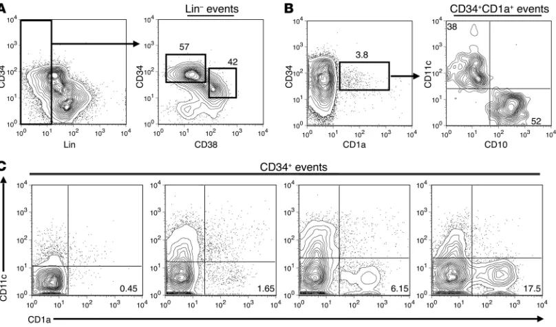
cells reside within the human tonsil. Dim expression of CD38 on CD34+ cells identifies multipotent progenitor populations in the bone marrow, thymus, and umbilical cord blood, and increased expression of CD38 on CD34+ cells is associated with differen-tiation (1, 3, 15–17). Analyzing magnetically enriched CD34+ tonsillar cells for the expression of CD34 and CD38, together with the lineage (Lin) antigens CD3, CD19, CD117, CD161, BDCA-2, CD11c, and CD1a, revealed that the human pediatric tonsil contains a population of CD34+CD38dimLin– cells as well as a CD34+CD38brightLin– subset. In general, the CD34+CD38dim subset tended to express higher surface density of CD34 than the CD34+CD38bright population (Figure 1A), suggesting that the
human tonsil may contain a multipotent hematopoietic progeni-tor minimally defined as CD34+CD38dimLin– as well as a more mature CD34+CD38brightLin– population.
Commitment to the T cell lineage in the human thymus is asso-ciated with the acquisition of CD1a on CD34+CD10+CD7+ thy- mocytes (1). However, CD1a expression is also canonically associ-ated with DC populations (18, 19). Within the human tonsil there was a small but consistent population of CD34+CD1a+ lympho-cytes (Figure 1B), which lacked CD117, CD161, BDCA-2, CD14, CD19, and CD3 (data not shown). However, when analyzed for the expression of CD10 and the DC marker CD11c, CD34+CD1a+ tonsillar cells expressed either CD11c or CD10 (Figure 1B). Sub-stantial donor-to-donor variation exists in the relative size of the CD34+CD1a+CD11c– subset in the tonsil (Figure 1C). In 24 pediat-ric donors, CD34+CD1a+CD11c– tonsillar cells constituted between 0.15%–19.4% of the total CD34+ population (mean, 3.82% ± 1.06%; median, 1.47%). No statistical correlation was found between the percentage of CD34+CD1a+CD11c– cells and the age (1–16 years) or gender of the donor.
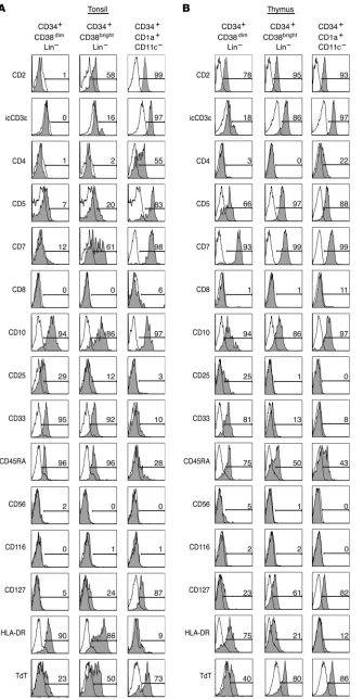
Tonsillar CD34+CD38dimLin–, CD34+CD38brightLin–, and
CD34+CD1a+CD11c– cells phenotypically resemble the
earli-est stages of thymocyte development. To determine whether or not the CD34+CD38dimLin–, CD34+CD38brightLin–, and CD34+CD1a+CD11c– cells identified in the tonsil represent extrathymic T cell precursors similar to those found in the thy-
mus, each subset from tonsil and thymus was extensively pheno-Figure 1
[image:3.585.84.488.83.321.2]typed by flow cytometry (Figure 2). In both the tonsil and thy-mus, CD34+CD38dimLin– cells expressed CD10, CD33, CD45RA, and HLA-DR as well as variable levels of CD25 and intracellular TdT. Similarly, CD34+CD38dimLin– cells from both tissues lacked expression of CD4, CD8, CD56, and CD116 (Figure 2). How- ever, there are distinct differences between tonsillar and thy-mic CD34+CD38dimLin– cells. Thymic CD34+CD38dimLin– cells expressed CD2 and most expressed CD5 and CD7, while a small fraction expressed the IL-7 receptor α (CD127) (Figure 2B). The same cells in the tonsil lacked CD2 and CD127, and only a small fraction expressed CD5 or CD7 (Figure 2).
CD34+CD38brightLin– cells from the tonsil and thymus are similar in that both expressed CD2, CD10, and low levels of CD45RA. Similarly, both populations lacked expression of CD4, CD8, CD56, and CD116. However, CD34+CD38brightLin– cells from the tonsil expressed variable levels of CD7 and CD5, and only small amounts of intracellular CD3ε (icCD3ε), whereas those from the thymus were all CD7+CD5+icCD3ε+. Further-more, tonsil CD34+CD38brightLin– cells expressed CD33 and vari-able levels of HLA-DR, which were largely absent on the same cells from the thymus (Figure 2). Despite the differences seen in CD34+CD38brightLin– cells, each subset represents a phenotype that is in many ways intermediate between CD34+CD38dimLin– cells and CD34+CD1a+CD11c– cells from the respective tissues. In the tonsil, intermediate levels of CD5, CD7, CD25, CD127, HLA-DR, and TdT on CD34+CD38brightLin– exemplify this.
The CD34+CD1a+CD11c– populations in the thymus and ton- sil are remarkably similar. Both expressed the early T cell mark-ers CD2, CD5, CD7, CD10, and icCD3ε. Furthermore, both demonstrated moderate levels of CD127 and intracellular TdT, an enzyme necessary for TCR gene rearrangement. Portions of the CD34+CD1a+CD11c– subset in both the tonsil and thymus expressed CD4, suggesting that some CD34+CD1a+ cells may be progressing to the immature single-positive stage, characteristic of normal thymocyte development (1). Like their thymic counter-parts, tonsillar CD34+CD1a+CD11c– cells did not express the GM-CSF receptor (CD116) or HLA-DR, consistent with the hypothesis that they are not DCs (Figure 2).
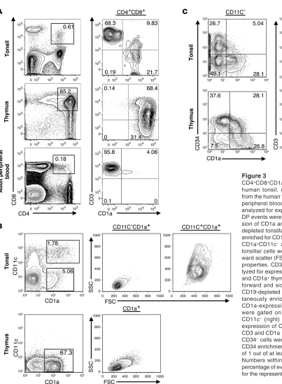
CD4+CD8+CD1a+ DP pre–T cells reside within the human tonsil. If
the human tonsil supports complete T cell development, it should contain DP pre–T cells. However, Nascimbeni et al. have identified peripheral blood CD3+ DP T cells that have a mature memory function (20), indicating that CD4 and CD8 alone are not enough to distinguish DP lymphocytes as pre–T cells. CD34- depleted tonsillar or thymic cells and total mononuclear periph-eral blood cells were analyzed for expression of CD4, CD8, CD3, and CD1a. Indeed, a population of CD4+CD8+ cells resides within the human tonsil, and it was divided into CD1a+ and CD1a– cells. The tonsillar CD1a– DP cells uniformly expressed CD3, whereas the CD1a+ DP cells variably expressed CD3, suggesting they con-tain CD3– T cell precursors (Figure 3A). Furthermore, thymic DP cells expressed CD1a with similar CD3+ expression as seen on the tonsillar CD1a+ subset (Figure 3A). Conversely, blood DP cells were CD1a–CD3+ (Figure 3A), consistent with the findings of Nascimbeni et al., who found that DP memory T cells lack CD1a (20). These findings suggest that expression of CD1a can distinguish among distinct subsets of DP lymphocytes. Further-more, its expression on a fraction of CD4+CD8+CD3+/– tonsillar cells, similar to that in the thymus, supports the hypothesis that pre–T cells reside within the tonsil.
In the thymus, CD1a is expressed throughout T cell devel-opment until its loss on naive CD3+ T cells (1). To investigate the relationship between CD34+CD1a+CD11c– tonsillar cells and potential downstream T cell intermediates, CD19– ton-sillar cells were enriched for CD1a-expressing cells. Like the CD34+CD1a+ subset described in Figure 1, the CD34–CD1a+ fraction of the human tonsil can be divided into 2 distinct populations based on its expression of the DC marker CD11c (Figure 3B). CD34–CD11c–CD1a+ tonsillar cells displayed low forward and side scatter properties, suggesting they are small, agranular lymphocytes, whereas the CD11c+CD1a+ subset dis-played higher forward and side scatter (Figure 3B). In contrast to those in the tonsil, CD34–CD1a+ cells in the human thymus were largely CD11c– and displayed low forward and side scat-ter (Figure 3B). These data suggest that CD11c expression in the human tonsil is useful for distinguishing large CD1a+ DCs from what we believe to be a novel population of small CD11c– CD1a+ lymphocytes.
Tonsillar cells were next enriched for cells expressing CD34, CD1a, or a combination of both and analyzed for the expression of CD11c, CD34, CD1a, and CD3. It appears that both CD1a+ thy-mic and tonsillar cells lose CD34 expression as they increase CD1a surface density (Figure 3C, left). Furthermore, when the enriched cells were gated on total CD34–CD11c– events, a similar progres-sion from CD1a+CD3– to CD1a+CD3+ cells was seen in both tis-sues (Figure 3C, right). As CD1a expression is seen on thymocytes, spanning from CD34+ precursors all the way to CD3+ near-mature T cells (1), these data suggest that a similar pattern describes puta-tive T cell developmental intermediates of the tonsil.
Tonsillar CD34–CD1a+CD11c– cells express T cell antigens in patterns
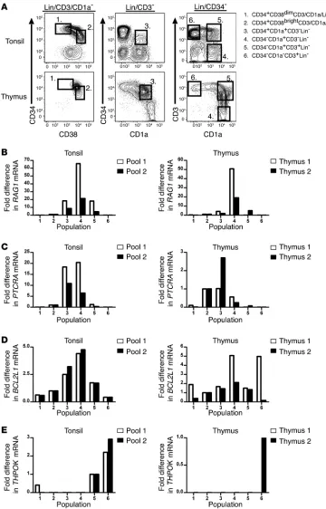
Expression of T cell development–associated genes in putative tonsillar T cell precursors. Specific gene expression patterns in the human thymus can be used to track the development of T cell precursors as they progress from multipotent cells toward mature T cells. For example, RAG1 and
PTCRA , both of which are required for suc-cessful TCR rearrangement, are expressed early in T cell development and peak at the CD34+CD1a+ and DP pre–T cell stages (3, 6). Similarly, expression of BCL-X has been detected in both murine and human CD4+CD8+ DP thymocytes, and the BCL-XL (also known as BCL2L1) isoform has been shown to be critical for providing neces-sary antiapoptosis signals in murine DP cells (21, 22). In contrast, THPOK is a transcription factor that is essential for development of mature CD4+ cells, and its expression in the thymus and periphery is largely restricted to CD3+ cells (23).
[image:5.585.46.381.82.726.2]Thus, we used real-time RT-PCR to assess expression of these 4 genes within putative T cell precursor populations in the human tonsil. Six distinct populations were sorted from human tonsil and thymus, as shown in Figure 5A: (a) CD34+CD38dimLin–, (b) CD34+CD38brightLin–, (c) CD34+CD1a+CD3–, (d) CD34–CD1a+CD3–, (e) CD34–CD1a+CD3+, and (f) CD34–CD1a–CD3+ cells. The com-plete gating strategy and representative sort purities are shown in Supplemental Figure 2, and the relative frequencies of each tonsillar subset are shown in Supple-mental Table 1. In duplicate experiments, the levels of RAG1 (Figure 5B), PTCRA (Fig-ure 5C), and BCL2L1 (Figure 5D) mRNA were highest in either population 3 or 4 of the tonsil. Expression of THPOK mRNA, on the other hand, was largely restricted
Figure 2
to the CD3+ populations 5 and 6 (Figure 5E). These patterns of gene expression were found to be similar to the patterns noted in the comparable thymic populations (Figure 5, B–E).
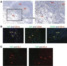
In situ localization of tonsillar CD34+TdT+ and CD1a+TdT+ cells. TdT+ cells have been identified in the human tonsil near the fibrous scaffold (8–11). However, they have yet to be characterized with regard to origin, hematopoietic lineage, or function. Our iden-tification of CD1a+CD11c–TdT+ cells in the tonsil suggests that the previously identified TdT+ cells may be extrathymic T cell
[image:6.585.47.450.72.621.2]precursors. Paraffin-embedded pediatric tonsils were analyzed by immunohistochemistry for the presence of TdT, CD34, CD1a, CD11c, and Rag1. We confirmed that TdT+ cells are largely restricted to the extrafollicular regions near the fibrous scaf-fold of the tonsil (Figure 6A). Further, these cells did not express CD11c (data not shown) but did variably coexpress CD34, CD1a (Figure 6B), and/or nuclear Rag1 (Supplemental Figure 3). Fur-thermore, CD34+CD1a+ cells were easily identified in the same region of the tonsil fibrous scaffold (Figure 6B). TdT+ cells were
Figure 3
identified in both pediatric and adult tonsil. Analyses performed in adult reactive lymph nodes revealed that rare TdT+ cells could be identified, but no coexpression of CD1a or CD34 was observed (data not shown). To further examine the microenvironment of tonsillar TdT+ cells, we used immunohistochemistry to analyze the expression of the notch ligands delta-like 1 (DL1) and delta-like 4 (DL4) within the tonsil fibrous regions, as notch ligands are essential for complete T cell development (24–27). Both pro-teins were easily identified (Figure 6C), further supporting the hypothesis that the tonsil may contribute to extrathymic T cell development. Taken together, these data suggest that the TdT+ cells previously identified in the tonsil belong to a program of extrathymic T cell differentiation and localize to a specific micro-environment near the tonsillar fibrous scaffold.
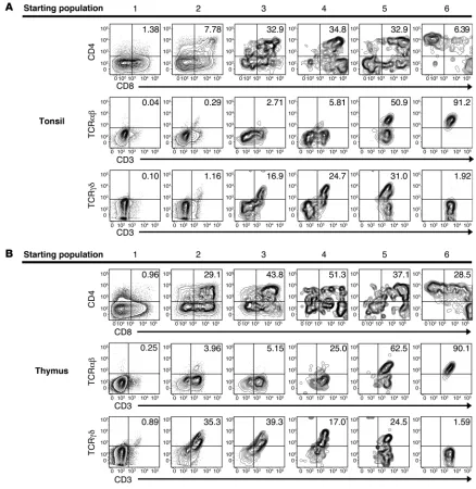
Ex vivo T cell differentiation potential of putative extrathymic T cell pre-cursors . To determine whether the putative extrathymic T cell pre-cursors have the capacity to differentiate into mature T cells, the 6 populations were sorted from the tonsil and thymus, as shown in Figure 5A and Supplemental Figure 2, and were cultured on the OP9-DL1 cell line with flt3 ligand (FL) and IL-7. After 26 days, the cells were harvested and analyzed by flow cytometry. To exclude the GFP+ OP9-DL1 cells, harvested cells were gated on GFP–CD45+
[image:7.585.43.543.84.402.2]events. Populations 1–3 from the tonsil and thymus expanded 93– 270 fold, whereas populations 4–6 only expanded 1–6 fold (Sup-plemental Table 2). All 6 populations from the tonsil and thymus gave rise to CD3+ cells. In the tonsil and thymus, the percentage of GFP–CD45+ cells that coexpress CD4 and CD8 increased as the cells progressed from population 1 to population 4 (Figure 7). Further-more, in both the tonsil and thymus, the percentage of GFP–CD45+ cells that express CD3 increased as one progressed from popula-tion 1 to population 6, as did the percentage of CD3+ cells that coexpress a TCR (Figure 7 and Supplemental Figure 4, A and B). When we compared the relative ability of each tonsillar population to generate mature T cells with that of the same populations in the thymus, we found that population 2 and 3 cells from the thymus generated a significantly higher proportion of CD3+ cells than did populations 2 and 3 from the human tonsil (population 2, 51.9% ± 7.0% from the thymus vs. 4.1% ± 1.12% from the tonsil, P < 0.0001; population 3, 57.23% ± 4.8% from the thymus vs. 32.36% ± 8.1% from the tonsil, P = 0.0044; Supplemental Figure 4A). Similarly, we found that thymic populations 2 and 3 generated significantly more CD3+ cells that coexpressed a TCR (either TCRαβ or TCRγδ) than did the same populations in the tonsil (population 2, 39.3% ± 8.5% from the thymus vs. 1.41% ± 0.6% from the tonsil, P < 0.0001;
Figure 4
population 3, 44.1% ± 7.2% from the thymus vs. 20.3% ± 5.7% from the tonsil, P = 0.0041; Supplemental Figure 4B). No statistical dif-ference was observed in the percentage of GFP–CD45+ cells that were CD3+ or CD3+TCR+ when we compared tonsillar populations 1, 4, 5, or 6 with those same populations in the human thymus. Several controls were performed to rule out contamination from
[image:8.585.43.403.82.644.2]mature tonsillar T cells as being responsible for the observed out-growth of CD3+ cells from cultures of populations 1–4. For exam-ple, CD19+ tonsillar cells were similarly sorted and cultured on the OP9-DL1 cell line. After 26 days, no CD3+ cells were generated from the CD19+ tonsillar cells. Thus, these data demonstrate that CD34+CD38dimLin–, CD34+CD38brightLin–, CD34+CD1a+CD3–, and CD34–CD1a+CD3– tonsillar cells (populations 1–4) contain differ-entiation potential for T cell development ex vivo.
Figure 5
Quantification of gene expression in tonsillar precursor cells by real-time RT-PCR. (A) Tonsillar cells were depleted of CD19+ cells, enriched for CD34+ and CD1a+ cells, and gated on the 6 popula-tions shown. (B–E) Expression of the genes RAG1, PTCRA,
BCL2L1, and THPOK in 6 popu-lations of human tonsillar and thymic cells. Cells were sorted from 8 tonsil donors, and 2 pools were generated for each popu-lation consisting of cells from 4 of the 8 donors (see Methods). Cells were sorted from 2 thymic donors. (B–D) For analysis of
To gain further insight into the kinetics of T cell development from tonsillar precursor cells, we sorted populations 1–6 from the tonsil and cultured them on the OP9-DL1 cell line with exo-genous FL and IL-7 for either 7 or 14 days. Whereas a few of the GFP–CD45+ progeny were CD3+ at days 7 or 14, only a very small percentage of these CD3+ cells coexpressed a TCR (Supplemen-tal Figure 4, C and D). This suggests that TCR expression is an event that occurs later in T cell development and confirms that the appearance of mature T cells at 26 days is not simply an artifact of contamination by tonsillar CD3+TCR+ cells. Furthermore, in 26-day cultures with FL and IL-7 on the OP9 stromal cell line, which lacks notch ligand expression, populations 1–4 from the tonsil failed to generate CD3+TCR+ T cells. Tonsillar populations 5 and 6, on the other hand, maintained a population of CD3+TCR+ cells in these conditions (data not shown).
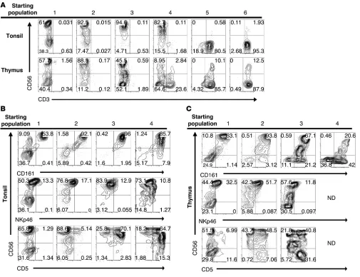
Ex vivo NK cell differentiation potential of putative extrathymic T cell precursors . Human NK cell development has been studied exten- sively in human tonsils and lymph nodes (12–14, 28–31). To exam- ine whether extrathymic T cell precursors could contribute to ton-sillar NK cell development as well, the 6 populations identified in Figure 5A were cultured with FL, c-kit ligand (KL), IL-3, IL-7, and IL-15 on the OP9 stromal cell line. Whereas cultures of popula-tions 5 and 6 from both tissues remained exclusively CD3+ (Figure 8A), populations 1–3 from both the tonsil and thymus gave rise to CD56+CD3– NK cells. Population 4 cells from the tonsil gave rise to CD56+CD3– cells in 3 out of 3 experiments, whereas thymic population 4 cells gave rise to CD56+CD3– cells in 1 out of 5 exper-iments, although no population 4 cells survived in 3 out of these 5 thymic experiments. The resulting CD56+ cells derived from tonsil and thymus expressed CD161 and low levels of NKp46 (Figure 8, B and C), along with variable expression of CD5, the latter of which increased from less than 10% in tonsillar populations 1 and 2 to more than 50% in populations 3 and 4.
To further investigate the T and NK cell differentiation capa-bilities of putative extrathymic T cell precursors in the tonsil, single cells were sorted from tonsillar populations 1–4 into wells containing OP9-DL1 cells and the cytokines FL, IL-7, and IL-15. The individual cells were cultured in this system for 14 days. After culture, each well was harvested and examined for the presence of CD5+CD3– T cell precursors, CD3+ T cells, or CD56+CD3– NK cells (Supplemental Table 3). Between 16% and 67% of the wells plated sustained CD45+ cells at the time of harvest. This assay demon-strates that all 4 populations have the potential to differentiate into CD5+CD3– T cell precursors and CD56+CD3– NK cells and that there was an increasing tendency for a single cell to generate CD3+ cells as one progressed from population 1 through popula-tion 4. Occasionally, single cells from populations 2 and 3 gave rise to wells containing both CD3+ T cells and CD56+CD3– NK cells.
Discussion
[image:9.585.42.317.82.353.2]We have identified 5 subsets of lymphocytes which we believe to be novel within the human tonsil: (a) CD34+CD38dimLin–, (b) CD34+CD38brightLin–, (c) CD34+CD1a+CD11c–, (d) CD34– CD1a+CD3–CD11c–, and (e) CD34–CD1a+CD3+CD11c– cells. We have provided phenotypic, gene expression, and functional evi-dence that these subsets represent intermediates in a program of extrathymic T cell development. Tonsil CD34+CD38dimLin– and CD34+CD38brightLin– cells share many phenotypic properties of the same cells identified in the thymus and have differentiation potential to develop into T cells and NK cells. Remarkably, the CD34+CD1a+CD11C– cells identified in the tonsil display a phe-notype that is virtually identical to that of thymic CD34+CD1a+ T cell precursors and display consistent T cell developmental capabilities. Similarly, CD34–CD1a+CD11c– cells of the tonsil highly resemble CD34–CD1a+ cells of the thymus, in that they can be divided into 2 populations based on the expression of CD3,
Figure 6
they coexpress CD4 and CD8, and they retain T cell developmen-tal potential. Furthermore, in addition to identifying CD1a+TdT+ extrathymic T cell precursors in human tonsil, we have localized the bulk of this population near fibrous scaffolding, suggesting that it resides in a distinct microenvironment. Finally, we have demonstrated that CD34+/–CD1a+CD11c– cells from the thymus and tonsil are not committed to the T cell lineage but retain ex vivo differentiation potential to also develop into NK cells.
While previous reports have identified a role for murine intes-tinal lymphoid tissue in T cell development (32–35), the few studies investigating human extrathymic T cell differentiation
are difficult to interpret. It is known that uncommitted hema-topoietic progenitors with T cell differentiation capabilities reside within the bone marrow (6, 36, 37) and cord blood (24, 38). Furthermore, tonsillar TdT+ cells of unclear etiology and pre–Tα+ cells within bone marrow and neonatal intestine have been identified (4, 7–9). However, a complete description of
[image:10.585.67.505.83.533.2]T cell differentiation in an extrathymic human tissue has not been provided. Our work suggests that T cell development occurs in the human tonsil, and future studies should address what proportion of the total CD3+ tonsillar pool is derived with-in the tonsil compared with that from the thymus.
Figure 7
It is intriguing to think of a scenario in which both the thy-mus and tonsil are seeded by the same bone marrow–derived CD34+CD38dimCD45RA+ progenitor that gives rise to NK cells and T cells in each tissue. However, of the 5 populations identified, we found the most substantial differences when comparing the CD34+CD38dimLin– and CD34+CD38brightLin– populations from the tonsil and thymus. Specifically, thymic CD34+CD38dimLin– cells largely expressed the T cell–associated antigens CD2, CD7, and CD5, whereas tonsillar CD34+CD38dimLin– cells did not. Thus, it is possible that these populations represent distinct progeni- tors that independently seed the thymus and tonsil. Alternative-ly, it is possible that the thymus and tonsil are seeded by similar CD34+CD38dim progenitors but that the phenotype of these enter-
ing cells is rapidly altered by the thymic or tonsillar microenviron-ment, as is seen in the mouse (39, 40). As demonstrated in Figure 2B, while a majority of thymic CD34+CD38dimLin– cells are CD7+, a few lack CD7 (4%–17%). Indeed, recent reports suggest that a CD34+CD45RA+CD10+CD7– cell seeds the thymus after birth, and a CD34+CD45RA+CD10+CD7+ precursor has yet to be identi-fied in postnatal bone marrow or peripheral blood (5, 6). Thus, it is possible that both tissues are seeded by the same circulating CD34+CD38dimCD10+CD7– hematopoietic progenitor and that this cell is capable of giving rise to downstream T cell developmen-tal intermediates in each tissue.
[image:11.585.42.547.82.470.2]Regardless of whether the 2 tissues are seeded by the same pro-genitor, it is intriguing that TdT+ cells within the tonsil aggregate in foci, surrounding fibrous scaffolding. Were the trafficking of T cell precursors to the tonsil a random event associated with a
Figure 8
“leaky” thymus, one would assume that the cells would perfuse the tonsil in a scattered arrangement in the interfollicular zone near mature T cells. However, the consistent aggregation of T cell precursors near the fibrous regions of the tonsil suggests that this space serves a unique anatomical function in supporting extrathy-mic T cell precursor homing and/or maturation. This hypothesis is further supported by the identification of Notch ligands, which are required for complete T cell differentiation, within the same regions of the human tonsil. The capsule of the tonsil is highly invaginated into crypts, which create a large surface area important for foreign antigen exposure. Thus, the localization of T cell precursors to this fibrous region may play a role in mucosal immunity. Alternatively, the thymus is segregated to support different phases of thymocyte develop-ment in anatomically distinct microenvironments (41–44), and our finding of tonsillar CD1a+TdT+ cells near the fibrous scaf-folding, but not in the T cell–rich interfollicular zones, suggests that perhaps extrathymic T cell development in the tonsil is also anatomically divided to provide the proper microenvironment for each phase of maturation. In the thymus, the least mature pro- genitors enter the thymus at the junction between the outer cor-tex and the inner medulla. These cells migrate outward until they reach the subcapsular zone, which marks the division between the fibrous thymic capsule and the cortex, and then they travel inward toward the medulla to complete differentiation (43, 44). Future studies should investigate whether the microenvironment of the tonsillar fibrous scaffold is analogous to the subcapsular zone of the thymus, in terms of supporting T cell development, or whether it provides a unique signal directing the homing of extrathymic precursors to this region of the tonsil. This report also provides evidence for the expression of key genes involved in TCR rearrangement within these tonsillar popula-tions. Specifically, we found that populations 2–5 express mRNA for RAG1, which is necessary for initiation of TCR rearrangement (45), and for PTCRA, which is a molecular indicator that successful TCRβ rearrangement has occurred (3). The expression of these 2 genes, along with TdT, suggests that somatic rearrangement of the TCR may occur in the human tonsil.
In 1998, Spits et al. reported that CD34+CD1a+ thymocytes gen- erate only small numbers of NK cells (46). Since then it has gen-erally been held that loss of CD34 and acquisition of CD1a are associated with T cell commitment (1, 46, 47). Our findings con-firm and extend their work in the thymus, in that we found that CD34+CD38dim, CD34+CD38bright, and CD34+CD1a+ thymocytes and tonsil cells readily generate NK cells and that tonsillar CD34– CD1a+CD11c– cells also retain NK cell potential. Furthermore, some CD56+CD3– NK cells generated in our ex vivo cultures coexpressed CD5, and there was a relative increase in CD56+CD5+CD3– prog-eny generated as one progressed from CD34+CD38dimLin– cells to CD34–CD1a+CD11c– cells. This suggests that the generated NK cells are not merely a result of starting populations that were impure at sorting but that a unique CD5+ progenitor may be giving rise to the CD56+CD3– cells in cultures initiated with the CD34+CD1a+CD11c– and CD34–CD1a+CD11c– subsets. Our finding that all 4 popula-tions from the tonsil generate CD56+CD3– NK cells at the single cell level as well substantiated these results. It has become evident that the human tonsil is a significant site of NK cell development (12–14, 28–31). However, whether or not differentiating CD34+/– CD1a+CD11c– cells that also express TdT contribute to the physi-ological NK cell differentiation within this tissue remains unclear. That the tonsil does support T cell development could have sig- nificant clinical and scientific implications. First, the role of tonsil-lar T cell development in human disease is unknown. That T cells can develop extrathymically in this tissue raises the possibility that the tonsil may actually support the generation of autoreactive T lymphocytes or even contribute to malignant transformation. Fur-thermore, the role of extrathymic T cell development in the tonsil during inflammation should be of future interest. In this study, we have obtained pediatric tonsils from patients undergoing routine tonsillectomy. While it can be presumed that the patients are afe-brile at the time of surgery, the possibility that repeat or sustained inflammation contributes to the presence of extrathymic T cell development should be further explored. Alternatively, the identification of an extrathymic reservoir of human T cell precursors in normal individuals could suggest clinical opportunities to enhance T cell regeneration in situa- tions of thymic defect. For example, T cell deficiency is charac-teristic in patients with poor thymic function due to complete DiGeorge syndrome, chemoablation or radioablation, or surgical thymectomy (48–52). Indeed, the efficient regeneration of viable thymic tissue after total body irradiation for bone marrow trans-plant continues to be a significant clinical challenge (49). In the future it may be possible to harvest tonsillar T cell precursors or healthy tonsil stroma via tonsillectomy in order to augment T cell regeneration after transplant in the same individual. While these possibilities remain speculative at the moment, the identification of a program of extrathymic T cell development in the human tonsil is a unique finding with potential implications for human health and disease. Methods
Human samples. Human tonsils and thymuses were obtained from Nationwide Children’s Hospital (Columbus, Ohio, USA). Tonsils were obtained from pediatric patients undergoing tonsillectomies, whereas thymuses were obtained from children undergoing thoracic surgery. Par-affin-embedded adult tonsils and reactive lymph nodes were obtained from the Biospecimen Shared Resource at The Ohio State University, and adult peripheral blood was obtained from the American Red Cross (Columbus, Ohio, USA). Mononuclear cells were obtained from ton- sils and thymuses after manual disaggregation and Ficoll-Paque cen-trifugation. For experiments wherein tonsillar cells were enriched for CD34-expressing cells only, total cells were depleted of CD19+ and CD3+
cells by magnetic depletion and were enriched for CD34+ cells using an
indirect CD34 Selection Kit (Miltenyi Biotec). For experiments in which tonsillar cells were enriched for CD1a+
cells only, total cells were deplet-ed of CD19+ cells and then magnetically enriched for CD1a-expressing
cells (CD1a magnetic beads, Miltenyi Biotec). To simultaneously enrich for CD34- and CD1a-expressing tonsillar cells, CD19-depleted mono-nuclear cells were subjected to positive selection using the indirect CD34 Selection Kit (Miltenyi Biotec) and CD1a magnetic beads (Miltenyi Bio-tec) concurrently. To isolate thymic T cell progenitors, total thymic cells were subjected to positive CD34 selection using the indirect CD34 Isola-tion Kit (Miltenyi Biotec).
analyzed using FlowJo software (Treestar Inc). Cell sorting was performed on a FACSAria (BD). The complete gating strategy and a representative purity assessment are shown in Supplemental Figure 2. Purities of sorted populations were routinely more than 95% pure.
Immunohistochemistry. Immunohistochemistry was performed as described previously (12, 31, 53) using the UltraView Universal system (Ven-tana Medical). Αnti-TdT (1:100), α-CD1a (1:10), α-CD34 (1:100), α-CD11c (1:100), α-DL1 (1:300), and α-DL4 (1:300) were purchased from Abcam, and α-Rag1 (1:10) was purchased from Santa Cruz Biotechnology Inc. For immunohistochemistry involving α-DL1 and α-DL4, antigen retrieval was performed for 30 minutes prior to staining. For immunohistochemistry involving α-Rag1, antigen retrieval was performed for 30 minutes, along with digestion in Ventana Protease 1 (1:100) for 4 minutes. Images were obtained using a DP 12 camera, a BX50 microscope, and UPLANF1 objec- tives (Olympus). DAB- and fast-red–stained sections were digitally convert-ed to fluorescent green and red, respectively, using the Nuance FX system (Cambridge Research & Instrumentation).
Differentiation assays. Cultures were performed in α-MEM plus l -gluta-mine (Gibco) supplemented with 20% fetal bovine serum and penicillin G plus streptomycin (100 μg/ml; Gibco), with 500–5,000 cells per well. Medi-um was supplemented with the following cytokines as indicated: human recombinant FL (100 ng/ml; Miltenyi Biotec), KL (100 ng/ml; Amgen), IL-3 and IL-7 (10 ng/ml; Miltenyi Biotec), and IL-15 (1 nM; Miltenyi Biotec). T cell differentiation assays were performed as described previously (13, 24) on OP9-DL1 cells (a gift from J.C. Zúñiga-Pflücker, University of Toronto, Toronto, Ontario, Canada) with IL-7 and FL. NK cell differentiation assays were performed on OP9 cells (a gift from J.C. Zúñiga-Pflücker) with FL, KL, IL-7, IL-3, and IL-15. For single cell cultures, individual cells from popu-lations 1–4 of the human tonsil were sorted directly into 59 to 60 wells containing OP9-DL1 cells, FL, IL-7, and IL-15 for 14 days. Medium was changed every 4 to 5 days.
Real-time RT-PCR. Tonsillar populations 1–6 were sorted from 8 tonsil donors, as shown in Figure 5A and Supplemental Figure 2. From these, we generated 2 pools for each population, with each pool containing cells from 4 of the 8 donors and approximately 3,500 cells. 5,000 cells from pop-ulations 1–6 were sorted from 2 thymus donors for comparison. RNA was extracted using the RNeasy Micro Kit (Qiagen), and real-time RT-PCR was performed as previously described (30), using ABI TaqMan primer/probe sets for PTCRA (probe Hs00300125_m1), RAG1 (probe Hs00172121_m1),
BCL2L1 (probe Hs00236329_m1), and THPOK (probe_Hs01035470_m1). Expression levels were normalized to 18S expression and were analyzed using the comparative ΔδCT method (54).
Statistics . To investigate whether age or gender correlates with the per-centage of CD34+CD1a+CD11c–
cells, a Spearman correlation and a 2-sample, 2-tailed t test were used. In reporting the percentages of CD3+
T cells generated in T cell differentiation assays, the mean percentage ± SEM of GFP–CD45+ cells was listed. For comparison of CD3+ cells
generated from populations in the human thymus and those from the tonsil, the linear mixed effects models were used to take account of the correlation among observations from the same donor. After Holm’s adjustment for multiple comparisons, P values of equal to or less than 0.005 were considered significant.
Study approval. Tonsillar, thymic, and peripheral blood samples were obtained with approval of these experiments from The Ohio State Uni-versity Comprehensive Cancer Center Institutional Review Board. As all human samples were obtained as deidentified tissue, informed consent was not required.
Acknowledgments
This work was supported by NCI grants (CA95426 and CA68458). The authors acknowledge the use of the bioreposito-ry of Nationwide Children’s Hospital and the Biostatistics, Flow Cytometry, Microscopy, Leukemia Tissue Bank, Biorepository, and Biospecimen Shared Resources of The Ohio State University Comprehensive Cancer Center. We thank Ventana Medical Sys-tems for providing reagents for the immunohistochemistry. Received for publication December 16, 2010, and accepted in revised form January 11, 2012. Address correspondence to: Michael A. Caligiuri, 521B James Cancer Hospital, 300 W. 10th Ave., Columbus, Ohio 43210, USA. Phone: 614.293.7521; Fax: 614.293.3132; E-mail: Michael. [email protected]. 1. Blom B, Spits H. Development of human lymphoid cells. Annu Rev Immunol. 2006;24:287–320. 2. Staal FJ, Weerkamp F, Langerak AW, Hendriks RW,
Clevers HC. Transcriptional control of T lympho-cyte differentiation. Stem Cells. 2001;19(3):165–179. 3. Dik WA, et al. New insights on human T cell devel- opment by quantitative T cell receptor gene rear-rangement studies and gene expression profiling. J Exp Med. 2005;201(11):1715–1723.
4. Klein F, et al. T lymphoid differentiation in human bone marrow. Proc Natl Acad Sci U S A. 2003;100(11):6747–6752. 5. Doulatov S, Notta F, Eppert K, Nguyen LT, Ohashi PS, Dick JE. Revised map of the human progeni-tor hierarchy shows the origin of macrophages and dendritic cells in early lymphoid development. Nat Immunol. 2010;11(7):585–593. 6. Six EM, et al. A human postnatal lymphoid progen-itor capable of circulating and seeding the thymus. J Exp Med. 2007;204(13):3085–3093.
7. Williams AM, et al. Intestinal alpha beta T cells differentiate and rearrange antigen receptor genes in situ in the human infant. J Immunol. 2004;173(12):7190–7199.
8. Strauchen JA, Miller LK. Terminal deoxynucleo-tidyl transferase-positive cells in human tonsils. Am J Clin Pathol. 2001;116(1):12–16.
9. Strauchen JA, Miller LK. Lymphoid progenitor cells in human tonsils. Int J Surg Pathol. 2003;11(1):21–24.
10. Sening W, Lisner R, Niedobitek G. Rare detection of phenotypically immature lymphocytes in Hashi-moto thyroiditis and rheumatoid arthritis. J Auto-immun. 2004;22(2):147–152.
11. Meru N, Jung A, Baumann I, Niedobitek G. Expres-sion of the recombination-activating genes in extrafollicular lymphocytes but no apparent rein-duction in germinal center reactions in human tonsils. Blood. 2002;99(2):531–537.
12. Freud AG, et al. A human CD34(+) subset resides in lymph nodes and differentiates into CD56bright natural killer cells. Immunity. 2005;22(3):295–304. 13. Freud AG, et al. Evidence for discrete stages of
human natural killer cell differentiation in vivo. J Exp Med. 2006;203(4):1033–1043.
14. Freud AG, Caligiuri MA. Human natural killer cell development. Immunol Rev. 2006;214:56–72. 15. Hao QL, Shah AJ, Thiemann FT, Smogorzewska
EM, Crooks GM. A functional comparison of CD34 + CD38- cells in cord blood and bone mar-row. Blood. 1995;86(10):3745–3753.
16. Hao QL, Zhu J, Price MA, Payne KJ, Barsky LW, Crooks GM. Identification of a novel, human multilymphoid progenitor in cord blood. Blood. 2001;97(12):3683–3690.
17. Res P, et al. CD34+CD38dim cells in the human thymus can differentiate into T, natural killer, and dendritic cells but are distinct from pluripotent stem cells. Blood. 1996;87(12):5196–5206.
18. Indrasingh I, Chandi G, Jeyaseelan L, Vettivel S, Chandi SM. Quantitative analysis of CD1a (T6) positive Langerhans cells in human tonsil epithe-lium. Ann Anat. 1999;181(6):567–572.
19. Liu YJ. Dendritic cell subsets and lineages, and their functions in innate and adaptive immunity. Cell. 2001;106(3):259–262. 20. Nascimbeni M, Shin EC, Chiriboga L, Kleiner DE, Rehermann B. Peripheral CD4(+)CD8(+) T cells are differentiated effector memory cells with antiviral functions. Blood. 2004;104(2):478–486.
21. Boise LH, et al. bcl-x, a bcl-2-related gene that func-tions as a dominant regulator of apoptotic cell death. Cell. 1993;74(4):597–608.
22. Chao DT, Korsmeyer SJ. BCL-XL-regulated apoptosis in T cell development. Int Immunol. 1997;9(9):1375–1384.
23. Tokunaga T, et al. Regulation of Th-POK and Runx3 in T cell development in human thymoma. Autoimmunity. 2009;42(8):653–660.
24. La Motte-Mohs RN, Herer E, Zuniga-Pflucker JC. Induction of T-cell development from human cord blood hematopoietic stem cells by Delta-like 1 in vitro. Blood. 2005;105(4):1431–1439.
25. De Smedt M, et al. Active form of Notch imposes T cell fate in human progenitor cells. J Immunol. 2002;169(6):3021–3029.
JC. Maintenance of T cell specification and differ-entiation requires recurrent notch receptor-ligand interactions. J Exp Med. 2004;200(4):469–479. 27. Van de Walle I, et al. An early decrease in Notch
activation is required for human TCR-alphabeta lineage differentiation at the expense of TCR-gam-madelta T cells. Blood. 2009;113(13):2988–2998.
28. Cupedo T, et al. Human fetal lymphoid tissue- inducer cells are interleukin 17-producing precur-sors to RORC+ CD127+ natural killer-like cells. Nat Immunol. 2009;10(1):66–74.
29. Crellin NK, Trifari S, Kaplan CD, Cupedo T, Spits H. Human NKp44+IL-22+ cells and LTi-like cells constitute a stable RORC+ lineage distinct from conventional natural killer cells. J Exp Med. 2010;207(2):281–290.
30. Hughes T, et al. Interleukin-1beta selectively expands and sustains interleukin-22+ immature human natural killer cells in secondary lymphoid tissue. Immunity. 2010;32(6):803–814.
31. Hughes T, et al. Stage three immature human natural killer cells found in secondary lymphoid tissue constitutively and selectively express the TH17 cytokine interleukin–22. Blood. 2009; 113(17):4008–4010.
32. Oida T, et al. Role of gut cryptopatches in early extrathymic maturation of intestinal intraepithe-lial T cells. J Immunol. 2000;164(7):3616–3626. 33. Ishikawa H, Saito H, Suzuki K, Oida T, Kanamori
Y. New gut associated lymphoid tissue “crypto-patches” breed murine intestinal intraepithelial T cell precursors. Immunol Res. 1999;20(3):243–250. 34. Suzuki K, et al. Gut cryptopatches: direct evidence
of extrathymic anatomical sites for intestinal T lymphopoiesis. Immunity. 2000;13(5):691–702.
35. Guy-Grand D, et al. Extrathymic T cell lymphopoi- esis: ontogeny and contribution to gut intraepi-thelial lymphocytes in athymic and euthymic mice.
J Exp Med. 2003;197(3):333–341.
36. Haddad R, et al. Dynamics of thymus-coloniz-ing cells during human development. Immunity. 2006;24(2):217–230.
37. Hao QL, et al. Human intrathymic lineage com-mitment is marked by differential CD7 expression: identification of CD7- lympho-myeloid thymic progenitors. Blood. 2008;111(3):1318–1326.
38. Awong G, Herer E, Surh CD, Dick JE, La Motte-Mohs RN, Zuniga-Pflucker JC. Characterization in vitro and engraftment potential in vivo of human progenitor T cells generated from hematopoietic stem cells. Blood. 2009;114(5):972–982.
39. Krueger A, Garbe AI, von Boehmer H. Phenotypic plasticity of T cell progenitors upon exposure to Notch ligands. J Exp Med. 2006;203(8):1977–1984. 40. Schwarz BA, Sambandam A, Maillard I, Harman
BC, Love PE, Bhandoola A. Selective thymus set- tling regulated by cytokine and chemokine recep-tors. J Immunol. 2007;178(4):2008–2017.
41. Lind EF, Prockop SE, Porritt HE, Petrie HT. Map-ping precursor movement through the postnatal thymus reveals specific microenvironments sup- porting defined stages of early lymphoid develop-ment. J Exp Med. 2001;194(2):127–134.
42. Bousso P, Bhakta NR, Lewis RS, Robey E. Dynam-ics of thymocyte-stromal cell interactions visualized by two-photon microscopy. Science. 2002;296(5574):1876–1880.
43. Petrie HT, Zuniga-Pflucker JC. Zoned out: func-tional mapping of stromal signaling microen-vironments in the thymus. Annu Rev Immunol. 2007;25:649–679.
44. Klein L, Hinterberger M, Wirnsberger G, Kyewski B. Antigen presentation in the thymus for positive selection and central tolerance induction. Nat Rev Immunol. 2009;9(12):833–844. 45. Spanopoulou E, et al. Localization, interaction, and RNA binding properties of the V(D)J recombi-nation-activating proteins RAG1 and RAG2. Immu-nity. 1995;3(6):715–726. 46. Spits H, et al. Early stages in the development of human T, natural killer and thymic dendritic cells. Immunol Rev. 1998;165:75–86.
47. Weerkamp F, et al. Human thymus contains multipotent progenitors with T/B lymphoid, myeloid, and erythroid lineage potential. Blood. 2006;107(8):3131–3137.
48. Prelog M, et al. Thymectomy in early child-hood: significant alterations of the CD4(+) CD45RA(+)CD62L(+) T cell compartment in later life. Clin Immunol. 2009;130(2):123–132. 49. Hollander GA, Krenger W, Blazar BR. Emerging
strategies to boost thymic function. Curr Opin Phar-macol. 2010;10(4):443–453.
50. Markert ML, et al. Transplantation of thymus tis-sue in complete DiGeorge syndrome. N Engl J Med. 1999;341(16):1180–1189.
51. Fischer A, et al. Naturally occurring primary defi-ciencies of the immune system. Annu Rev Immunol. 1997;15:93–124.
52. Sauce D, et al. Evidence of premature immune aging in patients thymectomized during early childhood. J Clin Invest. 2009;119(10):3070–3078. 53. Fehniger TA, et al. CD56bright natural killer cells
are present in human lymph nodes and are acti-vated by T cell-derived IL-2: a potential new link between adaptive and innate immunity. Blood. 2003; 101(8):3052–3057.