Content Based Image Retrieval by Supervised
Metric Learning
S. Himabindu Sri1, Dr. A. Sharada2,
M.Tech Student Department of Computer Science and Engineering, G. Narayanamma Institute of Technology &
Science, Ambedkar Nagar, Shaikpet, Hyderabad, Telangana, India.
Professor, Department of Computer Science and Engineering, G. Narayanamma Institute of Technology & Science,
Ambedkar Nagar, Shaikpet, Hyderabad, Telangana, India.
ABSTRACT: Content-based Image Retrieval [CBIR] is a pursuit innovation that could help medical analysis by recovering/retrieving and introducing prior revealed cases that are identified with the one being analyzed. To recover significant cases, CBIR frameworks rely upon administered figuring out how to delineate level image substance to abnormal state analytic ideas. Be that as it may, the comment by medical specialists for preparing and assessment reasons for existing is a troublesome and tedious assignment, which limits the directed learning stage to particular CBIR issues of all around characterized clinical applications. This system proposes another strategy that naturally takes in the similitude between the few exams from printed separations extricated from radiology reports, in this manner effectively diminishing the quantity of explanations required. Our technique initially construes the connection between patients by utilizing data recovery procedures to decide the printed separates between quiet radiology reports. These separations are hence used to administer a metric learning calculation that changes the image space likewise to printed separations. CBIR frameworks with various image depictions and diverse levels of medical explanations were assessed, with and without supervision from printed separations, utilizing a database of PC tomography outputs of patients with interstitial lung diseases. The proposed strategy reliably enhances CBIR mean normal exactness, with enhancements that can achieve 38%, and more stamped picks up for little explanation sets. Given the general accessibility of radiology reports in image chronicling and correspondence frameworks, the proposed approach can be extensively connected to CBIR frameworks in various restorative issues, and may encourage the presentation of CBIR in clinical practice.
KEYWORDS: Metric Learning, CBIR, Content Based Image Retrieval, PC Tomography, Computer-Aided-Design [CAD].
I. INTRODUCTION
Content Based Image Retrieval - CBIR is a pursuit worldview that chooses cases from a image gathering that have a comparable substance to a question image. The expansive number of exams in Picture Archiving and Communication Systems [PACS] spurred the investigation of CBIR frameworks for Computer-Aided-Design [CAD], where given an undiscovered exam, CBIR can be utilized to help the indicative procedure via naturally recovering beforehand announced important exams. This empowers radiologists to think about cases and check kindred radiologists' decisions, which may enhance the analysis, especially for unpracticed radiologists and complex symptomatic issues.
of structures, or with—not generally very much characterized or promptly quantifiable-deviations of typical appearance of structures.
This contrast between abnormal state radiologic ideas and low-level CBIR visual portrayals is the alleged semantic crevice, which is the primary motivation behind why CBIR advances are not utilized as a part of clinical practice. To diminish the semantic crevice, CBIR analysts connected regulated learning with a specific end goal to gather a connection between low-level components and conceptual restorative portrayals, from comments given by therapeutic specialists.
Metric learning is a well known directed learning strategy for CBIR, where the goal is to take in a metric, which composes therapeutic exams as nearly as conceivable to subjective evaluations on likeness of exams that were beforehand gathered from restorative specialists. As in different types of supervision, the execution of metric learning relies upon an agent dataset. The many-sided quality of symptomatic issues, the normal intra-and between client fluctuation, and the changed appearance of restorative images, prescribes the utilization of a substantial arrangement of comments. In any case, since therapeutic explanations are costly, most research bunches just approach confined arrangements of comparative/disparate exam sets given by likeness evaluations, class names, and pertinence input.
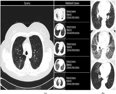
Fig. 1 (a) Example of a CAD interface for ILD within a CBIR framework. On the left the undiagnosed query exam, while on the right the retrieved related cases. (b) Slices of CT exams from patients with ILD. In the upper and middle slices are two patients with interstitial pulmonary fibrosis, respectively, in an early and late stage, and on the bottom a
patient with a NSIP in an early stage.
Our approach utilizes data retrieval techniques, by building up literary separations between cases, in light of the recurrence of terms in the patient reports. These separations are then used to manage a metric learning CBIR framework, which is accordingly connected in undiscovered exams where reports are not yet accessible. Since radiology reports are straightforwardly created by therapeutic specialists, and contain a specialized portrayal of exam determinations, discoveries, and conclusions, report recovery is not influenced by the semantic hole, as is certified by its expansive use in clinical practice. In addition, radiology reports are regularly present for all exams in most healing center PACS, guarantying the accessibility of expansive and a la mode datasets in all radiology conclusion issues.
Therefore, radiology reports can't be considered as solid as medicinal explanations. In this paper, we assess if, and to which degree, the utilization of literary separations to direct CBIR frameworks enhances CBIR execution. For an assortment of CBIR arrangements, our assessment analyzes utilize versus nonuse of supervision from literary separations, on a database of PC tomography filters from an ILD CAD issue. ILD are an arrangement of various lung issues that, in spite of having differing causes, are typically viewed as together in light of the fact that they all influence the lung interstitium.
These difficulties achieve the degree that much of the time other clinical components, for example, clinical history or histological exams, are important to deliver a dependable determination. Fig. 1[b] indicates three cuts of three patients with ILDs. In the upper and center cuts are two patients with interstitial aspiratory fibrosis, individually, in an early and late stage, and on the last a patient with a Non-Specific Interstitial Pneumonia [NSIP] in an early stage. It represents the variety in visual appearance inside the same subtype [up and center], and the visual closeness that can happen between various ILD subtypes [up and base cuts]. Subsequently, ILD subtype recognizable proof is viewed as a complex radiological issue, requiring particular chest radiologists with years of experience. Moreover, since some ILD sorts are uncommon, inhabitants, or general radiologists in nearby doctor's facilities may have not seen enough exams to have the capacity to deliver a dependable conclusion.
This roused the improvement of ILD CBIR CAD frameworks all things considered they enable radiologists to upgrade their involvement with the investigation of cases beforehand revealed by particular radiologists. With the goal of recognizing and portraying ILD designs, CAD frameworks have been produced for the programmed examination of anomalous Volumes Of Interest [VOI] inside the lung parenchyma.
These VOI are either physically portioned by a radiologist, or relying upon a programmed division of the lung taken after by a division in volumes. The division of the lung is critical as to isolate the lung from other anatomical structures that are of little pertinence to ILD analysis. Considering the examples are heterogeneous in their appearance, the portrayal of each VOI is regularly in view of set elements, of shifted nature, and crosswise over various scales. The list of capabilities ordinarily incorporates force, vital for e.g., for low-lung growths, emphysema] and high-weakening examples consolidation, ground glass [GG], and surface descriptors, imperative for designs that relate to a consistent redundancy of some anomalous structure, for example, honeycombing, emphysema, or Crazy Paving [CP].
Distinctive classes of surface descriptors have been assessed for the issue, without a complete conclusion regarding its relative benefits. Since the list of capabilities typically contains differing sorts of elements over a few scales, the subsequent element space is ordinarily unpredictable. Thus, it is regular to decide the significance of every area of the element space to each example, either utilizing unsupervised learning by isolating the component space into a codebook, or administered gaining from manual comments created by radiologists.
Since manual explanations are hard to gather, the arrangement of preparing illustrations utilized by directed frameworks is regularly bound to a modest bunch of examples, commented on in a couple of dozen cases, restricting their effectivity. Portrayals of the whole output can be accomplished by summing the number, or region of each kind of VOI.
II. MATERIALS AND RELATED STRATEGIES
Materials
(a) Dataset: Gathered two types of datasets. They are- 1. Training database 2.Testing database. Training dataset
consists of 21 lung CT scan examines and respective 21 descriptions. Which are used to train the metric learning algorithm. Testing dataset consists of 534 lung CT scan images from Lung Data Image Consortium (LDIC) , which are used to test the metric learning algorithm .
(b) Terminology: The marking of each term in the accumulation as indicated by a therapeutic class. Our phrasing was
built by a radiologist from all terms in the reports of the dataset portrayed in the past segment.
equivalent words, terms emphases, and condensings to a solitary term. A rundown with all groupings is utilized as a part of the equivalent word channel.
(c) Medical Annotation of VOI: A moment database, to be utilized by the image handling chain, is made out of 24 CT
checks from a similar number of patients. They were gained on a Philips Mx8000 IDT and a Philips Brilliance iCT scanner (Philips Medical Systems, Best, and The Netherlands). Utilizing the device portrayed in [21], an assistant radiologist commented on all VOI for all sweeps as per tissue designs: diminished thickness, union, honeycombing, GG, CP, NSIP design, nodular example, lastly, inhomogeneous when more than one class of example was available. All residual VOI were viewed as ordinary lung tissue.
Strategies
(a) Overview: This paper presents the utilization of separations among radiology reports to administer a metric learning
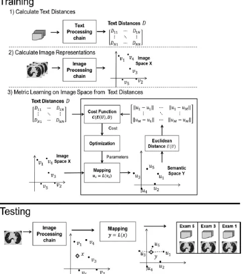
calculation in image space. The goal is to adjust for the nonattendance of master explanations in restorative CBIR frameworks, via naturally gathering the relations between exams from radiology reports. In this area, we detail the techniques utilized as a part of our approach. As shown in Fig. 2, our philosophy includes a preparation arrange and a testing stage. The preparation arrange has as info all PACS exams, and yields a mapping capacity, later utilized as a part of the testing stage.
It can be deteriorated into three stages: 1) content separation computation among patients utilizing a, 2) extraction of sweep portrayals in image space X for all exams utilizing a image handling chain, and 3) taking in the metric on which the separations among the image portrayals are like content separations. It ought to be seen that the goal of the depicted framework is to gain specifically from a database in clinical setting, which does not regularly contain manual explanations, and is heterogeneous in its securing parameters and ILD subtype circulation, the basic situation in a PACS. We accept that the metric can be deteriorated into a mapping, that changes a point from image space to what we name as semantic space, trailed by an Euclidean metric.
Amid step 3, a streamlining technique dynamically adjusts the mapping parameters, with a specific end goal to limit a cost work that measures the vicinity between the separations in the semantic space and the content separations, beforehand ascertained in step 1. Testing utilizes the mapping built in the preparation organize for each undiscovered output to discover in the semantic space Y the most comparative arrangement of exams. The following segments detail each stage appeared in Fig. 2.
(b) Text Distances: In this area, we depict the few option portrayals utilized as a part of the content handling chain.
Our content preparing chain utilizes standard procedures from data recovery, all in view of a measurable examination of terms recurrence, utilizing the cosine separate between the term recurrence converse archive recurrence (tf-idf) score. tf-idf is a vector portrayal that scores the significance of a term as per its recurrence in the content, weighted by the quantity of reports, where the term is available (see [11] for points of interest). tf-idf ascribes higher significance to terms that are visit in the content and, along these lines, vital to depict it, however occasional in the gathering, bringing down the score of regular words. It is usually utilized as a part of data recovery to assemble writings as indicated by its subjects [11].
For our situation, the computation of tf-idf score requires five stages:
(a) Concatenation, where all reports from a similar patient are linked into a solitary content; in this manner, permitting to consider a separation between patients rather than a separation between exams. This limits the inclination from follow-up exams and biopsies, which are normal in ILD databases, and have short reports without an unmistakable portrayal of the patient and its condition.
(c) Lower case, where all tokens are lowercased (e.g., "Exam" to "exam").
Fig. 2 Schematic of a supervised metric learning process from radiology reports for a CBIR system. In the top layers the training stage, which is subdivided into three steps. In the bottom layer the testing stage.
(d) Synonym channel, where equivalent words and term varieties are gathered to a typical term as indicated by the equivalent word list portrayed in Section III-A2 (e.g., "knobs" and "nodular" to "knob").
(e) Term score, where each term is scored by its tf-idf score.
The matrixWis the same for all patient vectors. We tried five options for W.
(a) text1: all terms are similarly weighted.
(b)text2: terms set apart as superfluous are expelled from the score (zero weighted) and all others are similarly weighted. 3) text3: terms are weighted by conclusion (2), discoveries (1.5), life structures (1), intensifiers (0.5), others (0.5), and unimportant (0). 4) text4: terms are weighted by determination (2) and discoveries (1), and all others (0). 5) text5: terms are weighted by conclusion (1) and all others (0). These options continuously ascribe bigger pertinence to term classes identified with restorative ideas, and were picked as to permit an assessment of the significance of each term class to ILD recovery. The yield of the content portrayals is a N × N separate grid D, where N is the quantity of patients.
(c) Image Representation: The image handling chain, which is the premise of the CBIR portrayal, changes the crude Hounsfield units (HU) values into important medicinal portrayals.
The division of the lung is completely programmed and utilizes the calculation depicted in Sluimer et al. [3]. It begins by extricating the fundamental aviation routes by the area developing from a seed point distinguished naturally from the HU, shape, and position properties. The area developing procedure stops when the span of the aviation routes develops suddenly, showing a hole to the interstitium. Another locale developing procedure, beginning from the most minimal HU in the bronchi, fragments the parenchyma, having as a ceasing standard an edge dictated by the Otsu strategy.
It utilizes seed focuses in light of neighborhood maxima or minima with a base separation of five voxels. A volume developing calculation is then connected until the point that volumes impact in view of an acknowledgment decides that similarly considers a segment in light of the separation to the seed point and a part in light of the distinction in HU esteems to the VOI mean. This strategy looks at positively to a settled piece division as far as the homogeneity of the VOI, albeit no critical distinction in the execution of the image handling chain was found between the two frameworks.
We base our area descriptors of every individual VOI on a 3-D adaptation of the arrangement of elements utilized as a part of Sluimer et al. [7], which was appropriate for lung surfaces portrayal. The components are nearby mean, standard deviation, skew, and kurtosis of each VOI for 21 separated images. The separated images incorporate Gaussian, Laplacian, and first-and second-arrange Gaussian subordinates in the three anatomical planes. The standard deviations of the Gaussian channels are 0.5, 1, 4, and 8. The standard deviations were chosen by experimentation to cover the diverse lung structure scales, and are equivalent for every anatomical plane. This approach goes for speaking to each VOI by multiscale surface channels, a typical approach in medicinal image portrayal. As alluded the initial four phases are situated in past work in the examination of ILD lung designs.
Four distinct strategies were assessed to condense all VOI as a single vector: • MEAN
• HIST • UNSUP • CLASS
Related Study
Medicinal CBIR frameworks utilizing both content and picture exam depictions have as of now been investigated in the past in the multimodal frameworks [1], [3]. Image CLEF has a therapeutic picture recovery track since 2004, which has both content and picture portrayals [3]. Clinical patient data was likewise beforehand utilized as a component for CBIR [3], [9]. Such methodologies utilize message as a component and don't consider the relations between the picture and content portrayals, and can consequently not be connected when content is absent in the inquiry, which speaks to the run of the mill utilize case for a CBIR framework. Our investigation is nearer to the range of cross-media recovery, where the goal is to discover a portrayal for a picture in content space (and the other way around) to permit questions over the two modalities [1].
As a result of the distinctive goal, cross-media recovery neither considers the distinctions in semantic estimation of the picture and the content portrayals, nor was the incorporation amongst content and master explanations at any point considered. A specific illustration which is near our approach is the examination portrayed in Slaney et al.
III. EXPERIMENTAL RESULTS
Laplacian and gaussian filtering:
Laplacian is a derivative filter which is used to find the edges in the image. These filters are very sensitive to noise.
Segmentation:
It is the process of dividing the image into number of segments. The goal of segmentation is to change the representation of the image into something that is more meaningful and easier to analyze.
Features Extraction:
Metric learning:
Metric learning is the task of learning a distance function between number of objects. Similarity learning is an area of supervised metric learning. It is closely related to classification which is to learn similarity from different examples based on similarity function. It measures how similar two objects are.
The similarity can be calculated by using Euclidean distance function. It is the normal straight linear distance between two objects Euclidean space. With this distance it becomes a metric space.
In our approach, we have four different stages: Mapping - ui = L(vi)
Metric - E(U)ij = || ui − uj ||
Costfunction-
Optimization- It updates the parameters of mapping L.
Euclidean Distance function:
Calculating Cosine similarity:
Optimization of the class labels:
Best optimized Relevant Image:
Retrieving the relevant Report:
Supervised Learning Precision and Recall Performance:
Supervised and Unsupervised Methods Performance Comparison:
IV. CONCLUSION
With an end goal to enhance CBIR medical portrayals, this system examined the utilization of radiology reports to direct CBIR frameworks. The displayed technique utilizes content separations between exam reports to oversee a metric learning calculation in the exam image space. We contrasted our approach and conventional CBIR frameworks, in light of visual portrayals and on master explanations utilizing a database of ILD CT. In both cases, our approach reliably expanded CBIR execution for the tried image depictions. Since radiology reports are typically accessible in all healing facility PACS, and since comes about propose it is advantageous for a wide scope of CBIR setups and image depictions, our approach can be connected to an assortment of image recovery applications also, consequently, add to the presentation of CBIR innovation into a clinical setting.
REFERENCES
[1] R.Datta, D. Joshi, J. Li, and J. Z.Wang, “Image retrieval: Ideas, influences, and trends of the newage,” ACMComput. Surveys, vol. 40, no. 2, pp. 1–60, 2008.
[2] C. B. Akgul, D. L. Rubin, S. Napel, C. F. Beaulieu, H. Greenspan, and B. Acar, “Content-based image retrieval in radiology: Current status and future directions,” J. Digital Imag., vol. 24, no. 2, pp. 208–222, Apr. 2010.
[3] A. Kumar, J. Kim, W. Cai, M. Fulham, and D. Feng, “Content-based medical image retrieval: A survey of applications to multidimensional and multimodality data,” J. Digital Imag., vol. 26, no. 6, pp. 1025–1039, Dec. 2013.
[4] A. M. Aisen, L. S. Broderick, H. Winer-Muram, C. E. Brodley, A. C. Kak, C. Pavlopoulou, J. Dy, C.-R. Shyu, and A.Marchiori, “Automated storage and retrieval of thin-section CT images to assist diagnosis: System description and preliminary assessment,” Radiology, vol. 228, no. 1, pp. 265–270, Jul. 2003.
[5] J. G. Dy, C. E. Brodley, A. Kak, L. S. Broderick, and A. M. Aisen, “Unsupervised feature selection applied to content-based retrieval of lung images,” IEEE Trans. Pattern Anal. Mach. Intell., vol. 25, no. 3, pp. 373–378, Mar. 2003.
[6] B. Andre, T. Vercauteren, A. M. Buchner, M. B. Wallace, and N. Ayache, “Learning semantic and visual similarity for endomicroscopy video retrieval,” IEEE Trans. Med. Imag., vol. 31, no. 6, pp. 1276–1288, Jun. 2012.
[7] L. Yang, R. Jin, L. Mummert, R. Sukthankar, A. Goode, B. Zheng, S. C. Hoi, and M. Satyanarayanan, “ A boosting framework for visualitypreserving distance metric learning and its application to medical image retrieval,” IEEE Trans. Pattern Anal. Mach. Intell., vol. 32, no. 1, pp. 30–44, Jan. 2010.
[8] M. Rahman, P. Bhattacharya, and B. C. Desai, “A framework for medical image retrieval using machine learning and statistical similarity matching techniques with relevance feedback,” IEEE Trans. Inf. Technol. Biomed., vol. 11, no. 1, pp. 58–69, Jan. 2007.
[9] A. Depeursinge, D. Racoceanu, J. Iavindrasana, G. Cohen, A. Platon, P.-A. Poletti, and H. Muller, “Fusing visual and clinical information for lung tissue classification in high-resolution computed tomography,” Artif. Intell. Med., vol. 50, no. 1, pp. 13–21, Sep. 2010.
[10] K. J. Dreyer, M. K. Kalra, M. M. Maher, A. M. Hurier, B. A. Asfaw, T. Schultz, E. F. Halpern, and J. H. Thrall, “Application of recently developed computer algorithm for automatic classification of unstructured radiology reports: Validation study,” Radiology, vol. 234, no. 2, pp. 323– 329, Feb. 2005.
[11] C. D. Manning, R. Prabhakar, and S. Hinrich, Introduction to Information Retrieval, 1st ed. New York, NY, USA: Cambridge Univ. Press, Jul. 2008.
[12] S. J. Bourke, “Interstitial lung disease: Progress and problems,” Postgraduate Med. J., vol. 82, no. 970, pp. 494–499, Aug. 2006.
[13] W. R. W. Webb, N. L. Muller, and D. P. Naidich, High-Resolution CT of the Lung, 4th ed. Philadelphia, PA, USA: LippincottWilliams &Wilkins, Nov. 2008.
[14] American Thoracic Society and European Respiratory Society, “Idiopathic pulmonary fibrosis: Diagnosis and treatment, international consensus statement,” Amer. J. Respir Crit. Care Med., vol. 161, no. 2, pp. 646–664, Feb. 2000.