Archives • 2020 • vol.1 • 174-185
EFFECT OF THE ADMINISTRATION OF ZINNIA PERUVIANA ON NASAL
COLONIZATION AND CUTANEOUS INFECTION BY METHICILLIN-RESISTANT
STAPHYLOCOCCUS AUREUS IN A MODEL MOUSE
Echenique, D. R.
1*; Aguilera Merlo, C.
2; Cruceño, A.M.
2; Mattana, C. M.
1; Satorres, S. E.
11
Área Microbiología e Inmunología, Facultad de Química, Bioquímica y Farmacia. Universidad Nacional de San Luis.
2
Área Morfología. Facultad de Química, Bioquímica y Farmacia. Universidad Nacional de San Luis. Ejército de los Andes 950. San Luis. Argentina
Abstract
Staphylococcus aureus is a bacterium that colonizes skin and human mucous membranes, being the anterior nostrils the main reservoir, which leads to risk of transmission and dissemination of this microorganism. The resistance of this bacterium to multiple antibiotics makes imminent the need to search for new therapeutic options including folk medicine. The spectrum of infections caused by S. aureus is wide, with skin and soft tissue infections being among the most prominent. Host factors and expression of virulence factors in bacteria, as resistance to methicillin, may influence S. aureus carriage and pathogenesis of infections. The aim of this study was to analyze the effect of the administration of
Zinnia peruviana organic extract on nasal colonization and cutaneous infection by methicillin-resistant S. aureus (MRSA), using a mouse model. The administration of the Z. peruviana extract with MRSA resulted in a higher bacterial counts in nasal infection and showed a cytotoxic effect in the histological study, while an anti-staphylococcal effect was observed in the cutaneous infection after the administration of this extract.
Introduction
Staphylococcus aureus is a Gram-positive bacterium that causes a high incidence of disease and death both in the community and in hospital settings. In addition, many S. aureus isolates have demonstrated resistance to multiple antibiotics, which represents a constant cause for concern [1]. S. aureus is also a frequent colonizer of human mucosa and skin. Although colonization of the anterior nares with S. aureus bears a commensal relation with the healthy human host in daily life, susceptible individuals who carry this bacterium have a high probability of acquiring serious infections [2, 3]. A further major risk related to this microorganism lies on the transmission and dissemination in hospital environments [1, 4, 5].
S. aureus is characterized by its enormous capacity of adaptation to antibiotics, acquiring mechanisms of resistance to the majority of them, in particular to methicillin. Since the occurrence of methicillin-resistant S. aureus (MRSA) in England in 1960, the incidence of infections caused by this microorganism has been steadily increasing in most countries, and due to the multiresistance that these strains frequently present, only few antibiotics have remained effective for these infections [6]. This has made it imperative to find new therapeutic options [7, 8]. In this respect, information gathered from traditional medicine or plant research has gained considerable interest in recent years. At present, ethnobotany, also known as folk medicine, is the main source for the development and research of natural drugs. Plant extracts or essential oils obtained from plants containing bioactive molecules with antibacterial action, which have the potential to evade current resistance mechanisms, represent a great contribution as an emerging alternative for the treatment of different infectious diseases [9, 10]. In particular, medicinal plants presenting active compounds with anti-staphylococcal properties may offer an important alternative to reduce the spread and transmission of MRSA strains.
The genus Zinnia is composed of about 20 annual and perennial plant species that belong to the
Asteraceae family. Zinnia peruviana (L.) is a native plant known as "Chinita del Campo", an annual erect herbaceous, 70-100 cm tall, with ovate or elliptical
leaves, which is widespread in central and northern Argentina [11]. Although this species is widely used in folk medicine as a hepatoprotective, antiparasitic, antifungal and antibacterial agent, as well as in the treatment of malaria and stomachache [12, 13], there is little information on the bioactivity of the organic extracts of this plant.
In order to study the influence of the virulence factors of S. aureus on nasal portation and skin infections, a mouse model was used. Mice are often the first choice as a model for pathogenesis and immunity studies because of their well-defined strains [14].
The aim of this study was to analyze the effect of the administration of the organic extract from the autochthonous plant Zinnia peruviana on nasal colonization and cutaneous infection caused by methicillin-resistant S. aureus.
Materials and Methods
Bacterial strains
The methicillin-resistant S. aureus ATCC 43300 (MRSA) strain was used for this study.
Mice
Male BALB/c mice of 6 to 8 weeks of age were used. The animals, provided by the bioterium laboratory of the National University of San Luis (UNSL), were maintained with ad libitum access to sterile water and food. The animal model experimentation protocol was supervised and approved by the Commission for the Use of Laboratory Animals of the National University of San Luis. All experiments were repeated twice under the same conditions.
Infections
Nasal infection
Mice were infected by intranasally instilling 20 μl (10 μl in each nostril) of a bacterial suspension of 1x10⁸ CFU/ml using a sterile pipette. Before the sacrifice of the mice by the physical method of cervical dislocation, blood was extracted from the submandibular vein. Then the nares and the internal organs (spleen and lung) were removed in sterile form. Each experiment was performed in groups of
Cutaneous infection
To evaluate the cutaneous infection by S. aureus, the hair was shaved in a 3 x 4 cm area on the dorsal back of the mice. Subsequently, 50 μl of a 1x10⁸ CFU/ml bacterial suspension were injected subcutaneously into the shaved area. The animals were observed for 3 to 5 h after injection to ensure their survival and the injury area size and morphology were recorded daily. Blood was extracted from the submandibular vein prior to the sacrifice of the animals by cervical dislocation. Cutaneous lesion and the internal organs (spleen and kidney) were removed with sterile scissors.
Sample processing
Nasal homogenate: the nasal homogenate was prepared in eppendorf tubes containing 500 μl of physiological solution. 50 μl from the initial homogenate and 50 μl of the first base 10 dilution were seeded into mannitol salt agar plates for subsequent colony counting.
Cutaneous lesion homogenate: the homogenate of the cutaneous lesion was prepared in eppendorf tubes containing 1000 μl of SF. Subsequently, 100 μl of base 10 dilutions were seeded into mannitol salt agar plates for subsequent colony counting.
Blood: 50 μl of blood were placed into mannitol salt agar plates and incubated at 37°C for 24-48 h. Subsequently, the colonies were counted and expressed in CFU/ml of blood.
Histological study: both the nostrils and the skin lesions were fixed in Bouin's liquid for approximately 12-24 hours. Subsequently, the tissues were dehydrated in alcohols of increasing concentration and included in paraffin. Sections 3-4 μm thick were obtained with a Microm HM 325 rotating microtome and stained according to the Hematoxylin-Eosin (H-E) staining technique.
Administration of organic extracts
Nasal infection: the nasal infection was implemented for three consecutive days. The first day, eight BALB/c mice were infected with MRSA strain ATCC 43300. The second day, 30:70% ethyl acetate: n-hexane (AcOEt:HEX) Z. peruviana extract in a concentration of 8000 µg/ml was administered in the nares of half the mice, with the other half
being used as control. The third day all mice were sacrificed. The nares and internal organs (spleen and lung) were removed and homogenized for bacterial quantification.
Cutaneous infection: One group of mice was subcutaneously inoculated with a 1:1 v/v mixture of 1x10⁸ CFU/ml of the MRSA ATCC 43300 strain and 30:70% ethyl acetate: n-hexane (AcOEt: HEX) Z. peruviana extract in a concentration of 8000 μg / ml. Another group of mice was inoculated with an equal concentration of the MRSA strain only (control without extract). On the second day post infection all the mice were sacrificed. The cutaneous lesion and internal organs (spleen and kidney) were removed and homogenized for bacterial quantification. Tissue lesions observed in nasal and cutaneous samples were analyzed under light microscopy.
The plant extracts were kindly provided by Dr. Carlos Tonn from the Organic Chemistry Area, Faculty of Chemistry, Biochemistry and Pharmacy, UNSL.
Statistical Analyses
The mean values of the different groups were compared using Student's t-test. Values of p<0.05 were considered statistically significant. Statistical analyzes were performed using GraphPad Prism 5.0 software.
Results
Administration of Z. peruviana extracts in nasal infection
Bacteriological study
Administration of the 30:70% AcOEt / n-hexane Z. peruviana extract in the nostrils of BALB/c mice infected with S. aureus ATCC 43300 resulted in a higher bacterial counts obtained from the nasal homogenates (* p = 0.016) (Fig. 1).
Histological study
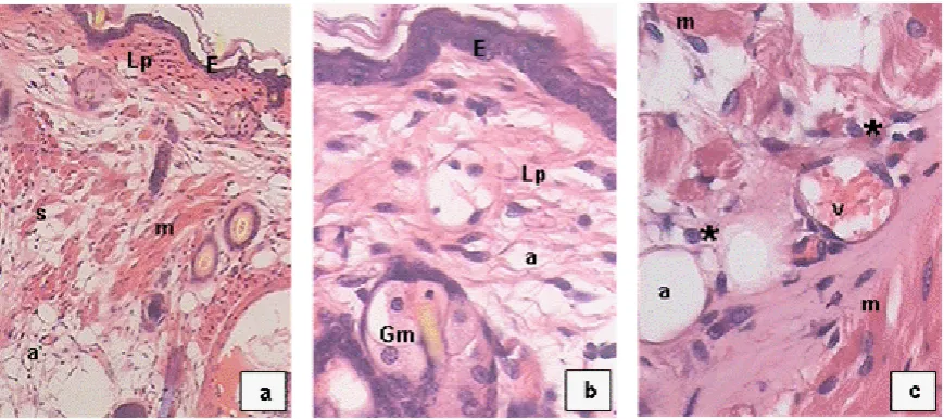
In mice infected with MRSA only, the mucosa presented a keratinized flat stratified epithelium without alterations and a lamina propria with moderate presence of adipose cells. In addition, blood vessels exhibited normal morphology and low presence of leukocyte infiltration (Figs. 2a, 2b and 2c).
Mice infected with MRSA and subsequent administration of the Z. peruviana extract showed significant morphological changes in the nasal mucosa. The stratified epithelium was slightly eroded, and the connective stroma showed a marked increase in the number and size of undifferentiated mesenchymal cells and fibroblasts with alterations in nuclear morphology. The blood vessels showed hypertrophic endothelial nuclei. An important leukocyte infiltrate and clusters of connective cells included within histiocytes were also found. (Figs. 3a, 3b, 3c, 3d and 3e).
Administration of Z. peruviana extracts in the cutaneous infection
Bacteriological study
The joint cutaneous administration of the 30:70% AcOEt: HEX Z. peruviana extract with S. aureus ATCC 43300 in BALB/c mice caused no difference in the evolution of the lesions (size and inflammatory reaction) with respect to mice infected with MRSA only. However, significantly lower counts were found in the homogenates of the skin lesions of mice infected with the MRSA + extract mixture. (**
p = 0, 0076) (Fig. 4).
The study of the internal organs showed the passage of bacteria to the spleen and kidney in the infection by MRSA, obtaining a bacterial result of their homogenates of 20 CFU/organ. There was no passage of bacteria to the internal organs in the mice that received the plant extract. No bacteria were isolated from the blood in any of the experiments.
Histological study
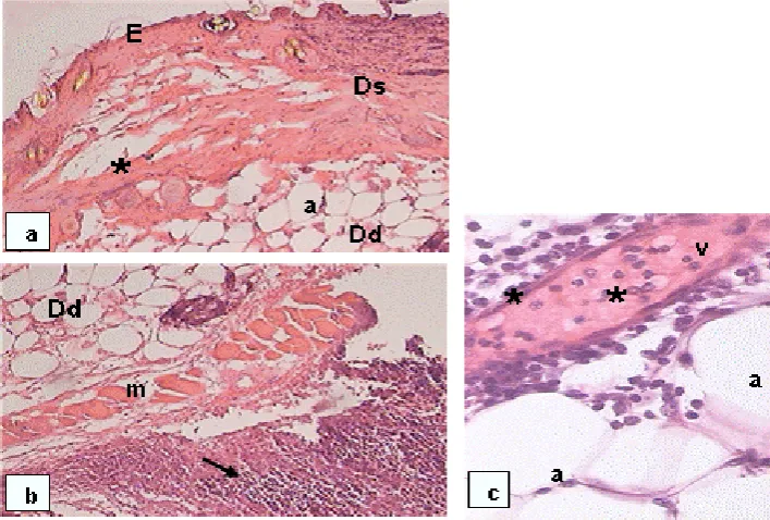
BALB/c mice infected with MRSA showed skin morphological alterations compared with non-infected animals. These alterations included thin epidermis and an important development of
adipose tissue throughout the dermis. In the deep dermis, an important inflammatory infiltrate with predominance of polymorphonuclear cells was found below a large development of adipose tissue. (Figs. 5a, 5b and 5c).
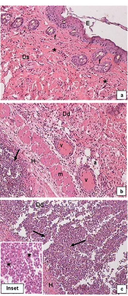
In mice treated with S. aureus ATCC 43300 + Z.
peruviana, histological sections showed better preserved epidermis and dermis. The epidermis presented a stratified epithelium constituted by several cellular layers. In the region of the superficial dermis an organized and compact connective tissue formed by abundant collagen fibers was observed. As opposed to mice infected with MRSA, no adipose cells were found in this group. In the deep dermis and hypodermis a marked inflammatory infiltrate with predominance of polymorphonuclear cells was observed. (Figs. 6a, 6b and 6c).
Discussion
Methicillin-resistant S. aureus involves a serious therapeutic problem, mainly in hospital environments. These microorganisms, which present broad patterns of resistance to antibacterial agents, have been the object of extensive research in recent years in order to find alternative treatments. The use of plant extracts and their active compounds may constitute a significant contribution to the advancement of new therapies in the cure of diseases produced by these microorganisms.
MRSA exhibited a flat keratinized stratified epithelium without alterations and a well-developed lamina propria.
The consumption of medicinal herbs has increased around the world due to their effectiveness, availability and general acceptance. In fact, approximately 80% of the general population, especially in developing countries, uses medicinal herbs for primary health care. For this reason, adverse reactions, possible interactions with other medications and toxicity risks are issues of great importance and interest [15].
Some authors have suggested that plants belonging to the Asteraceae family, a known source of triterpenoids and sesquiterpene lactones present cytotoxic activity, especially Z. peruviana. Fouche et al. observed that extracts of this plant present potent anti-cancer activity against human cell lines of leukemia, melanoma, as well in lung, colon, kidney, ovary and central nervous system cancer [16]. Nambo Camacho demonstrated the cytotoxic effect of Z. peruviana after the exposure of cell lines of biphasic lung cancer to organic extracts from the leaves of this plant, obtaining a high percentage of cell death. To investigate the cytotoxic mechanism, this author evaluated the interference activity on the enzyme topoisomerase I and proved that ethyl acetate extracts affected this enzyme activity [17]. The cytotoxicity of this family of plants was also detected in studies carried out by Bashyal et al., who pointed out that some compounds isolated from
Zinnia grandiflora, such as sesquiterpene lactones, showed a strong cytotoxicity against cancer cell lines of NCI-H460, MCF- 7, SF-268, and MIA Pa Ca-2 and normal human fibroblasts WI-38 [18].
On the other hand, Mattana et al. demonstrated genotoxic activity by 30:70% AcOEt/n-hexane Z. peruviana extract evidenced by the presence of chromosomal aberrations in Allium cepa root cells. They also observed DNA fragmentation by the “Comet” test, which evidenced DNA damage caused by this extract [19].
In this study, the greater infection originated by MRSA after the administration of the extract, demonstrated by an increase of bacterial counts in the nasal homogenates, was probably due to the
toxic effects of Z. peruviana, which were confirmed by the nasal mucosa histological studies.
Although S. aureus counts were higher when the
Z. peruviana extract was administered, no bacterial development was detected either in the blood culture or in lung and spleen. These results show that the infection remained localized.
The isolation and identification of the active principles of Z. peruviana extract would allow performing further in vivo experimental studies of nasal colonization to elucidate the potential antibacterial effect of these compounds.
In BALB/c mice infected with MRSA + Z. peruviana
extract, bacterial counts of the lesion homogenates were significantly lower than those in mice infected with MRSA only. However, no differences between the two mice groups were observed in the evolution and size of the lesions. In addition, no bacterial development was detected in the culture of blood, spleen and kidney in either of the groups. The histological studies showed the constitution of a well structured stratified epithelium with several cellular layers, as well as a superficial dermis that presented an organized and compact packing formed by abundant collagen fibers.
At present, there is little information about the in vitro antibacterial activity of Z. peruviana, and no studies have been found regarding the in vivo
antistaphylococcal activity of this plant. Some authors, such as Amani et al., demonstrated that aqueous and alcoholic extracts of Z. peruviana
showed different degrees of antibacterial activity against Gram-positive and Gram-negative microorganisms [20].
Phytochemical analyses from chromatographic studies of different extracts obtained from Z. peruviana organs (root, stem, leaf and flower) have detected the presence of terpenoid compounds [17]. Other authors have reported the presence of saponins, steroids, flavonoids, phenols and glycosides from different species of this family, such as Zinnia elegans [21]. All these compounds constitute an interesting group of biologically active phytochemicals.
glucuronides, catechol, phenols, among others, have antimicrobial properties [10, 22], which suggests that the presence of some of these active metabolites in Z. peruviana could be responsible, in part or in full, for the in vivo activity demonstrated by the lower bacterial count from the homogenates of the skin lesions observed in this study.
In agreement with our results regarding the skin histological analysis, Mahboubi et al. described a thin epidermis and a dermis with infiltrate of inflammatory cells in the control group of mice infected with MRSA, while in mice treated with herbal cream containing oils from Pelargonium graveolens and Oliveria decumbens, the epidermis and dermis were normal [23]. These authors also demonstrated the formation of a more compact dermis after the administration of medicinal herbs, causing the increase of collagen deposition, one of the main phases in the healing of wounds that is not observed in the natural infection.
It could be postulated that one or some of the active compounds of the Z. peruviana extract, in addition to having antimicrobial properties, may influence the process of collagenization and the healing of the wounds. However, more clinical and toxicological studies are required to demonstrate the efficacy of this extract in vivo.
On the other hand, the histological study of the natural subcutaneous infection showed a great proliferation of adipose tissue throughout the dermis, with numerous and large adipose cells. After administration of the Z. peruviana extract, adipocytes content decreased. In the non infected control mice, no adipose cells were observed.
Several authors have shown that dermal adipocytes contribute directly to the innate immune response after cutaneous infection by S. aureus, causing the proliferation and hypertrophy of mature adipocytes that lead to a large expansion of adipose tissue. As this tissue expands, mature adipocytes produce cathelicidin, an important antimicrobial peptide. Therefore, a local increase in subcutaneous adipocytes is an important host defense response against skin infections, suggesting the role of adipocytes as immunologically active cells [24, 25].
In the present work, although the lesion bacterial counts were lower when the plant extract was
added, a decrease in the adipose cells was also detected. Further studies should be carried out in order to elucidate whether any of the components of this extract was involved in the decrease of adipose tissue.
It is important to note that only in cutaneous infection a beneficial effect was obtained after the administration of Z. peruviana extract, demonstrated by a significant decrease in the bacterial count, and no alterations were detected in the different cells observed in the histological study. Thus, it can be inferred that the differences found both in the bacteriological and the histological studies after the application of the extract of Z. peruviana could be related to the different routes of administration.
These results suggest that ethnomedical drugs should be evaluated according to their route of administration and bioabsorbancy, in order to avoid their use when possible adverse effects, such as toxicity, allergies, and local hypersensitivity, are detected.
Several authors have highlighted the growing resistance of S. aureus to antibiotics and recent studies indicate the need to carry out ethno-medical studies in vivo, specifically related to MRSA infections.
The detection of the anti-staphylococcal activity of this plant species using an infection model in vivo
will contribute to the knowledge of their antibacterial properties and will open a way for future studies in the treatment of infections caused by S. aureus.
Conclusions
The administration of the extract of Z. peruviana
showed a better conserved epidermis and dermis with a greater deposition of collagen.
Acknowledgements
We appreciate language revision by staff from Instituto de Lenguas, UNSL.
Conflict of Interests
The authors declare that there is no conflict of interests regarding the publication of this paper.
References
1. Holtfreter S, Radcliff F, Grumann D, et al. Characterization of a Mouse-Adapted
Staphylococcus aureus Strain. PLoS ONE 2013; 8:1-10.
2. Naber C. Staphylococcus aureus Bacteremia: Epidemiology, Pathophysiology, and Management Strategies. Clin Infect Dis 2009; 48:231-237.
3. Soloaga R, Guelfand L. Diagnóstico
Microbiológico de Enfermedades
Infecciosas: de la teoría al caso clínico, parte 4. Cursos Asociación de Bioquímicos de la ciudad de Buenos Aires. 2016.
4. French G, Munns D, Jenison R, Hicke B. Direct detection of nasal Staphylococcus aureus carriage via helicase-dependent isothermal amplification and chip hybridization. BMC Res Notes. 2012; 5:430.
5. Rodríguez Tamayo E, Jiménez Quiceno J. Factores relacionados con la colonización por Staphylococcus aureus. IATREIA 2015; 28:66-77.
6. Peacock S, de Silva I, Lowy F. What determines nasal carriage of Staphylococcus aureus? Trends Microbiol 2001; 9:605-610.
7. May J. Time-Kill studies of tea tree oils on clinical isolates. J Antimicrob Chemother. 2000; 45:639-643.
8. Martin K, Ernst E. Herbal medicines for treatment of bacterial infections: a review of controlled clinical trials. J Antimicrob Chemother 2003; 51:241-246.
9. Buru A, Pichika M, Neela V, Mohandas K. In vitro antibacterial effects of Cinnamomum
extracts on common bacteria found in wound infections with emphasis on methicillin-resistant Staphylococcus aureus. J Ethnopharmacol 2014; 153:587-595.
10. Salaheen S, Peng M, Joo J, Teramoto H, Biswas D. Eradication and Sensitization of Methicillin Resistant Staphylococcus aureus
to Methicillin with Bioactive Extracts of Berry Pomace. Front Microbiol 2017; 8:253.
11. Barrie F. Report of the General Committee: 11. Taxon 2011; 60:1214.
12. Satorres S, Chiaramello A, Tonn C, Laciar A. Antibacterial activity of organic extracts from Zinnia peruviana (L.) against gram-positive and gram-negative bacteria. Emir J Food Agric 2012; 24:344-347.
13. Barboza G, Cantero J, Núñez C, Pacciaroni A, Espinar L. Medicinal plants: A general review
and a phytochemical and
ethnopharmacological screening of the native Argentine Flora. Kurtziana 2009; 34:7-365.
14. Arciola C, Campoccia D, Speziale P, Montanaro L, Costerton J. Biofilm formation in Staphylococcus implant infections. A review of molecular mechanisms and implications for biofilm-resistant materials. Biomaterials 2012; 33:5967-5982.
15. Alonso-Castro A, Domínguez F, Ruiz-Padilla A, et al. Medicinal Plants from North and Central America and the Caribbean Considered Toxic for Humans: The Other Side of the Coin. Hindawi 2017; 2017:1-28.
16. Fouche G, Khorombi E, Kolesnikova N, Maharaj V, Nthambeleni R, van der Merwe M. Investigation of South African plants for anticancer properties. PhOL 2006; 3:494-500.
18. Bashyal B, McLaughlin S, Gunatilaka A. Zinagrandinolides A-C, Cytotoxic δ-Elemanolide-Type Sesquiterpene Lactones from Zinnia grandiflora. J Nat Prod 2006; 69:1820-1822.
19. Mattana C, Cangiano M, Satorres S, Alcaráz L, Laciar A. Potential Genotoxicity of Zinnia peruviana extract. PhOL 2016; 2:72-80.
20. Amani S, Isla M, Vattuone M, et al. Antimicrobial activities in some Argentine medicinal plants. Acta Horticulturae 1999; 501:115-122.
21. Mohamed A, Ahmed F, Ahmed O. Hepatoprotective and Antioxidant Activity of Zinnia elegans Leaves Ethanolic Extract. IJSR 2015; 6:154-161.
22. Marathe N, Rasane M, Kumar H, et al. In vitro antibacterial activity of
Tabernaemontana alternifolia (Roxb) stem bark aqueous extracts against clinical isolates of methicillin resistant
Staphylococcus aureus. Ann Clin Microbiol Antimicrob 2013; 12:1-8.
23. Mahboubi M, Feizabadi M, Khamechian T, et al. The Effect of Oliveria Decumbens and
Pelargonium Graveolens on Healing of Infected Skin Wounds in Mice. World J Plast Surg 2016; 5:259-264.
24. Zhang L, Guerrero-Juarez C, Hata T, et al. Dermal adipocytes protect against invasive
Staphylococcus aureus skin infection. Science 2015; 347:67-71.
Figure 1. Average count of S. aureus ATCC 43300 obtained from nasal homogenates of BALB/c mice. Influence of Z. peruviana extract on nasal portation. *
p <0.05
Figure 4. Average count of S. aureus ATCC 43300 in BALB/c mice obtained from homogenates of skin lesions. Influence of the extract Z. peruviana on cutaneous infection. ** p<0.01