Scholarship@Western
Scholarship@Western
Electronic Thesis and Dissertation Repository
8-26-2015 12:00 AM
The Development of Cyclic RGD Peptides Stabilized Through
The Development of Cyclic RGD Peptides Stabilized Through
99mTc/Re(CO)3+
99mTc/Re(CO)3+
Aagam Patel
The University of Western Ontario
Supervisor Dr. Leonard Luyt
The University of Western Ontario Graduate Program in Chemistry
A thesis submitted in partial fulfillment of the requirements for the degree in Master of Science © Aagam Patel 2015
Follow this and additional works at: https://ir.lib.uwo.ca/etd
Part of the Inorganic Chemistry Commons, and the Radiochemistry Commons
Recommended Citation Recommended Citation
Patel, Aagam, "The Development of Cyclic RGD Peptides Stabilized Through 99mTc/Re(CO)3+" (2015). Electronic Thesis and Dissertation Repository. 3207.
https://ir.lib.uwo.ca/etd/3207
This Dissertation/Thesis is brought to you for free and open access by Scholarship@Western. It has been accepted for inclusion in Electronic Thesis and Dissertation Repository by an authorized administrator of
Integrated Article
by
Aagam Patel
Graduate Program in Chemistry
A thesis submitted in partial fulfillment of the requirements for the degree of
Master of Science
The School of Graduate and Postdoctoral Studies The University of Western Ontario
London, Ontario, Canada
ii
Abstract
Protein secondary structure can be mimicked by incorporating structural constraints into
peptides and this can be facilitated by metal coordination. The objective of this project is to
establish a chelation system with a cancer targeting peptide sequence, where the coordination
to a metal centre results in a cyclic metallopeptide. Cyclic RGD peptides are antagonists for
αvβ3 and other integrins, which are present during tumour angiogenesis. Arg-Gly-Asp (RGD)
sequence is employed in the peptide backbone, with an un-natural amino acid (3-Pal) and a
chelating molecule (pyridyl-triazole, pyta) present on opposite ends of the peptide sequence
to form a linear peptide sequence, pyta-RGD-3-Pal-NH2. The linear peptide was reacted with
[Re(OH2)3(CO)3]+ to form a cyclic system, with the pyridyl-triazole coordinating in a
bidentate fashion. The linear peptide and resulting cyclic metallopeptide were characterized
by high resolution mass spectrometry (HRMS), Circular Dichroism (CD) spectroscopy, one-
and two-dimensional 1H-NMR spectroscopy, and variable temperature (VT) NMR
spectroscopy, with purity > 90% as determined by HPLC. Computational studies on the
coordinated peptide suggested intra-molecular hydrogen bonding consistent with VT NMR
data. The linear peptide was successfully radiolabelled with Tc-99m demonstrating the
potential application as a SPECT (single photon emission computed tomography) imaging
agent for angiogenesis. This approach to adding a structural constraint, through metal-based
peptide cyclization, results in a metallopeptide where the metal is central to creating the turn
mimetic.
Keywords
Molecular imaging probe, Technetium-99m, metal-based peptide cyclization, cyclic RGD,
iii
Co-Authorship Statement
The computational study presented in Chapter 3 was completed by Dr. Jinqiang Hou, a
postdoctoral fellow in Dr. Leonard Luyt’s Lab.
iv
Acknowledgments
First of all, I would like to thank Dr. Len Luyt for the opportunity to work in his lab. Thanks
for all the support and motivation you provided over the last couple years.
Next, I would to thank Milan for teaching me peptide synthesis and being a great bud, in and
out of the lab. I would also like to thank Neha and Axie for putting up with me last couple
years, and giving helpful insights on the project.
Special thanks to all the lab members, past and present, in the Luyt lab. It’s been a pleasure
knowing and working with you guys. Cheers.
Finally, I would like to thank my family and close friends for all their love and support
v
Table of Contents
Abstract ... ii
Co-Authorship Statement... iii
Acknowledgments... iv
Table of Contents ... v
List of Tables ... viii
List of Figures ... ix
List of Schemes ... xi
List of Appendices ... xii
List of Abbreviations and Symbols... xiii
Chapter 1 ... 1
1 Introduction ... 1
1.1 Molecular Imaging ... 1
1.2 Targeting Entity ... 1
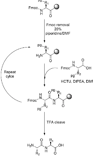
1.3 Solid Phase Peptide Synthesis (SPPS) ... 2
1.4 Structural Constraints... 4
1.5 Metal Induced Structural Constraints ... 6
1.6 Signaling Source ... 8
1.7 Single Photon Emission Computed Tomography (SPECT) ... 9
1.8 Technetium-99m as Signaling Source ... 10
1.9 Using 99mTc/Re(CO)3+ as a “2+1” Chelation System to Induce a Turn Conformation in a Linear Pentapeptide ... 12
1.10Integrin, αVβ3, as a Biological Target ... 13
1.11Thesis Scope ... 17
1.12 References ... 17
vi
Using 99mTc/Re(CO)3+ ... 23
2.1 Introduction ... 23
2.2 Results and Discussion ... 26
2.2.1 Synthesis of [Re(CO)3(OH2)3]OTf 12 ... 26
2.2.2 Synthesis of Ac-HRGDH-OH... 26
2.2.3 Characterization of Ac-HRGDH-OH ... 28
2.2.4 “2+1” Coordination Using Natural Amino Acids as Metal Chelators ... 29
2.2.5 “2+1” Mixed Coordination with an External Mono-Dentate Chelator ... 32
2.2.6 “2+1” Coordination with Un-natural Amino Acids as Metal Chelators ... 33
2.3 Conclusion ... 37
2.4 Experimental ... 37
2.4.1 Small Molecule Synthesis ... 38
2.4.2 Peptide Synthesis ... 38
2.4.3 Re(CO)3+ Coordination ... 41
2.5 References ... 42
Chapter 3 ... 45
3 A Turn Induced by 99mTc/Re (I) Tricarbonyl Coordination to Form Cyclic Metallopeptides ... 45
3.1 Introduction ... 45
3.2 Results and Discussion ... 46
3.2.1 Strategy ... 46
3.2.2 Peptide Synthesis ... 47
3.2.3 1H-NMR Analysis of pyta-RGD-3-Pal-NH2 ... 48
3.2.4 Re(CO)3+ Coordination ... 49
3.2.5 Characterization of [Re(CO)3(pyta-RGD-3-Pal-NH2)]OTf ... 50
vii
3.2.8 Computational Study ... 55
3.2.9 99mTc Labelling ... 56
3.2.10 Derivatives of [Re(CO)3(pyta-RGD-3-Pal-NH2)]+ ... 57
3.3 Conclusion ... 58
3.4 Experimental ... 58
3.4.1 Small Molecule Synthesis ... 59
3.4.2 Peptide Synthesis ... 60
3.4.3 Metal Coordination ... 62
3.4.4 Circular Dichroism (CD) spectroscopy ... 64
3.4.5 Computational Studies ... 64
3.5 References ... 65
Chapter 4 ... 69
4 Conclusion ... 69
4.1 References ... 71
Appendices ... 73
Appendix A: Chromatograms of selected compounds ... 73
Appendix B: 1H-NMR spectra of linear and Re(CO)3+ coordinated peptides ... 79
Appendix C: Computational study on Re(CO)3+, 3.5 ... 95
viii
List of Tables
Table 2.1 Calculated and observed m/z ratio of Ac-HRGDH-OH, 2.4. ... 27
Table 2.2 Reaction conditions used to coordinate Ac-HRGDH-OH with [Re(CO)3(OH2)3]+.30
Table 2.3 Increasing molar equivalent of pyridine led higher % of
[Re(CO)3(Ac-HRGDH-NH2)(py)]+. ... 33
Table 2.4 Number of dominant isomers displayed upon coordination with Re(CO)3+, for
2/3-Pal-RGD-3/4-Pal series ... 35
Table 3.1 Amino acids’ proton shifts (ppm) of the linear peptide, pyta-RGD-3-Pal-NH2 ... 49
Table 3.2 Amino acids’ proton chemical shifts (ppm) of the coordinated peptide,
pyta-RGD-3Pal-NH2-Re(CO)3+. ... 50
Table 3.3 Chemical shifts of amide protons expressed in numerical values, Δδ/ΔT (ppb/K)
ix
List of Figures
Figure 1.1 Schematic for a molecular imaging probe. ... 1
Figure 1.2 Solid phase peptide synthesis (SPPS) employing 9-fluorenylmethoxycarbonyl (Fmoc) strategy ... 3
Figure 1.3 Structural constraints introduced through different bond types; cilengitide, cyclization through amide bond, and octreotide, cyclization introduced by di-sulfide bonds . 6 Figure 1.4 Dominant isomer of Ac-HAAAH-NH2 coordinated with Pd2+, through N1 nitrogen atoms on both imidazole rings.18 ... 8
Figure 1.5 Decay scheme of 99Mo to 99Ru ... 10
Figure 1.6 Coordination of Ac-HAAAH with Re(CO)3+ through N1 and N3 nitrogen atoms of the imidazole rings, and carboxyl oxygen at the C-terminus.31... 13
Figure 1.7 Simple schematic of integrin consisting of α and β subunits. ... 14
Figure 1.8 Integrins exhibiting binding towards different membrane proteins.36... 15
Figure 1.9 Interactions between cyclic RGD peptide and integrin, αvβ3. The cyclic peptide (yellow) making important interactions (dashed lines) with the amino acid residues of αv (blue) and β3 (red) subunits.39 ... 15
Figure 1.10 Project design rationale ... 17
Figure 2.1 Schematic of pendant and integrated technetium-99m radiopharmaceutical designs ... 24
Figure 2.2 Model “2+1” chelation system ... 27
Figure 2.3 Linear Ac-HRGDH-OH peptide... 27
Figure 2.4 1H-NMR spectrum of Ac-HRGDH-OH in CD3OD. ... 28
x
Figure 2.7 Bidentate coordination at the C-terminal or N-terminal when using histidine
residue as a metal chelator ... 34
Figure 2.8 Bidentate coordination at the N-terminus using un-natural amino acids as a metal
chelators ... 35
Figure 2.9 LC-MS traces of 3Pal-RGD-3Pal-OH crude mixture and of an isolated isomer .. 37
Figure 3.1 Structures of pyridyl-triazole (pyta) and 3-pyridylalanine (3-Pal)………... 51
Figure 3.2 Partial 1H-NMR spectra (600 MHz, DMSO-d6, 25 oC) of the linear peptide and
coordinated peptide ... 51
Figure 3.3 Partial 1H-NMR spectra (600 MHz, DMSO-d6) of the linear peptide (top) and
coordinated peptide, [Re(CO)3(pyta-RGD-3Pal-NH2)]OTf (below). ... 52
Figure 3.4 Circular Dichroism (CD) analysis of linear peptide and Re(CO)3+ coordinated
peptide in H2O at 25 oC. ... 53
Figure 3.5 VT NMR analysis of the rhenium coordinated peptide in DMSO-d6 at 600 MHz.
... 54
Figure 3.6 Optimized structure of Re-coordinated peptide, 3.5, displaying intra-molecular
hydrogen bonding. ... 56
Figure 3.7 U-HPLC analysis showing correlations between γ trace of 99mTc(CO)3+ labelled
peptide and UV trance of Re(CO)3+ coordinated peptide. ... 57
xi
List of Schemes
Scheme 2.1 Synthetic route for [Re(OH2)3(CO)3]OTf, as 0.1 M solution. ... 7
Scheme 2.2 Synthetic approach for Ac-HRGDH-OH coordination with [Re(CO)3(OH2)3]+ and proposed coordination product. ... 29
Scheme 2.3 Synthetic approach towards mixed “2+1” Re(CO)3+ complexes, and a porposed coordination product ... 32
Scheme 2.4 Synthetic approach for Ac-3-Pal-RGD-3-Pal-OH coordination with [Re(CO)3(OH2)3]+, and proposed coordination structure ... 36
Scheme 3.1 Synthetic route for pyridyl-triazole (pyta) ... 47
Scheme 3.2 Synthesis of pyta-RGD-3Pal-NH2 employing SPPS ... 47
Scheme 3.3 Re(CO)3+ coordination of pyta-RGD-3-Pal-NH2 ... 49
xii
List of Appendices
Appendix A: Chromatograms ………..70
Appendix B: 1H- and COSY NMR spectra of linear and Re(CO)3+ coordinated peptides ... 77
xiii
List of Abbreviations and Symbols
2D: two-dimensional
2-Pal: 2-pyridylalanine
3-Pal: 3-pyridylalanine
4-Pal: 4-pyridylalanine
Ac: acetyl
Boc:tert-butoxycarbonyl
CD: circular dichroism
CT: computed tomography
CD3OD: deuterated methanol
DCM: dichloromethane
DIPEA:N,N-diisopropylethylamine
DMF:N,N-dimethylformamide
DMSO-d6: deuturated dimethyl sulfoxide
ECM: extracellular matrix
en: ethylenediamine
ESI: electrospray ionization
Fmoc: 9-fluorenylmethoxycarbonyl
gCOSY: correlation spectroscopy
xiv
hexafluorophosphate)
His: histidine
HPLC: high performance liquid chromatography
IC50: half-maximal inhibitory concentration
K: Kelvin
Kd: dissociation constant
keV: kilo electron volt
LC: liquid chromatography
M: molar
MIDAS: metal-ion dependent adhesion site
MRI: magnetic resonance imaging
MS: mass spectroscopy
NaOH: sodium hydroxide
nM: nano molar
NMR: nuclear magnetic resonance
OtBu: tert-butyl ester
OTf: trifluoromethanesulfonate
Pbf: 2,2,4,6,7-Pentamethyldihydrobenzofuran-5-sulfonyl
PBS: phosphate buffer saline
xv ppb: parts per billion
Py: pyridine
pyta: pyridyl-triazole
RGD: arginine-glycine-aspartic acid
ROE: rotating frame overhauser enhancement
ROSEY: rotating frame nuclear overhauser effect
RP: reverse phase
SPECT: single photon emission computed tomography
SPPS: solid phase peptide synthesis
TBME: tert-butyl methyl ether
TIPS: triisopropylsilane
TFA: trifluoroacetic acid
Trt: triphenylmethane
TSTU: (O-(N-succinimidyl)-1,1,3,3-tetramethyl uranium tetrafluoroborate)
US: ultrasound
Chapter 1
1
Introduction
1.1
Molecular Imaging
The methods used to evaluate and visualize biological events have changed over the
years. Different imaging techniques have been introduced that provide insight into the
biological processes taking place within our body. Molecular imaging is one such
technique and can be defined as the non-invasive characterization and visualization of
biological events taking place at the cellular and molecular level.1,2 Molecular imaging
allows diseased cells and normal tissue cells to be differentiated, by targeting those
biological receptors that are overexpressed in diseased cells, while present at low
concentrations in the normal tissue cells. Several factors are taken into account when
designing a molecular imaging probe (Figure 1.1), which include: a) an appropriate
biological target, b) a targeting entity with high affinity for the chosen biological target; a
good binding target would have a dissociation constant (Kd) in nM range, and c) an
appropriate signaling source that allows the target to be detected by an external imaging
modality.1
Figure 1.1 Schematic for a molecular imaging probe.
1.2
Targeting Entity
There are a number of different kinds of targeting entities that exist, ranging from small
have increasingly gained attention as a targeting component, as they have a number of
desirable properties.2,3 The advantages of using peptides as a targeting entity include: 1)
they can be readily synthesized and the monomeric amino acid residues can be
chemically modified; 2) there are many biological receptors that display good binding
affinity towards small peptides; 3) their low molecular weight and small size allow them
to penetrate tissue and tumours, as well as to be removed from the plasma and body
relatively quickly; 4) a variety of bi-functional ligands can be attached at a peptide’s
C-terminus and N-C-terminus; and 5) peptides display good tumour-background ratios.2-4 In
addition, small molecule drugs, and imaging molecules can be attached to peptides for a
variety of applications.5
The use of peptides as targeting entities also suffer from a number of disadvantages.
Firstly, the in vivo stability of short linear peptides is a major concern, as they readily
undergo degradation by proteases and peptidases.3, 6 Secondly, the flexible nature of
linear peptides allows them to bind to multiple receptors, which reduces their specific
binding towards the biological target.3, 6 Furthermore, the addition of an imaging source
can affect the peptide conformation and reduce the specific binding of the peptide.1
Despite these difficulties, structural constraints, such as through cyclization of the peptide
backbone, can resolve some of the difficulties seen with short linear peptides.
1.3
Solid Phase Peptide Synthesis (SPPS)
Peptides can be synthesized either in solution-phase or on a solid-phase support.
Solution-phase peptide synthesis is an efficient method; however, isolation, purification
and characterization steps are required after the addition of each amino acid, making this
a rather expensive and time consuming technique. On the other hand, solid phase peptide
synthesis (SPPS), introduced by Merrifield in 19637, is more efficient to construct large
and complex peptide sequences. With this technique, a peptide is constructed in a
stepwise manner, using an insoluble resin that fixes, the C-terminus of the peptide to the
resin. This allows the use of excess reagents, in order to ensure that the addition of each
amino acid proceeds to completion. When using this technique, the reagents and
filtering without affecting the peptide which remains attached to the resin. As a result,
this eliminates the need for a purification step after each amino acid coupling.
Figure 1.2 Solid phase peptide synthesis (SPPS) employing 9-fluorenylmethoxycarbonyl
(Fmoc) strategy
In SPPS, the peptide is constructed from the terminus to the N-terminus, with the
C-terminus attached to an insoluble resin. There are two common strategies involved with
SPPS based upon the protecting group being used, 9-fluorenylmethoxycarbonyl (Fmoc)
and tert-butoxycarbonyl (Boc). These strategies both involve orthogonal protecting
groups, which allows only the N-terminal protecting group of the peptide to be
selectively removed for the addition of the next amino acid, and the other amino acids
already coupled to the resin to remain protected. In the Fmoc strategy (Figure 1.2), the
-dimethylforamide (DMF). On the other hand, the resin, and the protecting groups on the
side chains of amino acid residues are acid labile. Once the Fmoc group is removed, the
next amino acid in the sequence is added in excess, in addition to the coupling reagent,
2-(6-chloro-1H-benzotriazole-1-yl)-1,1,3,3-tetramethylaminium hexafluorophosphate
(HCTU) and a base, N,N-diisopropylethylamine (DIPEA) in DMF. This cycle repeats
until the desired peptide is constructed. The peptide is cleaved off of the resin, and all of
the protecting groups are removed by vortexing the resin in an acidic solution containing
a carbocation scavenger, triisopropylsilane (TIPS). In suitable acidic conditions,
trialkylsilanes can act as hydrogen donors, and de-block the protecting groups from
additional reactions with the peptide.8 This results in a linear peptide that is purified with
reverse phase high performance liquid chromatography (RP-HPLC) and characterized by
mass spectroscopy (MS).
1.4
Structural Constraints
The exploration of peptide geometry has been of major interest in peptide chemistry.
Principal interest is the introduction of structural constraints within the peptide backbone,
in order to create effective probes for targeting biological receptors. Cyclized peptides are
to have a more rigid geometry, and therefore, when present in favorable conformations,
would result in better binding and selectivity towards the targeted receptors.5 Cyclization
of peptides results in reduced molecular conformations in solution as compared to their
linear counterparts. When these constrained peptides are used as targeting entities, the
limited number of conformations results in less competition for targeting biological
receptors. On the other hand, linear peptides undergo numerous conformations in
solution, and these conformations are present in very fast equilibrium. As a result, the
specific binding towards the targeted receptor is reduced, as different conformations
compete for interaction with the biological target.5, 9
Numerous examples have been presented, where introducing a structural constraint
results in better binding towards the targeted receptor. Kumar et al. reported cyclic
analogues of linear peptide sequence, Ac-CIYKYY, as an inhibitory agent against
phosphorylating proteins at the tyrosine site, and this inhibition slows down the
downstream signal transduction. This mechanism is used in an attempt to treat tumours.
Kumar et al. compared the linear peptide with cyclic heat-to-tail peptide analogues, and
found that the cyclic peptide (IC50 = 6.4 µM) had an inhibitory effect, 62.5-fold higher
than that of the linear peptide (IC50 = 400 µM), which is believed to be due to the
introduction of a structural constraint within the peptide backbone. They further reported
eight other cyclic derivatives of the same linear peptide sequence, using different
strategies to cyclize the peptide. Seven out of eight derivatives displayed better potency
against tyrosine kinase than the linear sequence.
Cyclization can be introduced using different strategies, such as head-to-tail cyclization,
and side-chain to side-chain cyclization. Cyclization can be presented by different bond
types, such as the formation of amide bond, disulfide bridges, metal coordination, and
several others. An example of where cyclization was introduced through amide bond
formation is cilengitide (Figure 1.3), a pentapetide cyclized from head-to-tail by the
formation of an amide bond.11 Cilengitide, c(RGDf(N-Me)V), contains a tripeptide
sequence, Arg-Gly-Asp (RGD), and is an antagonist for integrin, αvβ3. Numerous RGD peptides have shown good binding towards integrin, αvβ3. Vitronectin competitive assays displayed cilengitide (IC50 = 0.58 nM) to have 360-fold better binding towards αvβ3 than
the linear peptide sequence, GRGDSPK (IC50 = 0.58 µM).12Octreotide is an example
where cyclization is introduced through disulfide bonds, formed through side chain
interactions of cysteine residues. Octreotide is a synthetic octapeptide that mimics
somatostatin, a naturally occurring hormone; it binds to the somatostatin receptor. Both
linear and cyclic octreotide displayed binding towards somatostatin receptors; however,
Figure 1.3 Structural constraints introduced through different bond types; cilengitide,
cyclization through amide bond, and octreotide, cyclization introduced by di-sulfide
bonds
Cyclization has shown improved in-vivo stability of peptides. Pakkala et al. compared the
stability of linear and cyclic peptides targeting human glandular kallikrein (KLK2) with
the sequence GAARFKVWWAAG (KLK2b).5,14 The KLK2 protein is a highly specific
prostate serine protease that is overexpressed in aggressive prostate tumors. Targeting
this protein serves a potential therapeutic to reduce the metastasis of prostate cancer. As
mentioned earlier, the in-vivo stability of short linear peptides is a major concern, as they
are readily broken down through enzymatic degradation. Here, the stability of linear and
head-to-tail cyclized KLK2b peptides in trypsin and human plasma was compared.
Trypsin behaves as a protease, and cleaves amide bond following lysine or arginine
residues.The linear KLK2b peptide completely degraded after 30 minutes in trypsin;
however, only 57% of head-to-tail cyclized peptide degraded after 4 hours, according to
HPLC analysis. In addition, the linear peptide completely degraded after 30 minutes in
human plasma, while, the cyclized peptide was still intact after 24 hours. This study
demonstrated that stability of peptides can be achieved through head-to-tail amide
cyclization.
1.5
Metal Induced Structural Constraints
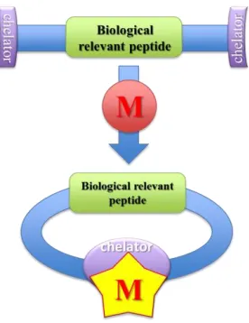
Cyclization can also be introduced through metal coordination. Metals have been shown
to exhibit structural constraints when introduced within the peptide sequences. This is
commonly accomplished by developing a metal clip that will coordinate to a metal. In
can be induced within the peptide backbone by metal coordination where the metal acts
as a central core around which a turn occurs. This is demonstrated by incorporating metal
chelators or metal chelating amino acids within the peptide backbone. A turn is an
element of secondary structure, and occurs when the peptide backbone that is in a random
coil sharply bends on itself, separated by a minimum of two amino acid residues. A turn
can be characterized based on the number of amino acids that are separating the two ends
of the cyclized peptide. Various types of turns include γ (i, i±2), β (i, i±3), α (i, i±4), α
helix (i, i±3.6); i represents the amino acid where the turn is created.17,18
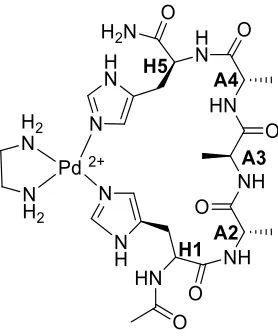
Hoang et al., demonstrated helicity within the peptide backbone upon metal
coordination.19 In their study, a linear peptide, in random coil, was cyclized and a turn
was induced upon metal coordination. The natural amino acid, histidine, was used to
coordinate with the metal center. The study reported that cyclization of linear
pentapeptide, Ac-HAAAH-NH2, through Pd(en)2+ core being coordinated through
nitrogen atoms of the imidazole ring on histidine residues, present at the opposite ends of
the pentapeptide. Three linkage isomers were observed when Ac-HAAAH-NH2 was
coordinated with Pd2+,which differed in the pair of imidazole nitrogen atoms bound to
the metal. The dominant linkage isomer was a 22-membered macrocyclic metallopeptide,
which had palladium (II) bound to the peptide through two N1 atoms of the imidazole
rings (Figure 1.4). From the ROSEY NMR spectra, ROE correlations were observed for
hydrogens of the imidazole rings on His1 and His5, which suggested that they were
present in close vicinity, and that Pd2+ was bound through the imidazole nitrogen atoms
of one peptide, instead of acting as a bridging metal for two different peptides. An upfield
chemical shift in the 1H-NMR spectra of CHα is characteristic for the presence of α
helicity in the peptide backbone.20 This upfield chemical shift was observed for all CHα
atoms present in the coordinated peptide backbone, which may suggest the presence of α
helicity. The Pd2+ complexes were able to establish a reverse turn through cyclization,
Figure 1.4 Dominant isomer of Ac-HAAAH-NH2 coordinated with Pd2+, through N1
nitrogen atoms on both imidazole rings.19
Albrecht et al. demonstrated activation of a biological relevant linear peptide sequence
upon metal coordination.21 They introduced a metal chelator, catechol, at the opposite
ends of a tripeptide sequence, Trp-Ala-Val. This tri-peptide sequence is the backbone of
Segetalins, cyclic peptides that possess estrogen-like activity.22 Upon coordination with
molybdenum, a turn was created, giving the tri-peptide sequence a conformational lock,
and making the linear tri-peptide sequence biologically relevant. This concept can be
used to develop imaging probes that employ metal clipped peptides whose biological
activity is activated, upon coordinating with an appropriate radiometal signaling source.
1.6
Signaling Source
The signaling source is an integral component of an imaging probe. It provides energy
that is detected by imaging modalities. More commonly used imaging modalities include
computed tomography (CT), magnetic resonance imaging (MRI), positron emission
tomography (PET), single photon emission computed tomography (SPECT), and ultra
sound (US). PET and SPECT imaging employ radioactive materials as their signaling
source. PET and SPECT share similar techniques that are based on the radiotracer
principle, which states that biological events are monitored by the movement of a
radiotracer circulating within the body. However, the pathway in which radiation is
In both PET and SPECT imaging the radiotracer is introduced within body fluids and is
allowed to circulate until it binds to the target receptor. In both types of imaging, γ-rays
are produced and detected by gamma cameras. The gamma camera most commonly
contains sodium iodide (NaI) crystals with small amount of thallium that converts the
γ-rays to photons. These photons are then captured by the photon multiplier (PM) tube,
which converts the photons to an electronic signal. This electronic signal is then
amplified and converted to a digital signal, which produces an image.23 PET and SPECT
isotopes are produced either by a cyclotron or a generator.
PET differs from SPECT in the way that γ-rays are emitted and subsequently detected by
gamma cameras. For example, PET isotopes, which include 18F, 68Ga, 64Cu, undergo β+
decay by emitting a positron. The positron travels a short distance before coming in
contact with an electron. This results in an annihilation event that produces two gamma
rays of 511 keV each, which travel in opposite directions (180o apart). These gamma rays
are detected by gamma cameras that are placed 360o around the body. In addition, the
life of PET and SPECT isotopes are different, PET radionuclides typically have
half-lives ranging from minutes to hours, while those of SPECT isotopes range from hours to
days.
1.7
Single Photon Emission Computed Tomography
(SPECT)
SPECT imaging employs commonly used radionuclides such as 99mTc, 111In, and 123I; all
radioisotopes decay by releasing γ-rays with different energies. The energies released
from SPECT radiotracers usually range from 100 – 300 KeV, which is strong enough to
penetrate the skin without harming other tissues. SPECT radiotracers releases a single
γ-ray that is detected by a gamma camera that circulates the whole body. SPECT
radionuclides are less expensive, low energy, and readily available. They are ideally
produced by on-site generators. SPECT radionuclides have longer half-lives ranging from
6 hours to 2.8 days, allowing for multiple syntheses, purification, and imaging of the
1.8
Technetium-99m as Signaling Source
Technetium-99m has been one of the more widely used radioactive metals to label
biomolecules. It is used as a signaling source for more than 70% of nuclear medicine
procedures.24 Technetium-99m has many desirable properties, some of which include: i)
easy availability, ii) low cost, iii) easy handling, iv), good imaging characteristics, and v)
absence of strong radiation such as α and β decay.399mTc is an artificial element that is
generated by a 99Mo/99mTc generator, in which 99Mo decays to 99mTc by β- decay. 99mTc is
a nuclear isomer of 99Tc present in a metastable state, which decays to 99Tc by emitting a
γ ray with an energy equal to 140 keV (Figure 1.5). The γ emission can be detected by
Single Photon Emission Computer Tomography (SPECT). 99mTc has a half-life of 6
hours, which is suitable for radiolabeling biomolecules, as well as acquiring images.
Figure 1.5 Decay scheme of 99Mo to 99Ru
The 99mTc metal can exist in various oxidation states; for example a +V oxidation state of
99mTc is observed in [99mTc=O]3+ and [99mTcN]2+, a +III oxidation state in 99mTc(III)
complexes, and a +I oxidation state in [99mTc(CO)3]+ complexes. All of these oxidation
states are the reduced form of [99mTcO4]-, where the oxidation state of 99mTc is +VII,
which is the form when eluted from the 99Mo/99mTc generator. The [99mTc(CO)3]+
three mono-dentate ligands. A ‘2+1’ chelation system is the combination of bi-dentate
and mono-dentate ligands attached to a metal.
The +1 oxidation state of 99mTc/Re(I) octahedral complexes, results in the d6 electrons
occupying degenerate t2g orbitals (dxy, dyz, and dxz) leaving the doubly degenerate eg
orbitals (dx2-y2, dz2) empty.25 Tc/Re (I) are usually referred to as a soft metals due to the
low charge to size ratio and their preference for soft ligands, because soft-soft
interactions are considered to be stronger than soft-hard interactions.
The 99mTc(CO)3+ core is a versatile building block for a variety of biomolecules. The
carbonyl groups are known to stabilize metals with low oxidation states, such as
[Mn(CO)6]+.25 According to the molecular orbital energy diagram of a carbonyl group,
the highest occupied molecular orbital (HOMO) is a 3σ orbital and lowest unoccupied
molecular orbitals (LUMO) are antibonding π (π*). The metal-carbonyl bond involves the
HOMO (3σ) participating in a sigma bond, whereas the LUMO (π*) form π bonds with
the metal center.25 The π bonding is achieved by donation of electrons from occupied d
orbitals of the metal to doubly degenerate empty π* orbitals of the carbonyl group; this
lowers the electron density on the metal center while increasing it on the carbonyl group.
Increase in the electron density helps stabilize the carbonyl group, and in return enhances
the sigma donation to the metal center.25 The π bonding can also be referred to as
π-backbonding.
The [99mTc(CO)3(OH2)3]+ complex is a convenient starting material for labeling various
biomolecules. Previously, the complex [99mTc(CO)3(OH2)3]+ was synthesized from
aqueous [99mTcO4]- in the presence of gaseous carbon monoxide, CO, and borohydride,
BH4-, as a reducing agent.26 However, the use of carbon monoxide is unsuitable for
developing radiopharmaceutical ‘isolink’ kits. Each ‘isolink’ kit contains a lyophilized
mixture of sodium carbonate, disodium boranocarbonate, sodium tartrate, and sodium
tetraborate.27 The compound K2[H3BCO2] has been reported to release CO in aqueous
solution at high temperatures.28,29 Alberto et al. employed [H3BCO2]2-, as a reducing
agent and a potential source for CO, to form a water soluble and water stable
an appropriate buffer solution and a complexing agent (sodium tartrate), for stabilizing
the intermediate complexes. Pitchumony et al. reported microwave assisted synthesis of
[99mTc(CO)3(OH2)3]+, with reduced reaction time to 3.5 minutes at 110 oC.31 According to
the reaction conditions described above, an octahedral complex was formed with the
carbonyls trans to the aqua ligands to provide a facial orientation. The aqua ligands can
be readily replaced with incoming biomolecules.
The 99mTc chemistry is performed at a microgram scale. To explore the coordination
chemistry, rhenium (185/187Re) is used as a non-radioactive analogue. Rhenium follows
technetium in Group VII of the periodic table. Elements in the same group share similar
physical and chemical characteristics. Thus, rhenium is a suitable metal to explore
coordination chemistry for radioactive technetium.
The 99mTc/Re(CO)3+ octahedral complexes commonly exists as facial isomers; however, a
report on meridional isomer has been published.32 Due to the strong bonding between the
metal center and the carbonyl groups, the trans aqua ligands are very labile and can be
easily substituted with incoming biomolecules. Electron density from nitrogen, oxygen,
and sulfur atoms of various ligands, can be donated to the empty d orbitals of
99mTc/Re(CO)3+ metal core, resulting in formation of σ bonds.
1.9
Using
99mTc/Re(CO)
3+as a “2+1” Chelation
System to Induce a Turn Conformation in a Linear
Pentapeptide
The 99mTc/Re(CO)3+ core has not been reported to create and stabilize a turn
conformation within a biologically relevant peptide. However, in 2014, Simpson reported
coordination of Ac-HAAAH with the Tc/Re(CO)3+ core to create a turn conformation.33
The 99mTc/Re (I) core was coordinated through imidazole nitrogen atoms of histidine
residues present at opposite ends of the peptide. This was based on a similar approach
employed by Hoang et al.19 The facially oriented octahedral complex was cyclized
through the oxygen atom of the carboxyl group, at the C-terminus. However, instead of
reported by Hoang et al.19, the peptide was coordinated through N1 and N3 nitrogen
atoms (Figure 1.6). The metallopeptide complex resulted in a 21 membered macrocycle,
and a small 7 membered ring. The peptide can be referred to as a “2+1” chelation system,
where a bidentate attack from imidazole nitrogen and carboxyl oxygen is observed at one
end, and a monodentate attack of imidazole nitrogen atom is observed on the other end.
Hydrogen bonding in the peptide backbone was reported by variable temperature (VT)
NMR and computational studies. Four hydrogen bonds were observed within the peptide
backbone. The radiolabeled peptide, with 99mTc(CO)3+, resulted in the identical dominant
isomer when coordination was demonstrated with Re(CO)3+.
Figure 1.6 Coordination of Ac-HAAAH with Re(CO)3+ through N1 and N3 nitrogen
atoms of the imidazole rings, and carboxyl oxygen at the C-terminus.33
1.10
Integrin α
Vβ
3, as a Biological Target
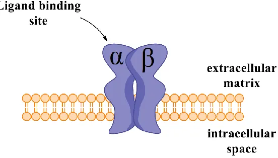
Integrins are heterodimeric type I transmembrane proteins that mediate cell and
cell-extracellular matrix (ECM) interactions. Integrin signaling regulates cell shape, cell
cycle, and cell migration.34 This is achieved by one of two ways: ‘outside-in signaling’
and ‘inside-out signaling’. For “inside-out signaling” to occur, an intracellular activator binds to the β subunit of the integrin, resulting in a conformational change, and increasing the binding affinity for the extracellular proteins. On the other hand, ‘outside-in
signaling’ involves a ligand binding to the integrin at the extracellular domain, resulting
in a conformational chance of the integrin structure. This subsequently promotes
cell polarity, cytoskeletal structure, and cell survival, while ‘inside-out signaling’ results
in cell invasion and migration. The detached cells result in apoptosis through a series of
events.35 Preventing the binding of extracellular membrane proteins to integrins leads to
cell death and suppresses the aggression of tumour such as metastasis and angiogenesis.36
The development of antagonists that prevent the binding of ECM proteins can serve as a
potential therapeutic for tumour suppression. Besides assessing integrins as therapeutic
targets, they also make good imaging biomarkers. The targeting of integrins allows for
the evaluation anti-angiogenic and anti-therapeutic agents, and also preliminary diagnosis
of cancer.34
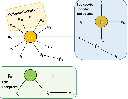
Figure 1.7 Simple schematic of integrin consisting of α and β subunits.
There exist 18α and 8β subunits that are non-covalently bonded to make up 24 different pairs of integrins. α and β subunits are non-homologous in structure, but similarities
within the subunits have been observed. Integrins are composed of three major units:
large extracellular domain, single spanning transmembrane domain, and short
cytoplasmic domain.
Integrins αvβ3, α5β1 and αvβ6 are present at low concentration in normal tissues, but are
overexpressed in tumour tissues. They are known to play an important role in tumour
angiogenesis, making them an attractive target for tumour imaging.34 Some of the ECM
proteins that bind to integrins include fibronectin, fibrinogen, vitronectin, collagens,
osteopontin, etc. They all share the same cellular recognition site, a tripeptide sequence,
towards the tripeptide sequence RGD (Figure 1.8). αvβ3 is a well-studied integrin; many
antagonists, such as cilengitide, and imagining probes, have been developed to target the
receptor. It is known to be overexpressed at multiple tumour sites, including prostate,
breast, ovarian, cervical, pancreatic, melanoma, and glioblastoma tumours.
Figure 1.8 Integrins exhibiting binding towards different membrane proteins.38
Xiong et al., published the first crystal structure of Arg-Gly-Asp cyclic ligand bonded to
the extracellular domain of the integrin αvβ3 (Figure 1.9).39 Interesting characteristics
about the tri-peptide sequence were observed. The side chains of the amino acids play an
important role in the binding of the RGD ligand to the intergrin. The side chain of
arginine, which contains a guanidinium group, was held together by a bidentate salt
bridge formed with Asp218 and Asp150. The arginine interaction occurs at the α subunit of
the extracellular domain. The glycine residue occupies the space between α and β
subunits. It makes numerous important hydrophobic interactions, including hydrophobic
interactions with the carbonyl oxygen of Arg216, of the α subunit. The side chain of
aspartic acid, a carboxylate group, makes vital interactions with amino acids of the β
subunit. One of the oxygen atoms of the carboxylate group forms hydrogen bonds with
amides of Tyr122 and Asn215. The other oxygen correlates with Mn2+ at the metal-ion
dependent adhesion site (MIDAS). The side chain of aspartic acid also makes
hydrophobic interaction with the beta carbon atom of Asn215. Thus, all three amino acids
Figure 1.9 Interactions between cyclic RGD peptide and integrin, αvβ3. The cyclic
peptide (yellow) making important interactions (dashed lines) with the amino acid
residues of αv (blue) and β3 (red) subunits (image adapted from Xiong et al.39)
Several linear and cyclic radiolabeled RGD peptides have been published for both
SPECT and PET imaging modalities.40,41 A 99mTc labelled linear peptide αP2
(RGDSCRGDSY) has been used to image metastatic melanoma in cancer patients.41,42
However, the lack of specificity, low binding affinity, and rapid degradation makes the
linear peptide less suitable for tumor imaging.41,43 On the other hand, cyclic RGD
peptides have shown to enhance binding affinity and improve stability. Examples of
cyclic radiotracers for PET imaging include, [18F]Galacto-RGD and
[18F]AH111585.40,44,45,46 Both cyclic RGD peptides are currently being evaluated as
non-invasive imaging agents for αvβ3 in cancer patients.40 The high cost, low tumor uptake,
and lack of preparative method for repetitive radio-synthesis, will limit the use of these
peptides as imaging entities.40 A 99mTc labelled cyclic peptide, 99mTc-NC100692,
containing RGD in the backbone, has displayed good binding towards integrin αvβ3. It
has also been used to identify malignant lesions in patients with breast cancer.40,47
is rapidly excreted from the body due to the lipophilic nature of the 99mTc chelator.40
Hence, there is a continuous need to develop new techniques for the designing
radiopharmaceuticals that allows for both quick and efficient ways to introduce
radionuclides, as well as to improve in-vivo stability and targeting capability.
1.11
Thesis Scope
Figure 1.10 Project design rationale
Prior work has demonstrated that 99mTc/Re(CO)3+ is able to establish a turn within a
linear peptide sequence. This results in a conformational lock and hydrogen bonding
within the peptide backbone. We now propose cyclization of a linear peptide, with a
biological relevant sequence, RGD, through 99mTc/Re(CO)3+. To coordinate with
99mTc/Re(CO)3+, use of natural and un-natural amino acids as well as metal chelators
were explored. The linear and cyclized peptides were characterized with LC-MS, 1
H-NMR, and 2D-NMR spectroscopy. The turn was characterized with Circular Dichroism
(CD), and Variable Temperature (VT) NMR spectroscopy.
1.12
References
1. James, M. L.; Gambhir, S. S., A Molecular Imaging Primer: Modalities, Imaging
2. Chen, K.; Chen, X. Y., Design and Development of Molecular Imaging Probes.
Curr Top Med Chem 2010,10 (12), 1227-1236.
3. Okarvi, S. M., Peptide-based radiopharmaceuticals: Future tools for diagnostic
imaging of cancers and other diseases. Med Res Rev 2004,24 (3), 357-397.
4. Danthi, S. N.; Pandit, S. D.; Li, K. C. P., A primer on molecular biology for
imagers: VII. Molecular imaging probes. Acad Radiol 2004,11 (9), 1047-1054.
5. Roxin, A.; Zheng, G., Flexible or fixed: a comparative review of linear and cyclic
cancer-targeting peptides. Future Med Chem 2012,4 (12), 1601-1618.
6. (a) Mansi, R.; Tesauro, D.; Pedone, C.; Benedetti, E.; Morelli, G., Peptide based
radiopharmaceuticals for imaging of two different receptors. Journal of Peptide Science
2006,12, 229-229; (b) Dong, C.; Liu, Z.; Wang, F., Peptide-based Radiopharmaceuticals
for Targeted Tumor Therapy. Curr Med Chem 2014,21 (1), 139-152.
7. Carpino, L. A.; Han, G. Y., 9-Fluorenylmethoxycarbonyl Function, a New
Base-Sensitive Amino-Protecting Group. J Am Chem Soc 1970,92 (19), 5748-&.
8. Pearson, D. A.; Blanchette, M.; Baker, M. L.; Guindon, C. A., Trialkylsilanes as
Scavengers for the Trifluoroacetic-Acid Deblocking of Protecting Groups in
Peptide-Synthesis. Tetrahedron Lett 1989,30 (21), 2739-2742.
9. (a) Ovchinnikov, Y. A.; Ivanov, V. T., Conformational States and
Biological-Activity of Cyclic Peptides. Tetrahedron 1975,31 (18), 2177-2209; (b) Blout, E. R.,
Cyclic-Peptides - Past, Present, and Future. Biopolymers 1981,20 (9), 1901-1912; (c)
Ramakrishnan, C.; Paul, P. K. C.; Ramnarayan, K., Cyclic-Peptides - Small and Big and
Their Conformational Aspects. J Bioscience 1985,8 (1-2), 239-251; (d) Deber, C. M.;
Madison, V.; Blout, E. R., Why Cyclic Peptides - Complementary Approaches to
Conformations. Accounts Chem Res 1976,9 (3), 106-113.
10. Kumar, A.; Ye, G. F.; Wang, Y. H.; Lin, X. F.; Sun, G. Q.; Parang, K., Synthesis
and structure-activity relationships of linear and conformationally constrained peptide
analogues of CIYKYY as src tyrosine kinase inhibitors. J Med Chem 2006,49 (11),
3395-3401.
11. Eskens, F. A. L. M.; Dumez, H.; Hoekstra, R.; Perschl, A.; Brindley, C.; Bottcher,
S.; Wynendaele, W.; Drevs, J.; Verweij, J.; van Oosterom, A. T., Phase I and
Cilengitide (EMD 121974), a novel inhibitor of the integrins alpha v beta 3 and alpha v
beta 5 in patients with advanced solid tumours. European Journal of Cancer 2003,39
(7), 917-926.
12. Mas-Moruno, C.; Rechenmacher, F.; Kessler, H., Cilengitide: The First
Anti-Angiogenic Small Molecule Drug Candidate. Design, Synthesis and Clinical Evaluation.
Anti-Cancer Agent Me 2010,10 (10), 753-768.
13. Patel, Y. C.; Murthy, K. K.; Escher, E. E.; Banville, D.; Spiess, J.; Srikant, C. B.,
Mechanism of Action of Somatostatin - an Overview of Receptor Function and Studies of
the Molecular Characterization and Purification of Somatostatin Receptor Proteins.
Metabolism 1990,39 (9), 63-69.
14. Pakkala, M.; Hekim, C.; Soininen, P.; Leinonen, J.; Koistinen, H.; Weisell, J.;
Stenman, U. H.; Vepsalainen, J.; Narvanen, A., Activity and stability of human
kalikrein-2-specific linear and cyclic peptide inhibitors. Journal of Peptide Science 2007,13 (5),
348-353.
15. (a) Ruan, F. Q.; Chen, Y. Q.; Hopkins, P. B., Metal-Ion Enhanced Helicity in
Synthetic Peptides Containing Unnatural, Metal-Ligating Residues. J Am Chem Soc
1990,112 (25), 9403-9404; (b) Torrado, A.; Imperiali, B., New synthetic amino acids for
the design and synthesis of peptide-based metal ion sensors. J Org Chem 1996,61 (25),
8940-8948.
16. Kelso, M. J.; Hoang, H. N.; Appleton, T. G.; Fairlie, D. P., The first solution
stucture of a single alpha-helical turn. A pentapeptide alpha-helix stabilized by a metal
clip. J Am Chem Soc 2000,122 (42), 10488-10489.
17. Gunasekaran, K.; Gomathi, L.; Ramakrishnan, C.; Chandrasekhar, J.; Balaram, P.,
Conformational interconversions in peptide beta-turns: Analysis of turns in proteins and
computational estimates of barriers. J Mol Biol 1998,284 (5), 1505-1516.
18. Kabsch, W.; Sander, C., Dictionary of Protein Secondary Structure -
Pattern-Recognition of Hydrogen-Bonded and Geometrical Features. Biopolymers 1983,22 (12),
2577-2637.
19. Hoang, H. N.; Bryant, G. K.; Kelso, M. J.; Beyer, R. L.; Appleton, T. G.; Fairlie,
(Ethylenediamine)Palladium(II): Effect of the Binding Mode on Peptide Conformation.
Inorganic chemistry 2008,47 (20), 9439-9449.
20. Wishart, D. S.; Sykes, B. D.; Richards, F. M., The Chemical-Shift Index - a Fast
and Simple Method for the Assignment of Protein Secondary Structure through
Nmr-Spectroscopy. Biochemistry-Us 1992,31 (6), 1647-1651.
21. Albrecht, M.; Stortz, P.; Weis, P., Mimicking the biologically active part of the
cyclopeptides segetalin A and B by "clipping" of a linear tripeptide derivative by metal
coordination. Supramol Chem 2003,15 (7-8), 477-483.
22. (a) Morita, H.; Yun, Y. S.; Takeya, K.; Itokawa, H.; Shiro, M.,
Conformational-Analysis of a Cyclic Hexapeptide, Segetalin-a from Vaccaria-Segetalis. Tetrahedron
1995,51 (21), 5987-6002; (b) Morita, H.; Yun, Y. S.; Takeya, K.; Itokawa, H.; Yamada,
K., Segetalin-B, Segetalin-C and Segetalin-D, 3 New Cyclic-Peptides from
Vaccaria-Segetalis. Tetrahedron 1995,51 (21), 6003-6014.
23. Saha, G. B., Fundamentals of nuclear pharmacy. 6th ed.; Springer: New York,
2010; p xviii, 409 p.
24. Technetium-99m Radiopharmaceuticals: Status and Trends. International Atomic
Energy Agency: 2009.
25. Atkins, P. W.; Shriver, D. F., Inorganic chemistry. 4th ed.; W.H. Freeman: New
York, 2006; p xxi, 822 p.
26. Alberto, R.; Schibli, R.; Egli, A.; Schubiger, A. P.; Abram, U.; Kaden, T. A., A
novel organometallic aqua complex of technetium for the labeling of biomolecules:
Synthesis of [Tc-99m(OH2)(3)(CO)(3)](+) from [(TcO4)-Tc-99m](-) in aqueous solution
and its reaction with a bifunctional ligand. J Am Chem Soc 1998,120 (31), 7987-7988.
27. Liu, G. Z.; Dou, S. P.; He, J.; Vanderheyden, J. L.; Rusckowski, M.; Hnatowich,
D. J., Preparation and properties of Tc-99m(CO)(3)(+)-labeled
N,N-bis(2-pyridylmethyl)-4-aminobutyric acid. Bioconjugate Chem 2004,15 (6), 1441-1446.
28. Malone, L. J.; Manley, M. R., Hydrolysis of Carbon Monoxide Borane. Inorganic
chemistry 1967,6 (12), 2260-&.
29. Malone, L. J.; Parry, R. W., Preparation and Properties of Boranocarbonates.
30. Alberto, R.; Ortner, K.; Wheatley, N.; Schibli, R.; Schubiger, A. P., Synthesis and
properties of boranocarbonate: A convenient in situ CO source for the aqueous
preparation of [(TC)-T-99m(OH2)(3)(CO)(3)](+). J Am Chem Soc 2001,123 (13),
3135-3136.
31. Pitchumony, T. S.; Banevicius, L.; Janzen, N.; Zubieta, J.; Valliant, J. F.,
Isostructural Nuclear and Luminescent Probes Derived From Stabilized [2+1]
Rhenium(I)/Technetium(I) Organornetallic Complexes. Inorganic chemistry 2013,52
(23), 13521-13528.
32. Banerjee, S. R.; Babich, J. W.; Zubieta, J., Site directed maleimide bifunctional
chelators for the M(CO)(3)(+) core (M=Tc-99m, Re). Chem Commun 2005, (13),
1784-1786.
33. Simpson, E. The Development of Metal-Organic Compounds for Use as
Molecular Imaging Agents. University of Western Ontario, 2014.
34. Desgrosellier, J. S.; Cheresh, D. A., Integrins in cancer: biological implications
and therapeutic opportunities. Nat Rev Cancer 2010,10 (1), 9-22.
35. Hynes, R. O., A reevaluation of integrins as regulators of angiogenesis. Nat Med
2002,8 (9), 918-921.
36. Brooks, P. C.; Montgomery, A. M. P.; Rosenfeld, M.; Reisfeld, R. A.; Hu, T. H.;
Klier, G.; Cheresh, D. A., Integrin Alpha(V)Beta(3) Antagonists Promote
Tumor-Regression by Inducing Apoptosis of Angiogenic Blood-Vessels. Cell 1994,79 (7),
1157-1164.
37. Ruoslahti, E.; Pierschbacher, M. D., New Perspectives in Cell-Adhesion - Rgd
and Integrins. Science 1987,238 (4826), 491-497.
38. Hynes, R. O., Integrins: Bidirectional, allosteric signaling machines. Cell 2002,
110 (6), 673-687.
39. Xiong, J. P.; Stehle, T.; Zhang, R. G.; Joachimiak, A.; Frech, M.; Goodman, S. L.;
Arnaout, M. A., Crystal structure of the extracellular segment of integrin alpha V beta 3
in complex with an Arg-Gly-Asp ligand. Science 2002,296 (5565), 151-155.
40. Zhou, Y.; Chakraborty, S.; Liu, S., Radiolabeled Cyclic RGD Peptides as
Radiotracers for Imaging Tumors and Thrombosis by SPECT. Theranostics 2011,1,
41. Liu, S., Radiolabeled multimeric cyclic RGD peptides as integrin alpha(v)beta(3)
targeted radiotracers for tumor imaging. Mol Pharmaceut 2006,3 (5), 472-487.
42. Sivolapenko, G. B.; Skarlos, D.; Pectasides, D.; Stathopoulou, E.; Milonakis, A.;
Sirmalis, G.; Stuttle, A.; Courtenay-Luck, N. S.; Konstantinides, K.; Epenetos, A. A.,
Imaging of metastatic melanoma utilising a technetium-99m labelled RGD-containing
synthetic peptide. Eur J Nucl Med 1998,25 (10), 1383-1389.
43. Liu, S.; Edwards, D. S., Fundamentals of receptor-based diagnostic
metalloradiopharmaceuticals. Top Curr Chem 2002,222, 259-278.
44. Morrison, M. S.; Ricketts, S. A.; Barnett, J.; Cuthbertson, A.; Tessier, J.; Wedge,
S. R., Use of a Novel Arg-Gly-Asp Radioligand, (18)F-AH111585, to Determine
Changes in Tumor Vascularity After Antitumor Therapy. J Nucl Med 2009,50 (1),
116-122.
45. Kenny, L. M.; Coombes, R. C.; Oulie, I.; Contractor, K. B.; Miller, M.; Spinks, T.
J.; McParland, B.; Cohen, P. S.; Hui, A. M.; Palmieri, C.; Osman, S.; Glaser, M.; Turton,
D.; At-Nahhas, A.; Aboagye, E. O., Phase I trial of the positron-emitting Arg-Gly-Asp
(RGD) peptide radioligand F-18-AH111585 in breast cancer patients. J Nucl Med 2008,
49 (6), 879-886.
46. Beer, A. J.; Haubner, R.; Goebel, M.; Luderschmidt, S.; Spilker, M. E.; Wester,
H. J.; Weber, W. A.; Schwaiger, M., Biodistribution and pharmacokinetics of the
alpha(v)beta(3)-Selective tracer F-18-Galacto-RGD in cancer patients. J Nucl Med 2005,
46 (8), 1333-1341.
47. Bach-Gansmo, T.; Danielsson, R.; Saracco, A.; Wilczek, B.; Bogsrud, T. V.;
Fangberget, A.; Tangerud, A.; Tobin, D., Integrin receptor imaging of breast cancer: A
proof-of-concept study to evaluate Tc-99m-NC100692. J Nucl Med 2006,47 (9),
Chapter 2
2
Employing Amino Acids to Cyclize Pentapeptides
Through “2+1” Chelation System Using
99mTc/Re(CO)
3+
2.1
Introduction
Structural constraints play an important role when designing peptides that mimic native
proteins as discussed in section 1.4. Short linear peptides, less than twelve amino acids,
display poor in-vivo stability as they are rapidly broken down through enzymatic
degradation.1 Linear peptides adapt numerous molecular conformations in solution and
these conformations compete to bind to a biological target, reducing the specific binding
of the original peptide.2 Structural constraints introduced through cyclization results in
limited molecular conformations, with fewer competing for the targeted receptor,
resulting in enhanced specific binding.2
Structural constraints can be introduced by different bonds types, including cyclization
through metal coordination. Short linear peptides, less than twelve amino acids, do not
have enough flexibility and energy to induce a secondary structure within a peptide
backbone. However, a metal clip can stabilize a turn upon metal coordination.1
Palladium, iridium, rhenium, and technetium have established and stabilized a turn within
the peptide backbone.3,4,5 Histidine residues have shown to coordinate with different
metal centers in native metallo-proteins, such as copper in hemocyanin,6 zinc in
thermolysin,7 and iron in hemoglobin.8 This is normally demonstrated by coordination of
nitrogen atoms on the imidazole ring with the metal center. Metal chelating di-histidine
residues are known to be present within protein secondary structure, affecting the overall
structure of the protein, such as His-X-His in a βstrand, His-X2-His in a reverse β turn,
and His-X3-His in a α-helix.9,10 Studies have shown that incorporating metal chelating
di-histidine residues enhances binding affinity, and also improves stabilization of the
protein. Kellis et al., incorporated di-histidine residues, His-X3-His, into the N-terminal α
iminodiacetate complex, the coordinated protein exhibited 24 times higher affinity than
the protein without the di-histidine residues. This study showed that incorporating a
suitable clip for a metal, such as histidine residues, not only improved stability of the
protein, but also enhanced its binding affinity.
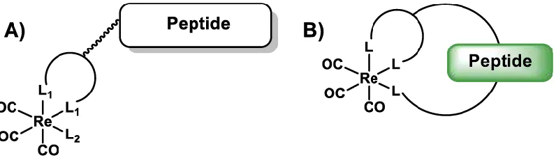
Targeted technetium radiopharmaceuticals generally consist of a biological peptide
sequence for the target receptor, a pharmokinetic modifier linker, and a bi-functional
chelator for the attachment of the metal. Technetium-99m can be introduced, either
through a pendant or an integrated design. The pendant design is a classical approach
where a bi-functional chelator is attached to the peptide, through a side chain of an amino
acid or the end terminus of the peptide, Figure 2.1a. An advantage of using this design is
that it does not interfere with the targeting entity; however, because the bi-functional
chelator is covalently attached to the peptide, the metal complex is placed at a position
that can be easily cleaved through hydrolysis.11 Also, due to the bulkiness of the chelator,
it could perhaps influence the conformation of the peptide and result in decreased binding
affinity to the receptor. Therefore, structural modifications of the metal complex must be
further explored to improve the binding affinity of the peptide. Numerous examples have
been provided for
Figure 2.1 Schematic of pendant and integrated technetium-99m radiopharmaceutical
designs
An integrated design introduces the metal complex directly into the peptide backbone,
approach ‘hides’ the metal complex within the peptide framework resulting in a more
stable, compact, and low molecular weight chelation system. Integration can be a result
of peptide cyclization or turn mimics, where the metal chelator is introduced into the turn
region of the peptide sequence (Figure 2.1b), or a chelator is formed when the peptide is
cyclized through metal coordination (Figure 2.1c). This approach eliminates the use of an
external bulky chelator resulting in low molecular weight radiopharmaceuticals.12
One of the more widely used technetium-99m core has been 99mTc(CO)3+. The
99mTc/Re(CO)3+ core has been shown to stabilize through a “2+1” chelation system.13 A
“2+1” chelation system is comprised of bi-dentate and mono-dentate ligands. There are two ways to introduce “2+1” chelation system, through a pendant design or an integrated
design. Figure 2.2 represents a simplistic model of “2+1” chelation systems. Figure 2.2a
represents a pendant design, where a bi-dentate chelator is attached to the peptide through
a spacer; on the other hand, Figure 2.2b is an integrated design, where the metal complex
is incorporated into the peptide framework.
Figure 2.2 Model “2+1” chelation system
Simpson reported cyclization of the pentapeptide, Ac-HAAAH-OH, through
99mTc/Re(CO)3+, with a mono-dentate coordination through imidazole nitrogen at the
N-terminus, and a bi-dentate chelation through a carboxyl oxygen and imidazole nitrogen at
the C-terminus.5 We can now replace the tri-alanine sequence with a biological relevant
peptide, RGD, to target receptor, integrin αvβ3, for imaging and therapeutic purposes. Here, efforts to cyclize RGD peptide in a “2+1” chelation fashion, with natural and
2.2
Results and Discussion
2.2.1
Synthesis of [Re(CO)
3(OH
2)
3]OTf
14The 99mTc/Re(CO)3+ species is one of the widely used metal cores for radiochemistry,
with the aqua ligands as a convenient starting point. The aqua ligands are very labile due
to the trans effect presented by the carbonyls. The aqua ligands can be easily displaced
with the incoming ligands. Commercially available bromopentacarbonyl rhenium, 2.1,
was reacted with silver triflate, [Ag(O3SCF3)], in freshly distilled dichloromethane
(DCM), under subdued light for three hours at room temperature. The reaction mixture
was gravity filtered to remove by-product salts, silver bromide (AgBr). The product was
precipitated with addition of hexanes, and slow evaporation resulted in
pentacarbonyltriflate rhenium (I), 2.2. Compound 2.2 was reacted with water under
microwave conditions to result tris-aqua tris-carbonyl rhenium (I) species, as a 0.1 M
solution, 2.3.
Scheme 2.1 Synthetic route for [Re(OH2)3(CO)3]OTf 14, as 0.1 M solution
2.2.2
Synthesis of Ac-HRGDH-OH
The peptide was constructed from C→N terminus on a solid phase support, with the
C-terminus attached to an insoluble resin. The synthesis of the linear peptide employed
9-fluorenylmethyloxycarbonyl (Fmoc) chemistry. The N-terminus of each amino acid was
already protected with the Fmoc group, and the side chain of amino acid residues were
protected with acid labile groups. After the removal of the Fmoc group, an amino acid
with a base and coupling reagent in N,N-dimethylformamide (DMF) were added. The
amino acid couplings were repeated until the desired peptide was constructed. The Fmoc
anhydride in DMF. The removal of protecting groups, and the cleavage of the peptide off
the resin was performed by vortexing the resin in a cleavage cocktail. The linear peptide
was precipitated with excess tert-butyl methyl ether (TBME) and centrifuged. The
centrifuged precipitates were dissolved in water and frozen over dry ice. The frozen
peptide was lyophilized to remove solvents. The peptide was purified with reverse-phase
preparative HPLC-MS (RP HPLC-MS), with a purity of >95%.
Figure 2.3 Linear Ac-HRGDH-OH, 2.4, peptide
Ac-HRGHD-OH Calculated (m/z) Observed (m/z)
[M+H]+ 663.66 663.34
[M+2H]2+ 332.33 332.25
Table 2.1 Calculated and observed m/z ratio of Ac-HRGDH-OH, 2.4, analyzed by
2.2.3
Characterization of Ac-HRGDH-OH
Figure 2.4 1H-NMR spectrum of Ac-HRGDH-OH, 2.4, in CD3OD.
The acetylated RGD peptide was characterized with HPLC-MS and 1H-NMR
spectroscopy. Table 2.1 reports the calculated and observed m/z ratio of the peptide, and
Figure 2.4 displays the 1H-NMR spectrum in CD3-OD. After analyzing the spectra,
interesting observations were made, with glycine appearing as a prochiral molecule and
the splitting of hydrogens were observed with a pair of doublets near 3.88 ppm. The same
prochirality was observed with other methylene groups, adjacent to CHα atoms, of other
amino acids. The methylene group of aspartic acid was observed to follow an ABM spin
system, where the methylene hydrogens (Hf) were coupled with each other and at the
same time were coupled with CHα (He). The arginine methylene groups were present
around 1.5–2.0 ppm, and coupling was hard to observe. The CHα atoms were spread
from 3.75–4.75 ppm. The CH hydrogen atoms of the imidazole rings were overlapped
downfield chemical shift when compared with Hb/n, due to the electron withdrawing
nitrogen atoms present on both side of the proton.
Figure 2.5 1H-NMR splits of Glycine and Aspartic acid in in CD3OD.
2.2.4
“2+1” Coordination Using Natural Amino Acids as Metal
Chelators
Following the coordination procedure reported by Simpson5, Ac-H1RGDH5-OH was
reacted with [Re(CO)3(OH2)3]+. The peptide was dissolved in water, [Re(CO)3(OH2)3]+
and concentrated base, 5M NaOH, were added to the solution. Upon completion of the
reaction, the solution was lyophilized to remove the solvent. LC-MS showed the
uncoordinated peptide left at the end of the reaction. There were numerous peaks at
different retention times that corresponded to the same m/z ratio of the peptide
coordinated with Re(CO)3+, which suggested that there might be linkage isomers present.
No dominant coordinated isomeric peak was observed. The reaction was optimized to
obtain a single dominant isomeric peptide. A generic scheme used for the coordination is
displayed in Scheme 2.2, and Table 2.2 reports various conditions used to optimize the
reaction. The peaks were compared based on the area under the curve, from the LC-MS
Scheme 2.2 Synthetic approach for Ac-HRGDH-OH, 2.4, coordination with
[Re(CO)3(OH2)3]+, and proposed coordination product.
# 0.1M
[Re(CO)3(OH2)3]+ (molar Eq.) 5M NaOH (molar Eq.) Temp (°C) Time (hours)
% coordinated vs uncoordinated
peptide
01 1.25 1.8 110
(micr-owave)
0.6 NR
02 1.20 1.0 rt 48 NR
03 1.20 1.0 rt 5 96
04 1.75 1.0 rt 2 56
05 1.75 0.6 rt 3 18
06 1.75 - 50 3.5 NR
07 1.65 1.0 rt 1 NR
08 0.90 7 rt 3 97
Table 2.2 Reaction conditions used to coordinate Ac-HRGDH-OH, 2.4, with
[Re(CO)3(OH2)3]+.
Analysis of the data suggested that increasing the molar equivalent of the Re(CO)3+
species led to a lower percentage of the coordinated peptide; this could be due to side
reactions taking place when excess Re(CO)3+ was used. Unfortunately, the masses
corresponding to the undesired side products were not observed from LC-MS analysis. It
was also noted that the concentration of the base plays an important role in peptide
coordination. The reaction proceeded to completion at a higher pH (~9-10); this could be