University of South Carolina
Scholar Commons
Theses and Dissertations
12-14-2015
Effects of Cell Adhesion Peptides, pH, and Matrix
Shape on Maintenance of Breast Cancer Stem Cells
in an Engineered Hydrogel Matrix
Leily Daneshian
University of South Carolina - Columbia
Follow this and additional works at:https://scholarcommons.sc.edu/etd
Part of theBiomedical Engineering and Bioengineering Commons
This Open Access Thesis is brought to you by Scholar Commons. It has been accepted for inclusion in Theses and Dissertations by an authorized administrator of Scholar Commons. For more information, please [email protected].
Recommended Citation
Daneshian, L.(2015).Effects of Cell Adhesion Peptides, pH, and Matrix Shape on Maintenance of Breast Cancer Stem Cells in an Engineered
Effects of Cell Adhesion Peptides, pH, and Matrix Shape on Maintenance of
Breast Cancer Stem Cells in an Engineered Hydrogel Matrix
by
Leily Daneshian
Bachelor of Science Shahed University, 2012
_______________________________________________
Submitted in Partial Fulfillment of the Requirements
For the Degree of Master of Science in
Biomedical Engineering
College of Engineering and Computing
University of South Carolina
2015
Accepted by:
Esmaiel Jabbari, Director of Thesis
Michael Gower, Reader
Michael Shtutman, Reader
ii
© Copyright by Leily Daneshian, 2015
iii
Dedication:
iv
Acknowledgments
First, I would like to thank my academic advisor, Prof. Esmaiel Jabbari, for all of his
support, encouragement, and time during my MSc studies. I would also like to express
my appreciation to my MSc committee, Michael Gower and Michael Shtutman, for their
v
Abstract
Metastasis, resistance to chemo- and radiotherapy, and eventual relapse has been
attributed to a tumor subpopulation known as cancer stem cells (CSCs). CSCs are
regulated in their tumor microenvironment by various factors. Synthetic hydrogels can
be used to investigate the effects of individual environmental factors on CSCs by
providing inert 3D matrices. In this thesis, poly ethylene glycol diacrylate (PEGDA)
hydrogel with 5kpa modulus has been used as a culture system to study the effect of; I)
integrin and heparin binding peptides, 2) pH, and 3) the shape of the microenvironment
on breast CSCs maintenance and tumorsphere formation in PEGDA. Human breast
cancer cells were encapsulated in PEGDA hydrogels and the effect of the peptides, pH,
and the shape of the environment on tumorsphere formation was investigated by
fluorescent microscopy, qRT-PCR and DNA content assay.
All peptides including RGD, RYD, IKLLI, LIGRKK, VAPG, WQPPRARI, and SPPRRARV
affected breast cancer cells by reducing their capability of sphere formation. Among
peptides, RGD, RYD, and WQPPRARI were the most effective peptides in reducing
sphere formation of breast CSCs.
Moreover, different shapes of micropatterned PEGDA including circle, square, and
vi
Breast CSCs formed spherical tumors regardless of the shape of the micropatterned
PEGDA and had the minimum surface area for a given volume. Furthermore, breast CSCs
showed more resistance to acidic pH compared to non-stem breast cancer cells and
vii
Table of Contents
Dedication: ... iii
Acknowledgments ... iv
Abstract ... v
Table of Contents ... vii
List of Figures ... ix
Chapter 1. Introduction ... 1
1.1. Tumor Heterogeneity ... 1
1.2. CSCs and Their Microenvironment ... 3
1.3. CSC microenvironment and peptides... 3
1.4. CSC microenvironment and pH ... 16
1.5. CSC Microenvironment and Shape ... 19
1.6. Why Choosing PEGDA for Investigation on CSCs and Their Microenvironment? ... 20
Chapter 2. Results and Discussion ... 28
2.1. Results and Discussion for Peptide Experiment ... 28
2.2. Result and Discussion for pH Experiment ... 41
2.3. Results and Discussion for Matrix Shape Experiment ... 45
viii
3.1. PEGDA Synthesis ... 52
3.2. Peptide Synthesis and Characterization ... 53
3.3. Hydrogel Preparation and Modulus Measurement ... 57
3.4. Cell Culture and Encapsulation in the Hydrogel ... 57
3.5. PH Adjustment at 6.8 ... 58
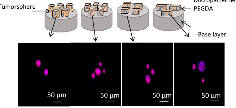
3.6. Micropatterning ... 58
3.7. Fluorescent Imaging ... 59
3.8. Determination of Tumorsphere Number and Size ... 60
3.9. DNA Extraction from PEGDA Hydrogel and Cell Number Measurement ... 60
3.10. RNA Extraction from PEGDA Hydrogel and qRT-PCR ... 61
Conclusion ... 63
ix
List of Figures
Figure 2.1. Comparison of tumorsphere formation in PEGDA gels conjugated with peptides. .... 30
Figure 2.2. Comparison of cell density, tumoresphere density and and sphere size of the cells in PEGDA gels conjugated with peptides.. ... 32
Figure 2.3. Comparison of cell density in 0.01 and 0.02 concentrations, and tumoresphere density in 0.02 of the cells in PEGDA gels conjugated with peptides ... 33
Figure 2.4. Comparison of CD44, ABCG2, and TGF β expressions of the tumor cells in PEGDA gels conjugated with peptides. ... 37
Figure 2.5. Comparison of normalized CD44, ABCG2, and TGF β expressions of the tumor cells encapsulated in PEGDA gels with 2% concentration of different conjugated peptides. ... 38
Figure 2.6. Comparison of tumorsphere formation in PEGDA gels, encapsulated in CSC medium with PH=7.4 and 6.8 ... 42
Figure 2.7. Comparison MDA-MB-231 and MCF10a cell numbers cultured in CSC with pH 7.4 and pH 6.8.. ... 43
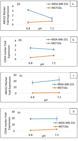
Figure 2.8. Comparison of CD44, and ABCG2 expressions of the cells in CSC medium with pH 7.4 and pH 6.8 ... 46
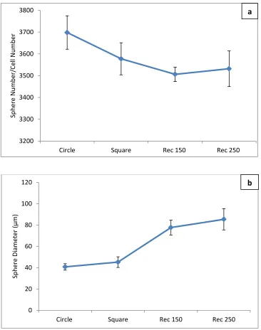
Figure 2.9. Comparison of tumorsphere formation in micropatterned PEGDA gels ... 47
Figure 2.10. Comparison of tumoresphere density/ and sphere size of the cells encapsulated in pmicroatterned PEGDA gels ... 48
Figure 2.11. Comparison of CD44, ABCG2, and TGF β expressions of the tumor cells in
1
Chapter 1
Introduction
Breast cancer is the second leading cause of cancer death in women. About 1 in 8 (12%)
women in the US will have invasive breast cancer during their life (American Cancer
Society 2015). Regardless of advancements in diagnosis/treatment of metastatic breast
cancer, the rate of death from this disease remains high. This is due to the fact that, the
available therapies are limited by the existence of therapy-resistant cancer cells. Thus,
metastatic breast cancer is an irrepressible disease by current treatment approaches.
That means, further investigation needs to be performed on the breast cancer research
area. Therefore, this research has been designed to achieve a better insight about
these therapy-resistant cancer cells (cancer stem cells) behaviors and interactions in
their microenvironment. Hopefully, the collected information will open up a new useful
path toward the eradication of metastatic breast cancer.
1.1. Tumor Heterogeneity
Tumor heterogeneity refers to the existence of different cells within tumors or between
tumors. Tumor cells are different in morphology, metabolism, proliferation, and ability
2
heterogeneity: “Cancer stem cell” model and “clonal evolution” model (Shackleton et
al. 2009)Cancer Stem Cell (CSC) model: CSCs are a subpopulation of cancer cells that
form tumors. They self-renew and differentiate to other cancer cells, which are not able
to form tumors. The idea that CSCs contribute a small population of cancer cells comes
from this view that if almost all of the cancer cells would proliferate extensively and
metastasize throughout the body, then all of them should be eradicated by available
therapies. In reality, current cancer treatments are truly able to remove most of the
cancer cells in the body, however the observation of cancer relapse illustrates that there
should be a small population of cancer cells left in the body which initiate the disease
later (Reya et al. 2001). CSCs are capable of self-renewing and differentiating. The
heterogeneity that has been observed between differentiated cells refers to differences
between cancer stem cells that they have originated from. The difference between
cancer stem cells usually arise from epigenetic changes simultaneous with natural
selection of advantageous genetic mutated cancer stem cells (Shackleton et al. 2009).
This model proposes that CSCs behave the same as normal stem cells. They undergo
epigenetic changes and differentiate to cells that are phenotypically different and have
limited proliferation. These differentiated cancer cells make the majority of cells in a
tumor (Shackleton et al. 2009).
Cancer stem cell model has been observed in multiple tumor types such as leukemias
(Campbell et al. 2008), breast cancer(Jabbari et al. 2015), and prostate cancer(Alvarado
3
spheres in NOD/SCID mice, and also specific markers for these tumorigenic cells have
been identified (Campbell et al. 2008, Jabbari et al. 2015, Alvarado et al. 2005).
1.2. CSCs and Their Microenvironment
Breast cancer stem cells exist in an environment (niche) which is responsible for the
maintenance of specific stem cell properties like self-renewal and remaining in an
undifferentiated state. The population in niche contains both stem cells and surrounding
differentiated cells. Biochemical signals that breast cancer stem cells get from the
interaction with ECM components and neighbor cells have critical roles in maintenance
of them. Cancer stem cells niche is responsible for the control of the essential pathways
that have critical roles in determination of stem cells destiny. Critical pathways such as
STAT, Notch, and Wnt have been recognized in CSCs niche while some features of
cancers like hypoxia and angiogenesis regulate these pathways. Investigating on
processes within breast CSCs niche can provide a better understanding of these CSCs for
prevention and treatment of metastasis breast cancer.
1.3. CSC microenvironment and peptides
The extracellular matrix (ECM) is a collection of extracellular molecules secreted by cells
that provide structural and biochemical support to the surrounding cells. Cell adhesion,
and cell-to-cell communication are common functions of the ECM (Abedin and King
4
Fibronectin, laminin and elastin are 3 components of ECM. These proteins which are
long chains of amino acids, and peptides derived from these proteins which are short
chains of amino acids are well known for mediating cell adhesion. It has been shown
that the occurrence of breast cancer is concurrent with changes in these proteins
expression, degradation and expression of their binding receptors.
VAPG (derived from elastin), IKLLI (derived from laminin), RYD (derived from
streptavidin), and RGD (derived from fibronectin) are integrin binding peptides and
WQPPRRARI and SPPRRARV (derived from fibronectin) are heparin binding peptides.
Integrins and heparan sulfate proteoglycans are receptors that play critical roles in
development of metastasis breast cancer as they activate focal adhesions mainly focal
adhesion kinase (FAK). FAK is an intracellular non-receptor tyrosine kinase. It has been
shown that FAK is highly overexpressed in breast cancers and it has key roles in
promoting tumorigenesis and metastasis (Luo and Guan 2010).
Therefore, we chose these cell binding peptides from different domains of these
proteins to investigate their effects on breast cancer stem cells maintenance in PEGDA.
The importance of using peptides over proteins is due to difficulties such as protein
denaturation and degradation, and problems with protein absorption. Peptides are part
of the ECM proteins which have similar stimuli of proteins while they are more stable
5
Some of the most recent usage of peptides in breast cancer research has been reviewed
in the following 5 parageraphs.
Patched receptor binding peptides have shown to have a growth inhibitory effect in
tumors with activated hedgehog signaling (Smith et al. 2014). Remarkable growth
inhibition has been observed in breast cancer cell lines treated with patch-blocking
peptides (Smith et al. 2014).
Cancer research has been illustrated that connexin 43 is effective in proliferation,
differentiation, and migration of breast cancer cell. There are drugs available related to
this but there is a lack of knowledge in specificity of these agents. In a study, α-connexin
carboxyl-terminal (ACT1) peptide, which modulates connexin 43 has been tested in
breast cancer. The peptide is able to regulate the connexin 43 activity in breast cancer
to sustain connexin 43 -mediated gap junctional activity which cause the decrease of
malignant progression. ACT1 peptide also is able to enhance the activity of lapatinib and
tamoxifen (Grek et al. 2015).
L-peptide has showed to bind to a wide variety of cancers including breast cancers.
Treatment of mice with breast cancer patient derived xenografts (PDX) with
L-peptide-conjugated lipodox (LD-L) has been illustrated to result in greater suppression of tumor
growth than lipodox (LD) alone (Lee et al. 2015).
Cell surface nucleolin is known to be overexpressed in cancer cells and also it is a marker
6
common receptor among breast CSC and non-stem cancer cell (non-SCC), a group of
researchers functionalized liposomes with the nucleolin-binding F3 peptide which
targeted both nucleolin-overexpressing putative breast CSC and non-SCC. An in vivo
assay showed that surface nucleolin overexpression could be related to the triple
negative breast cancer cells which potentially connect the nucleolin expression to the
stem-like properties in triple negative breast cancer cells (Fonseca et al. 2015) .
Proliferating cell nuclear antigen (PCNA) is one of the important regulators in the DNA
replication and repair process. A peptide (caPeptide) as a mimic of PCNA has been
synthesized and delivered into cells using a nine-arginine linking mechanism.
R9-cc-caPeptide displayed cytotoxicity in MDA-MB-436, a triple-negative breast cancer cell
line. R9-cc-caPeptide has also been resulted in blocking the association of PCNA with
chromatin (Smith et al. 2015).
1.3.1. Importance of Fibronectin and its Peptides
Fibronectin (FN) is one of the important ECM glycoprotein that exists in fibrillar form in
all tissues during life. Its formation is a cell-mediated process and is essential for life. FN
fibrils form linear and branched meshworks in order to connect neighboring cells to
each other. FN is a multidomain molecule that has different domains for interacting with
other ECM proteins including other FN proteins, cell receptors, and glycosaminoglycans
(GAGs). This arrangement of domains allows FN to bind to cells and molecules at the
same time. FN has binding sites for collagen/gelatin, heparin, fibrinogen, heparin sulfate
7
2010). It plays a major role in cell adhesion, growth, migration, and differentiation
(Pankov and Yamada 2002). Altered fibronectin expression, degradation, and
organization have been associated with a number of diseases, including breasr cancer
and fibrosis (Williams et al. 2008). Observing tumors and tumor-derived cell lines have
been attributed to the decreased fibronectin expression, increased
fibronectin degradation, and/or decreased expression of fibronectin-binding receptors
such as α5ß1 integrins (Hynes 1990). So far, the effects of several peptides derived from
different domains of fibronectin such as WQPPRARI (Hettick, Ruwona, and Siegel 2009,
Van Den Heuvel, Jefferson, and Jacobs 2005, Yun, Kim, and Jang 2013, Woods et al.
1993, Hoesli et al. 2014, Mooradian et al. 1993, Ouchani et al. 2012, Wilke and Furcht
1990, Sagnella et al. 2005, Björklund and Koivunen 2005, Garagorri et al. 2008),
SPPRRARV (Mooradian et al. 1993, Sagnella et al. 2005), LIGRKK (Hettick, Ruwona, and
Siegel 2009, Tong 2000), RGD (Wierzba et al. 1995, Fischbach et al. 2009, Naghdi et al.
2014, Panda et al. 2010), and RYD (Murray et al. 2002, Knight 2001, Guo et al. 2005) on
different cell lines behavior such as cell adhesion, proliferation and migration have been
studied. In the following seven paragraphs a brief summary of these studies has been
provided.
1.3.1.1. WQPPRARI Peptide
WQPPRARI is one of the well-known heparin binding peptides which is derived from the
COOH terminal heparin binding domain of fibronectin. This peptide is famous for its cell
8
Jacobs 2005, Yun, Kim, and Jang 2013, Woods et al. 1993). It has been shown that
WQPPRARI is able to improve umbilical vein endothelial cell adhesion, expansion, and
motility through focal adhesion formation and FAK activation (Hoesli et al. 2014);
enhance cell adhesion, spreading, and migration of rabbit corneal epithelial cells directly
(Mooradian et al. 1993); promote cell adhesion, spreading, and migration of normal and
leukemic progenitors through direct interaction with α4ß1 (Ouchani et al. 2012);
increase cell adhesion and spreading of human keratinocytes and saphenous vein
endothelial cells (Wilke and Furcht 1990); and improve human pulmonary artery
endothelial cell adhesion and spreading through local adhesion (Sagnella et al. 2005).
The WQPPRARI peptide is able to stimulate expression of MMP-1 and MMP-9 in
fibroblast plates on a fibronectin fragment, lacking the heparin binding domain. This
stimulation is mediated by α5β1 and α4β1 integrins (Björklund and Koivunen 2005). It
should be pointed out that, there is also a study on Keratocyte behavior in
three-dimensional photopolymerizable poly (ethylene glycol) hydrogels which used the
sequence WQPPRARI, tethered to hydrogels, and showed that it enhances adhesion,
spreading, and migration of corneal epithelial cells to the hydrogels (Garagorri et al.
2008).
1.3.1.2. SPPRRARV Peptide
The other COOH-terminal heparin-binding domain of fibronectin is SPPRRARVT. This
9
adhesion and spreading (but not RCE cell migration) (Mooradian et al. 1993); and also
human pulmonary artery endothelial cell adhesion and growth (Sagnella et al. 2005).
1.3.1.3. LIGRKK Peptide
KNNQKSEPLIGRKKT is another heparin-binding peptide that derived from the
COOH-terminal heparin binding domain of fibronectin which mediates cell adhesion for a
variety of cell types and promotes neurite outgrowth. The basic structural features
necessary for the activity have been identified in the COOH-terminal residues, LIGRKK
(Hettick, Ruwona, and Siegel 2009). This biologically ”active” sequence has been found
in several other heparin/heparan sulfate-binding peptides such as LIGRKK derived from
laminin, which helps fluoropolymer surfaces for the enhancement of nerve cells
interactions (Tong 2000).
1.3.1.4. RGD Peptide
RGD, another well-known peptide, is a sequence in extracellular matrix proteins such as
fibronectin, collagen, and laminin that mediates cell attachment by interacting with
proteins of the integrin family of cell surface receptors (Wierzba et al. 1995). It has been
shown that human breast cancer cells, MDA-MB-123, cultured in a hydrogel based
culture system coupled to RGD are able to secrete more interleukin 8 (IL-8) compare to
the cells cultured in a hydrogel based culture system without any conjugated peptide.
Up regulation of IL-8 is critical in control of tumor vascularization. Therefore, 3D RGD
coupled culture systems could regulate cancer cell angiogenic signaling, and controlled
10
Polyethylene glycol hydrogel (PEG) has been studied as a 3D culture system for neuron
cells as well. It has been reported that neurite outgrowth was improved in systems with
conjugating RGD to PEG polymer. Therefore, NSC survival, proliferation and
differentiation are enhanced when the cells are cultured in 3D-PEG–RGD compared to
3D-PEG environments (Naghdi et al. 2014).
Moreover, proliferation and growth of mammalian cells (HeLa and L929) in a 3D
environment with a dipeptide hydrogel chemically functionalized with a pentapeptide
containing Arg-Gly-Asp (RGD) motif has been investigated. The functionalized gel
exhibited enhanced cell growth promoting properties, and promoted 3D growth and
proliferation of cells for almost 2 weeks (Panda et al. 2010).
RGD peptide also has been used to examine the effect of substrate stiffness on melanoma cell
treatment responsiveness. Human cell lines derived from radial growth phase (WM35) and
metastatic melanoma (A375), PEG hydrogels as a cell culture system and PLX4032 as
pharmacological inhibitor were used. In this study, it was found that in A375 cells, matrix
elasticity did not alter cell morphology or apoptosis with PLX4032 treatment. But in WM35 cells,
matrix elasticity increased apoptosis and smaller focal adhesions on compliant substrates
(Tokuda, Leight, and Anseth 2014).
In a study, polyethylene glycol (PEG) hydrogels, attached to RGD was used and its ability
to support the growth of androgen-dependent LNCaP prostate cancer cells was
investigated. It was found that, the mechanical properties regulate the growth of LNCaP
11
formed tumor-like structures in 3D culture, with hypoxic and apoptotic cores (Sieh et al.
2012).
In another study, by using PEG, modified with RGD and another laminin derived peptide,
murine models of lung adenocarcinoma investigated. The focus was on how matrix can
influence epithelial morphogenesis of a metastatic cell line (344SQ). 344SQ
encapsulated in bioactive peptide-modified, matrix metalloproteinase–degradable PEG
hydrogels formed lumenized epithelial spheres. Changing matrix stiffness and peptide
concentrations affected epithelial morphogenesis, apoptosis , proliferation, and
expression of epithelial polarity markers (Gill et al. 2012).
Streptavidin is a biotin-binding tetrameric analogue of avidin, produced by the soil
bacterium Streptomyces avidinii. Streptavidin, like fibronectin, contains an RGD-like
sequence RYD, which promotes adhesion to the integrin receptor α5β1. This sequence,
Arg- Tyr-Asp-Ser (RYDS), exhibits structural homology to Arg-Gly-Asp-Ser (RGDS).
Binding of streptavidin to cell surfaces mediated through this RYDS domain, can be
inhibited by using fibronectin as well as RGD- and RYD-containing peptides (Murray et
al. 2002). Synthetic peptides containing such a sequence are able to mimic the
integrin-mediated binding of the entire protein(s).
1.3.1.5. RYD Peptide
It was proposed that RYD in ARRSPSYYRYDGAGPYYAMDY functions as an analogue to
RGD in fibrinogen. This peptide comprised the binding domain for the αIIbβ3 receptor. A
12
found to be an inhibitor of both fibrinogen and PAC-1 binding to activated platelets.
Exchanging RGD for RYD in the aforementioned peptide, increased its activity 10-fold
(Knight 2001). Also the sequence RYD has been introduced into the dendroaspin scaffold
in order to replace RGD. The RYD sequence produced a similar IC50 value to the RGD
sequence, in inhibiting A375-SM cell (β-3 integrin) adhesion to collagen (Guo et al.
2005).
1.3.2. Importance of Laminin and its Peptides
Basal lamina is mostly made of Laminin. This protein plays a critical role in cell
differentiation, migration, and adhesion, as well as cell phenotype and survival. Laminin
is linked to type IV collagen via entactin (Smith and Ockleford 1994), fibronectin
(Ockleford et al. 1993),and perlecan [28]. Moreover, this glycoprotein, Laminin, binds to
cell membranes through integrin receptors and other plasma membrane molecules,
such as the dystroglycan glycoprotein complex (Haralson, Hassell, and Streuli 1995).
Through these interactions, laminin causes cell attachment, differentiation, shape, and
movement (Haralson, Hassell, and Streuli 1995, Colognato and Yurchenco 2000).
As it has been shown in [30], laminin plays a critical role in regulating cancer cell
migration and facilitating tumor cell invasion. Laminin establishes one of the essential
components of basement membranes (BMs) as it is involved in cellular adhesion to BMs
and ECM. Invading tumor cells are capable of attaching to the matrix through specific
13
tumor cells release protease in the interstitial, consequently, promote BM disruption
and cell diffusion (Albrechtsen et al. 1981).
1.3.2.1. IKLLI Peptide
IKLLI is a sequence shown to be active in laminin (Fischbach et al. 2009). Several
peptides containing the IKLLI sequence in the α 1 chain of laminin-1 such as
CSRNLSEIKLLISRARK, EIKLLIS, and SEIKLLIS were found to mediate heparin binding and
cell adhesion of PC12 cells as well as promoting neurite outgrowth in these cells.
Furthermore, the CSRNLSEIKLLISRARK and SEIKLLIS sequences also mediated
proliferation in PC12 cells. As noted above, an IKLLI-containing peptide derived from the
laminin α 1 chain may be an active site of laminin and its cell adhesion maintenance may
be due to interaction with both integrin a3b1 and cell surface heparan sulphate
proteoglycan (TASHIRO et al. 1999).
These Neurons expressed integrin b1, beside the fact that the treatment of cultures
with an antibody against integrin b1 eliminated the protective effect of laminin.
Moreover, neurons maintained on laminin displayed a continued activation of the Akt
signaling pathway. The IKLLI-containing integrin-binding peptide is capabale of
mimicking the neuroprotective effect of integrin engagement by laminin. Due to this
14
1.3.3. Importance of Elastin and its Peptides
Degradation is a requirement for cancer progression. This is due to the fact that, ECM
degradation is vital to allow cell migration through its three-dimensional architecture
and also to generate ECM fragments. ECM proteolysis causes the release of matrix
fragments that exhibit proper biological activities. This degradation of ECM is coincident
with the degradation of Elastin, a major component of ECM that confers elasticity to
tissues (Panda et al. 2010). Protease-driven elastin degradation happens during
physiopathological processes such as cancer progression, which generates bioactive
elastin-derived peptides that are thought to contribute to tumor progression (Devy et al.
2010). In another words, Elastin peptides control proliferation, chemotaxis, and
protease expression.
1.3.3.1. VAPG Peptide
VGVAPG is an elastin-derived peptide shown to block ceramide-induced apoptosis in
human skin fibroblast cells. The elastin peptide treatment leads to activation of the
pro-apoptotic protein Bad, and caspase-9 (Cantarelli et al. 2009). As mentioned in [36],
elastin-derived peptides raise invasive capacities of lung cancer cells by
post-transcriptional regulation of MMP-2 and uPA.
The VAPG peptide sequence is repeated several times in human elastin and most likely it
is one of the breakdown products after the degradation of elastin. The VAPG elastin
peptides could bind to three identical receptors, namely; (i) galectin-3, (ii) integrin αvβ3,
15
increase the invasive potential of melanoma cells mostly by galectin-3 (Pocza, Falus, and
Darvas 2009).
In a study, VAPG peptide sequence attached to a hydrogel material, and its effects on
smooth muscle cell adhesion and spreading have been studied. The VAPG sequences
was specific for adhesion of smooth muscle cells while fibroblasts, endothelial cells, and
platelets cannot adhere to VAPG (Gobin and West 2003).
The effects of cell adhesion due to the VAPG peptide on vascular smooth muscle cells
were also examined in (Gill et al. 2012). These cells more strongly adhered to the
surfaces modified with adhesive ligands. In addition, cell migration was higher on
surfaces with the adhesive ligand than on control surfaces. Moreover, cell proliferation
was lower on adhesive surfaces. Likewise, in hydrogel which is functionalized with
VAPG, cell proliferation was lower in comparison with control groups (Gill et al. 2012).
Matrix protein synthesis by cells cultured on materials that was modified by cell
adhesion ligands, like the VAPG peptide, were examined in (Smith and Ockleford 1994).
While initial adhesion of smooth muscle cells, endothelial cells, and fibroblasts increased
on the higher density of peptides on surfaces, all cell types had less production of matrix
on the more highly adhesive surfaces. This result may actually pose limitations for the
use of bioactive materials, such as in tissue engineered scaffolds since matrix production
16
In this Study, the role of these integrins binding peptides (RYD, RGD, IKLLI, and VAPG)
and heparin binding peptides (WQPPRARI, SPPRRARV, LIGRKK, and IKLLI) on the
maintenance and behavior of breast CSCs encapsulated in PEGDA has been investigated.
1.4. CSC microenvironment and pH
Tumor microenvironment is extremely acidic (pH ~6.8) compared to normal tissue (pH
~7.4) which affect tumorigenesis, angiogenesis and metastasis activity of cancer cells
exclusively (Song, Griffin, and Park 2006). It has been shown that tumor
microenvironment has lower pH and is more acidic in comparison with normal tissue,
and this is due to anaerobic and aerobic pathways. In a tumor, vascularization is not
homogeneous and adequate to feed enough nutrition, specifically oxygen to rapid
dividing cancer cells. Lack of oxygen is the main reason for acid production. In hypoxia,
cells undergo glucose uptake and glucose goes through glycolytic pathway instead of
respiratory pathway which causes the production of lactic acid and reduction of pH in
the microenvironment (Song, Griffin, and Park 2006, Tannock and Rotin 1989).
Moreover, under hypoxia condition, ATP hydrolysis also causes the acidification of
tumor microenvironment. On the other hand, it has been shown that in cancer cells,
glucose undergo glycolysis even in the presence of sufficient oxygen in order to produce
lactate.
Cancer cells require high amount of lactate because it helps them to escape from
immune cells. In the presence of lactate produced from tumor cells, T cells do not
17
of dendritic cell activated by antigen-specific autologous T-cell stimulation. Moreover, it
also inhibits monocyte migration and cytokine release (Kato et al. 2013).
Moreover, tumor cells undergo glycolysis even in sufficient oxygen condition because of
their advantage. The glycolysis pathway produces acid and the acidic extracellular
environment help cells to become invasive and proliferative (Gatenby and Gawlinski
2003).
On the other hand, it has been shown that the pentose phosphate pathway is highly
active in tumor cells. CO2 is one of the products of this pathway while there is a large
amount of carbonic anhydrase (CA) is also present in a tumor. CO2 can be processed to
H+ and HCO3- by CA as a catalyzer (Gatenby et al. 2006). Therefore, CO2 is another
important reason for acidic PH of tumor microenvironment (Helmlinger et al. 2002).
It has been shown that Acidic pH can induce EMT in some types of cancer cells such as
lung and melanoma cancer cell lines (Peppicelli et al. 2014, Suzuki et al. 2014).
Moreover, acidic pH has also been associated to the expression of some genes that are
contributed to metastasis of cancer cells such as metastasis of melanoma cells to the
lungs (Rofstad et al. 2006). Acidic pH increases the expression of some genes that are
involved with pro-metastatic factors. It has been shown that when melanoma cells
incubated in acidic culture medium, had a higher metastatic rate accompanied by
proteinase MMP-9 and NHE activation (Gatenby et al. 2006, Kato et al. 2013) and a
higher angiogenesis by acid induced production of VEGF-A and IL-8 (Gatenby et al.
18
The glycolytic activity in tumor cells enhances tumor invasion. Protons (H+) produced by
cancer cells diffuse (carried by a buffering agent) from tumor to the normal tissue
nearby by using transporter proteins such as Na+/H+ exchanger (NHE) ,and cause the
decrease of pH in normal tissue. It has been shown that the Na+/H+ exchanger type 1
(NHE1) was an important regulator of H+ efflux in breast cancer cells MDA-MB-231
(Stock et al. 2005).
Therefore the intracellular pH of cancer cells does not become acidic while it is slightly
alkaline and in fact this alkaline pH is suitable for cell proliferation. The acidic pH causes
normal cells to undergo cell p53-dependent apoptosis but some of the tumor cells
survive which are resistant to acidic pH, which is probably due to mutation in p53 or
other components of apoptosis pathway. Low pH causes the extracellular matrix to
degrade by proteolytic enzymes that highly active in low pH and produces by fibroblasts
and macrophages (Gatenby and Gawlinski 2003). Moreover, it enhances angiogenesis by
using acid induced vascular endothelial growth factor and interleukin 8, and inhibits
immune system to response to tumor antigen (Kato et al. 2013). Therefore tumor cells
become more invasive as they disrupt the environment, and provide a better condition
for them to proliferate. Cell-cell junctions of tumor cells become separated when cells
move in to their surrounding tissue. Acidic pH helps tumor cells to destruct the
adherence junction. This is by Src activation, that causes E-cadherin degradation
19
To wrap up, pH is one of the important factor in tumor microenvironment and although
the effect of pH has been studied on some cancer cell lines (Peppicelli et al. 2014, Suzuki
et al. 2014, Rofstad et al. 2006) , the effect of pH on breast cancer stem cells has net
been investigated yet.
Herein, we used PEGDA having modulus of 5 kpa to study the role of acidic pH (6.8) in
the maintenance of breast CSCs. Having a good understanding of the role of this factor
on regulation of CSCs can offer important information on the behavior of breast CSCs in
their tumor microenvironment.
1.5. CSC Microenvironment and Shape
Tumor transformation and metastasis is involved with changes in mechanical properties
of cells and cell’s microenvironment including mechanics, shape, and topology of ECM.
Cells are able to sense the rigidity (elastic resistance) of ECM and balance this force by
exerting contractile stresses. The balance force is very important in regulating the
structure, motility, proliferation, and differentiation of tumor cells. Cells exert this force
by the use of adhesion receptors (e.g., integrins), intracellular focal adhesions,
cytoskeletal networks, and molecular motors. Therefore, direct connection between the
extracellular matrix (ECM) and the intracellular environment can control fundamental
behaviors such as differentiation, morphology, motility changes, and alterations in cell
cycle which can contribute to tumor transformation, invasion, and metastasis (Kumar
20
that gene expression, self-renewal, and differentiation of malignant cells can be
affected by geometrical confinement. Confinement of human cervical carcinoma cells
changed the average tumorsphere size and cluster size affected MCF10A cells
proliferation (Jabbari et al. 2015).
Previously we have encapsulated MDA-MB-231 cells in circle shape 50, 75, and 250 µm
patterned PEGDA gels and incubated for 9 days in CSC medium. It was shown that as the
patterns became smaller the expression of breast CSCs became higher. Therefore, using
micropatterning and the confinement of breast CSCs can help to get a higher expression
of CSCs markers (Jabbari et al. 2015) which can be used in further studies on breast
CSCs. So far, breast cancer stem cells encapsulated in PEGDA or in micropatterned
PEGDA form tumors in spherical shape. Herein, we hypothesize that if the shape of
micropatterned changed, the shape of tumors may change as well. Therefore, we
designed photomask with circle, square and rectangle micropatterns in order to shape
the PEGDA and investigate the effect of them on maintenance of breast cancer stem
cells in PEGDA.
1.6. Why Choosing PEGDA for Investigation on CSCs and Their Microenvironment?
CSCs are a small population of tumor cells that are drug resistant, capable of
differentiation, metastasis, and self-renewal through specific pathways provided in the
CSCs niche. On the other hand, cancer development is a hard process to follow as it may
21
investigate cancer tumor formation and progression on the molecular level. This
molecular level investigation has been performed in two or three dimensional cell
culture systems.
1.6.1. 2D Cell Culture Systems
Classical-two dimensional cell culture systems have provided the majority of modern
cancer biology science. For instance, the most common substrates for supporting cell
growth have been made from polystyrene or glass and have been made in shape of a
flat two-dimensional (2-D) surface. However, the main problem for this 2-D system is
the assumption that body physiology can be correctly reproduced using a cellular
monolayer. Obviously, a eukaryotic cell cannot develop same properties on a
two-dimensional glass or polystyrene substrate compared to the 3D extracellular matrix
found in innate tissue. When cells are cultured in 2-D plates, they are attached to rigid
and flat substrates which cause cells to be polarized and get sheet-like morphology
(Alemany-Ribes and Semino 2014, Haycock 2011). These cells are exposed to excessive
nutrition and oxygen. Moreover, in 2-D cultures cells have different surface receptors'
orientation and clustering; therefore, they have different ECM secretion in
composition, configuration and amount and as a consequence, they don’t have normal
signaling that comes from natural ECM (Alemany-Ribes and Semino 2014). Also, their
cell growth rate, migration and apoptosis change in this classical culture systems (Chen
et al. 2012). For the purpose of CSCs culturing, suspension (non-adherent plates) are
22
Based on the above discussion, it can be concluded that, the culture of cells in 2-D
systems is certainly too simple and neglects many important parameters for
reproducing the cell and tissue environment, such as of mechanical cues, cell-matrix and
cell-cell communication (Haycock 2011).
To be more specific in cancer, metastatic cells are not adherent and cannot form tight
focal adhesions. Thus, 2-D cultures are not applicable for them. Furthermore, in multiple
passages of cancer cells, those that have rapidly proliferation are the target of natural
selection, whereas these rapidly proliferative cells are sensitive to therapies that target
rapidly dividing cells while this is not the same condition for all cells in a tumor
(Alemany-Ribes and Semino 2014).
Some parameters such as gradient of nutrients and growth factors as well as cell-cell
and cell-matrix communication which are known to play crucial roles in cancer initiation,
progression, and metastasis cannot be mimicked accurately in 2-D culture systems. As
an example, cancer cells which are cultured in 2-D plates are less malignant in
comparison with those under in vivo conditions (Kitai et al. 2005). Therefore, results
from drugs that are designed to target cell-cell interaction, epithelial to mesenchymal
transition (EMT), and CSC are not trustable(Chen et al. 2012).
1.6.2. Animal Models in Cancer Research
Animal models act as another alternative that are commonly used for the study
purposes of molecular pathways and drug reaction in cancer research. In these cases,
23
immunocompromised animals are used for the research purposes. However, animal
models may not satisfactorily reproduce the structures of human cancers in vivo (Yang
et al. 2013).
Animal models constitute a wide range of models for cancer study:
1- Ectopic xenografts of tumor-derived cell lines or tissue, embedded into
syngeneic or immunecompromised.
2- Orthotopic xenografts of tumor cell lines or tumor tissues are implanted within
the proper organ or tissue.
3- Germ-line transgenic and conditional transgenic models (GEMMs)
4- Primary human tumorgrafts
5- Carcinogen-promoterinduced multi-stage tumor models
A review of pros and cons of these methods are available (Ruggeri, Camp, and
Miknyoczki 2014).However, they are not explained here in details as describing them is
out of scope of this thesis. As some important and most common examples of
disadvantages related to these methods are lack of native tumor microenvironment,
limited engraftment rates, labor intense, time consuming, and ethically problematic
(Ruggeri, Camp, and Miknyoczki 2014, Sachs and Clevers 2014).
1.6.3. 3D Cell Culture Systems
To have a suitable cell culture system, it should provide a 3D matrix having tunable
mechanical properties with a capacity of co-culturing cells in order to provide cell-cell
24
Available 3D in vitro co-culture models satisfy these requirements (Kim 2005) which are
also necessary in CSC research.
In many 3-D models, cell lines or cells from dissociated tissues are implanted and
cultured in 3-D matrices in order to promote cell–cell interaction, adhesion, migration,
and in vivo–like morphogenesis. There has been a big difference in all aspects of cell
behavior; (i) cell shape, (ii) cell growth, (iii) gene expression, and (iv) the response to
stimuli between 2-D and 3-D culture systems. Based on these differences, increasing the
attraction of researches toward utilizing 3-D environments for the most recent
biomaterial directed stem cell manipulation researches can be better understand (Yang
et al. 2013).
In order to work in 3-D environments, in vitro or in vivo, biomaterial-based matrices
have been used as an important tool. These 3-D environments provide ideal matrices for
cell-cell and cell-matrix interaction. Moreover, their properties can be adjusted for
specific features such as desired fluid transport, delivery of bioactive molecules, and
induction of signal transduction. These properties are important since they direct cell
adherence, nutrient/waste transport, cell proliferation, cell differentiation, and in
cancer stem cells studies, tumor sphere formation. Most of these materials can be
modified in order to adjust all of the mentioned critical matrices characteristics.
There are a variety of synthetic and natural materials that have been used for studying
SC and CSCs behavior by manipulating biomaterial-based matrices properties. One of
25
network that can swell under biological conditions. With the ability of swelling,
hydrogels have high water content, therefore they can offer environments that are
closer to natural soft tissue in comparison with other polymeric materials. Additionally
they are more biocompatible as they are highly permeable for oxygen, nutrients, and
other water soluble metabolites. Therefore, hydrogels are ideal for cell encapsulation
(Albrechtsen et al. 1981, TASHIRO et al. 1999, Devy et al. 2010, Cantarelli et al. 2009,
Pocza, Falus, and Darvas 2009, Gobin and West 2003, Mann et al. 1999). Most
hydrogels can be made by photopolymerization under mild conditions with consistent
seeding of cells throughout the scaffold (Ifkovits and Burdick 2007, Chung and Park
2009, Slaughter et al. 2009).
Hydrogels are divided in to three main groups; (i) natural, (ii) synthetic and (iii)
synthetic/natural hybrid hydrogels. There are many types of natural polymers such as;
• Proteins: gelatin, collagen, Matrigel™, fibrin, silk, and lysozyme (Glowacki and Mizuno
2008, Sakai et al. 2009, Wang et al. 2006, Mol et al. 2005, Yan et al. 2008, Kleinman and
Martin 2005).
• Polysaccharides: hyaluronic acid (HA), agarose, dextran, and Chitosan (Leach et al.
2003, Denizli et al. 2004, Kuo and Ma 2001, Kim et al. 2008).
However, using natural biomaterials has some disadvantages. These disadvantages
include limitation in the adjustment of physical and chemical properties, difficulty in
26
pathogen/viral issues when isolating from different sources. Furthermore, there are
problems with isolating and studying cell response to the individual factors in the
microenvironment. This problem is associated with the fact that naturally derived
matrices have many interactions with cell surface receptors which interfere with cell
responses to specific factors (Yang et al. 2013).
In order to overcome the disadvantages of natural hydrogels, synthetic hydrogels have
been considered as a desirable alternative. Synthetic polymers have more reproducible
physical and chemical characteristics, which is important for the production of tissue
engineered scaffolds. One of these synthetic polymers is Poly ethylene glycol.
1.6.4. Using PEGDA with 5kpa Modulus for this Research
Poly ethylene glycol (PEG) polymer is investigated extensively because of its
specifications, including solubility in water and organic solvents, low protein adhesion,
nontoxicity, and nonimmunogenicity (Buxton et al. 2007, Beamish et al. 2010).
Additionally, the end hydroxyl groups of PEG molecules can be grafted with various
functional groups such as acrylate in order to create hydrogels (Zhu 2010).
Among the parameters in the microenvironment, stiffness (elastic modulus) plays a
crucial role in regulating cell function in 2-D and 3-D culture systems (Rehfeldt et al.
2007). As it has been shown in (Sachs and Clevers 2014, Ifkovits and Burdick 2007,
Chung and Park 2009), in 3-D culture systems, encapsulated stem cell differentiation,
and the balance of cell proliferation, and apoptosis can be directed by the stiffness of
27
As mechanical properties and composition of hard and soft tissues are different, cells
can sense and respond to the matrix stiffness in their natural environment by making
the proper ECM composition. Similarly, the proliferation, differentiation, migration, and
apoptosis of cancerous cells in the tumor tissue are regulated by matrix stiffness
(Discher, Janmey, and Wang 2005, Schrader et al. 2011, Verbridge, Chandler, and
Fischbach 2010).
In this thesis, the inert poly ethylene glycol diacylate hydrogel (PEGDA), in a certain
moduli based on previous studies, as a 3-D cell culture system has been used in order to
investigate the role of cell binding peptides, microenvironment pH, and matrix shape on
the maintenance of breast CSCs. It has been shown that only breast CSCs among breast
cancer cells can form tumor sphere in PEGDA (Jabbari et al. 2015). Having a good
understanding of the role of these environmental factors on regulation of CSCs can
provide significant information on the behavior of breast CSCs in their tumor
28
Chapter 2
Results and Discussion
2.1. Results and Discussion for Peptide Experiment
We have shown that cancer cells can form tumorspheres in the PEGDA gel with respect
to the gel modulus (Yang et al. 2013). MDA-MB-231 human breast carcinoma cells
formed cancer tumorspheres in the gel with an optimum modulus of 5 kPa, and the
sphere formation was correlated with the expression of CSC markers (Yang et al. 2013).
In order to determine if synthetic cell binding peptides derived from ECMs’ components,
attached to PEGDA gel, could affect the sphere formation of MDA-MB-231 cells in the
PEGDA gel, different peptides such as; (i) VAPG, (ii) IKLLI, (iii) WQPPRARI, (iv) SPPRRARV,
(v) LIGRKK, (vi) RYD, and (vii) RGD have been conjugated to the gel with a 5 kPa modulus
in concentrations of 1%, 2%, 4%, 6%, and 9% (mg/mg). Sphere formation in samples was
compared with the sphere formation of MDA-MB-231 cells encapsulated in PEGDA
polymer with a 5 kPa modulus and no peptide. To achieve this goal, different
measurements such as; fluorescent images, cell number, sphere number, sphere size,
29
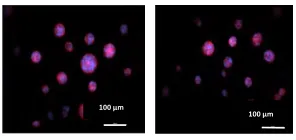
2.1.1. Comparison of Sphere Formation of Tumor Cells Encapsulated in PEGDA with Different Conjugated Peptides in Fluorescent Images
Figure 2.1 illustrates the effects of different peptides on the sphere formation of cells,
encapsulated in PEGDA gel in fluorescent images. As the concentration of peptides
increase, the ability of the cancer stem cells to form spheres decreases. That means,
there are a larger number of tumor spheres in groups with no peptide compared to the
1%, 2%, 4% concentrations. There was no sphere formation for all peptides in 6% and
9%.
As can be seen in all of the subfigures of Figure 2.1, larger spheres exist in the control
group. By the increase of the peptides concentratons to 1% and 2% the tumors sphere
size decreases gradually. In Figure 2.1, there are only four different concentrations (0%,
1%, 2%, and 4%) that have been shown. Concentrations of 6% and 9% are not shown
here as there was no tumorsphere formation in these concentrations for any of the
peptides.
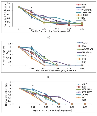
2.1.2. Comparison of Cell Density, Sphere Size, and Sphere Density of Tumorspheres for Different Peptides
The cell density for the cells encapsulated in PEGDA gel and incubated for 9 days in stem
cell culture medium are shown in Figures 2.2 (a). The cell density of MDA-MB-231 cells
encapsulated in PEGDA in the presence of all of the peptides decreased gradually as the
concentration of peptides increased, suggesting that peptides crosslinked to the PEGDA
30 VAPG IKLLI WQPPRARI SPPRRARV LIGRKK RYD RGD
Figure 2.1 Comparison of tumorsphere formation in PEGDA gels conjugated with
peptides. Illustrate of fluorescent images of the tumorsphere size and distribution for
MDA_MB_123 tumor cells encapsulated in PEGDA gels with different concentrations of VAPG, IKLLI, WQPPRARI, SPPRRARV, LIGRKK, RYD, and RGD peptides.
0% 1% 2% 4%
0% 0% 1% 1% 2% 2% 4% 4%
0% 1% 2% 4%
0% 1% 2% 4%
0% 1% 2%
200 μm
4%
200 μm 200 μm
200 μm
0% 1% 2% 4%
100 μm 100 μm 100 μm
100 μm 100 μm 100 μm 100 μm 100 μm
100 μm
100 μm 100 μm
100 μm 100 μm
100 μm
100 μm 100 μm 100 μm
100 μm 100 μm 100 μm 100 μm 100 μm
31
are able to decrease the proliferation of tumor cells and the rate of this decrement
increased with respect to the concentration increment.
The cell density was normalized with respect to the control groups without any peptides
for all of the peptides. The cell density decreased significantly for all peptides with a
concentration of 2% or more (mg/mg). Figure 2.3 (a) Displays the comparison of cell
density for the peptides used in a 0.02 concentration. The cell density of the samples
with a 2% peptide concentration decreased to 0.738469±0.059071, 0.372907±0.068205,
0.16477±0.071641, 0.52825±0.044298, 0.150917±0.065204,
0.362006962±0.044315007, and 0.296782544±0.117010065 per cell density of the
control groups, for VAPG, IKLLI, WQPPRARI, SPPRRARV, LIGRKK, RYD, and RGD,
respectively. Cell density decreased significantly for all of the peptides in this
concentration with respect to the control group. Moreover, there was a significant
difference in cell density between peptides and their scrambled ones, whereas cell
density did not change considerably with respect to the control group for scrambled
peptides, meaning that the effect of reducing cell number is specific for the peptides. In
comparison with 2%, a 1% peptide concentration showed less success for reducing the
cell number density with respect to the control groups, shown in figure 2.3. (b). There
was no significant difference between cell number of cells encapsulated with peptides in
32 (a) (b) (c)
Figure 2. 2 Comparison of cell density, tumoresphere density and and sphere size of
the cells in PEGDA gels conjugated with peptides. Illustrate the effect of the peptides
on normalized cell density (a), tumorsphere density (b) and sphere size distribution (c) for MDA-MB-123 tumor cells encapsulated in PEGDA gel and incubated in CSC medium for 9 days.
0 0.2 0.4 0.6 0.8 1 1.2
0 0.01 0.02 0.04 0.06 0.09
N o rm aliz ed Ce ll De n sity
Peptide Concentration (mg/mg polymer)
VAPG ILKKI WQPPRARI SPPRRARV LIGRKK RYD RGD 0 0.2 0.4 0.6 0.8 1 1.2
0 0.01 0.02 0.04 0.06 0.09
N o rm aliz ed Sp h ere De n sity
Peptide Concentration (mg/mg polymer )
VAPG IKLLI WQPPRARI SPPRRARV LIGRKK RYD RGD 0 0.2 0.4 0.6 0.8 1 1.2 1.4
0 0.01 0.02 0.04 0.06 0.09
N o rm aliz ed Sp h ere Siz e
Peptide Concentration (mg/mg polymer)
33
(a)
(b)
(c)
Figure 2.3 Comparison of cell density in 0.01 and 0.02 concentrations, and tumoresphere density in 0.02 of the cells in PEGDA gels conjugated with peptides. Representative the effect of the peptides in 0.02 concentration (a) and 0.01 concentration (b) on normalized cell density, and tumorsphere density in 0.02 concentration (c) for MDA-MB-123 tumor cells encapsulated in PEGDA gel and incubated in CSC medium for 9 days.
0 0.2 0.4 0.6 0.8 1 1.2 N o rm aliz ed Ce ll De n sity Peptides Peptide Scrambled Peptide 0.02 0 0.5 1 1.5 N o rm aliz ed Sp h ere De n sity Peptides Peptide
Scrambled Peptide0.01
0 0.2 0.4 0.6 0.81 1.2 1.4 SN o rm aliz ed p h ere De n sity Peptides Peptide
34
The variation in the cancer tumorspheres density has similar trends with respect to cell
density. As the concentration of the conjugated peptides increased, the cell density
decreased, meanwhile the cancer tumor spheres density reduced as well. In Figure 2.2
(b), the sphere density was normalized with respect to the control groups for all the
peptides. The highest densities of sphere were within samples with no peptide
concentration. In a 2% concentration, shown in figure 2.3 (c), all of the peptides reduced
the ability of cancer stem cell in order to form tumorspheres considerably with respect
to the control group. The cell number density decreased to 0.43628± 0.050591,
0.19286± 0.031074, 0.431222± 0.016621, 0.275115± 0.129692 , 0.196425± 0.046058,
per cell density of the control groups, for VAPG, IKLLI, WQPPRARI, SPPRRARV, LIGRKK,
respectively. There were no spheres for RYD and RGD in this concentration. On the
other hand, the scrambled peptides were not able to have the same effect on the
cancer cells; not only the tumorspheres densities did not have a major difference with
respect to the control, but also they had a significant variance with the density in
relative peptides samples. In addition, in a 1% concentration did not show any
significant effects on the decrease of tumor density with respect to the control groups
and scrambled peptides except RYD peptide, shown in Figure 2.3 (b). In all of the 4%
concentrations of the conjugate peptides except one, SPPRRARV, as well as higher
concentrated ones the cancer stem cells were unable to form tumorspheres. This was
also true for the scrambled peptides as there were no sphere formation in high
concentrations, proposing that the prevention of the cancer stem cells from forming
35
number was significantly reduced in 4% and higher concentrations for both peptides
and scrambled ones which shows that the reduction is through nonspecific interactions.
Human breast cancer stem cells formed larger tumorspheres in groups with no peptide
after 9 days of culturing in the gel. This has been shown in Figure 2.2 (c). The average
sphere size of groups with different peptides and concentrations were normalized with
respect to the control groups. The average sphere size of the cells encapsulated in
PEGDA with conjugated peptides having 2% concentration decreased to
0.633892042±0.076894548, 0.526900821±0.030774561, 0.459564566±0.076799477,
and 0.509764726±0.075889146 per average sphere size in control groups for VAPG,
IKLLI, WQPPRARI, SPPRARV, and LIGRKK, respectively. In addition, there was no sphere
formation for tumor cells encapsulated in PEGDA with conjugated RYD and RGD having a
2% concentration. The average sphere size did not change significantly for scrambled
peptides in a 2% concentration showing that peptides are able to reduce the size of
spheres. Moreover, in comparison with 2%, the average size of sphere also did not
change fundamentally for both peptides and scrambled ones in a 1 % concentration
with respect to the control group with no peptide except for RYD.
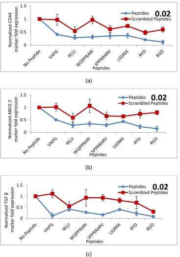
2.1.3. Comparison of CD44, ABCG2, and TGF β Expressions of Tumor Cells Encapsulated in PEGDA with Different Conjugated Peptides
Figures 2.4. (a)- (c) shows the expressions of breast CSC markers CD44, ABCG2, and TGF
β for the encapsulated cells, normalized with respect to the control group. CD44 is a cell
36
in cell proliferation, cell differentiation, cell migration, angiogenesis, presentation
of cytokines, chemokines, and growth factors to the corresponding receptors, and
reducing of proteases at the cell membrane, as well as in signaling for cell survival. All
these biological properties are critical to the physiological activities of cancer cells (Eibl
et al. 1995).
ABCCG2 (ATP-binding cassette sub-family G member 2) is a membrane-associated
protein encoded by ABCG 2 gene. ABCG2 protein is part of the superfamily of
ATP-binding cassette (ABC) transporters. ABC proteins transport several molecules across
extra- and intra-cellular membranes. Therefore, ABCG2 referred to as the Breast Cancer
Resistance Protein. This protein functions as a xenobiotic transporter which probably
play a role in multi-drug resistance to chemotherapeutic agents (Allikmets et al. 1996).
TGF-β (Transforming growth factor beta ) is a secreted protein that mostly
controls cellular proliferation and differentiation (Schoenhoff et al. 2009). Normally it is
acting through its signaling pathway, stops the cell cycle at the G1 stage to prevent
proliferation, cause differentiation, or stimulate apoptosis. In many cancer cells, parts of
the TGF-β signaling pathway are mutated, therefore, TGF-β no longer controls the cell
proliferation. As a consequence, the cancer cells and surrounding stromal cells
37
(a)
(b)
(c)
Figure 2.4 Comparison of CD44, ABCG2, and TGF β expressions of the tumor cells in
PEGDA gels conjugated with peptides. Illustrate the effect of peptides on the
normalized CD44 (a), ABCG2 (b), and TGF β (c) marker fold expressions of MDA-MB-123 cells encapsulated in PEGDA gel for 9 days in CSC medium.
-0.2 0 0.2 0.4 0.6 0.8 1 1.2
0 0.01 0.02 0.04 0.06 0.09
N o rm aliz ed CD 44 Ma rk er Fold E xp re ss ion
Peptide Concentration (mg/mg polymer)
VAPG IKLLI WQPPRARI SPPRRARV LIGRKK RYD RGD -0.2 0 0.2 0.4 0.6 0.8 1 1.2
0 0.01 0.02 0.04 0.06 0.09
N o rm aliz ed ABC G 2 Ma rk er Fold E xp re ss ion
Peptide concentration (mg/mg polymer)
VAPG IKLLI WQPPRARI SPPRRARV LIGRKK RYD RGD -0.2 0 0.2 0.4 0.6 0.8 1 1.2
0 0.01 0.02 0.04 0.06 0.09
N o rm aliz ed T G F β Ma rk er Fold E xp re ss ion
Peptide Concentration (mg/mg polymer)
38
(a)
(b)
(c)
Figure 2.5 Comparison of normalized CD44, ABCG2, and TGF β expressions of the tumor cells encapsulated in PEGDA gels with 2% concentration of different conjugated
peptides. Representative of the comparison of the effect of peptides in 2%
concentration on the normalized CD44 (a), ABCG2 (b), and TGF β (c) marker fold expressions of MDA-MB-123 cells encapsulated in PEGDA gel for 9 days in CSC medium.
39
surrounding stromal cells, immune cells, endothelial and smooth-muscle cells. It causes
immunosuppression and angiogenesis, which makes the cancer more invasive. TGF-β
also changes effector T-cells, which under normal condition attack cancer with an
inflammatory (immune) reaction, into regulatory (suppressor) T-cells, which turn off the
inflammatory reaction (Epstein et al. 2000).
After 9 days of incubation, CD44, ABCG2, and TGF β expression levels in the cells
encapsulated in PEGDA with no peptide was significantly higher than the level of
expressions in the cells encapsulated in PEGDA conjugated with peptides.
Figures 2.5. (a)- (c) shows the comparison of normalized expressions of breast CSC
markers CD44, ABCG2, and TGF β for the tumor cells, encapsulated in PEGDA with a 2%
concentration of different peptides for 9 days in CSC medium. As shown in Figure 2. 5.
(a), (b), and (c) for all of the peptides with 2% concentration, there is a significant
decrease in CD44, ABCG2, and TGF β expression levels of cancer cells with respect to the
control group. On the other hand, in groups with 2% concentration of respective
scrambled peptides, there is no major difference in the levels of CD44 and ABCG2
expression of cells with respect to the control group while there is a significant variance
between CD44 and ABCG2 expression of cells encapsulated with each peptide and its
scrambles one. In a 1 % concertation, WQPPRARI and RYD were more effective on
reducing the markers expression among the peptides. In a 2% concentration WQPPRARI,
RYD and RGD were more effective on reducing the markers expression among the
40
the markers expression significantly, suggesting that the reduction is not specific to the
peptides.
2.1.4. Comparison of the Effect of Conjugated Peptides on Sphere Formation of Tumor Cells Encapsulated in PEGDA for 9 Days in CSC Medium
Results for the measurements of cell and sphere density together with marker
expressions illustrated that in a 0.01 concentration of conjugated peptides, RYD and
WQPPRARI were the most effective peptides in reducing tumor sphere formation. In a
0.02 concentration, WQPPRARI, RGD, and RYD were the most effective peptides in
reducing the sphere formation among peptide. Plus, 4% and higher concentrations are
too high amounts of peptides which change the PEGDA matrix and cause nonspecific
interactions with breast cancer stem cells.
2.1.5. How Cell Adhesion Peptides Affect the Sphere Formation of Tumor Cells Encapsulated in an Inert PEGDA System?
When MDA-MB-231 cancer cells are cultured in the inert PEGDA hydrogel matrix, there
is a population of these cells that have high expression of CD44 cell surface glycoprotein.
This population of cells interacts with each other to grow and divide, and eventually
form a tumorsphere. These cells which have breast cancer stem cells properties are
non-adherent and through the cell-cell interaction instead of the cell-matrix interaction
survive in the matrix; however, conjugating cell binding peptides to the matrix will cause
a competition for the cells to interact with each other or with the matrix. By binding of