Analysis of a novel MRI Based Brain Tumour Classification Using
Probabilistic Neural Network (PNN)
Tasnim Azad Abir*, Jinat Ara Siraji, Eftekhar Ahmed
Department of Electronics and Communication Engineering, Khulna University of Engineering & Technology, Khulna, Bangladesh
ABSTRACT
Now-a-days the most horrendous curse to mankind is cancer. But if we can detect the tumour at an early stage, at least we can make an attempt to cure it. MRI is perhaps the most popular non-invasive imaging diagnostic technique which does not use any radio-active energy harmful for body tissue. Pre-processing of MRI images helps the detection method to eliminate computational complexity. The redundant pixels of an image can be removed by DCT. Feature values can reveal certain properties of an image. GLCM (Gray Level Co-occurrence Matrix) is an efficient way to extract the feature values. These feature values can be used as the input of a PNN structure. PNN is widely used in classification and pattern recognition problems. The prototype designed has desired accuracy, specificity and sensitivity.
Keywords: Brain tumour, MRI, GLCM, Feature Extraction, Classification, Accuracy.
I.
INTRODUCTIONThe cause of growing tumour cell is not known yet. Tumour refers to a special diagnostic theory of medical science. Cells at any place in the body swell with abnormal or discomforting slowly or rapidly [4], or in a word tumour, the transformation of the main body cells or the addition of new cells. Sometimes it grows slowly and sometimes abrupt fast growth of tumour causes cancer.
Magnetic resonance imaging (MRI) is currently a diagnostic imaging technique for the early detection of any abnormal changes in tissues and organs [4]. MRI (Magnetic Resonance Imaging) is an extraordinary Diagnostic Tool of the era. Through this, pictures of different parts of the body can be taken. The gel that is used when doing the MRI is not harmful for body tissue, which is one of the biggest advantages of this technique. It is a fast, non-invasive technique.
In this paper, we have classified the brain tumour into three types, [4] which are- benign, pre-malignant and malignant tumour. Benign tumour is the preliminary stage where the cell grows following a pattern and it is curable. Pre-malignant tumour refers as an abrupt growth of cell which can’t be pre-determined. The malignant tumour is the most harmful and actually known as the cancer cell.
DCT helps to decrease the redundancy of same types of pixel values of an image. GLCM (gray level co-occurrence matrix) decreases the level of the image matrix. Texture features are extracted from this GLCM. Each of the feature value represents some particular spatial properties of the tumour.
II.
METHODOLOGYA. Database
The raw pixel values of tumour image are taken from figshare.com.
B. Methodology
A brain tumour is a collection, or mass, of abnormal cells in your brain. Human skull, which encloses the brain, is very rigid. Any growth inside such a restricted space can cause problems. Brain tumours can be cancerous (malignant) or noncancerous (benign). When benign or malignant tumours grow, they can cause the pressure inside your skull to increase. This can cause brain damage, and it can be life-threatening.
Brain tumours are categorized as primary or secondary. A primary brain tumour originates in your brain. Many primary brain tumours are benign. A secondary brain tumour, also known as a malignant brain tumour, occurs when cancer cells spread to your brain from another organ, such as your lung or breast. An MRI is different from a CT scan because it doesn’t use radiation, and it generally provides much more detailed pictures of the structures of the brain itself.
The most common treatment for malignant brain tumours is surgery. The goal is to remove as much of the cancer as possible without causing damage to the healthy parts of the brain. While the location of some tumours allows for easy and safe removal, other tumours may be located in an area that limits how much of the tumour can be removed. Even partial removal of brain cancer can be beneficial.
Risks of brain surgery include infection and bleeding. Clinically dangerous benign tumours are also surgically removed. Metastatic brain tumours are treated according to guidelines for the type of original
cancer. Surgery can be combined with other treatments, such as radiation therapy and chemotherapy. Physical therapy, occupational therapy, and speech therapy can help to recover after neurosurgery.
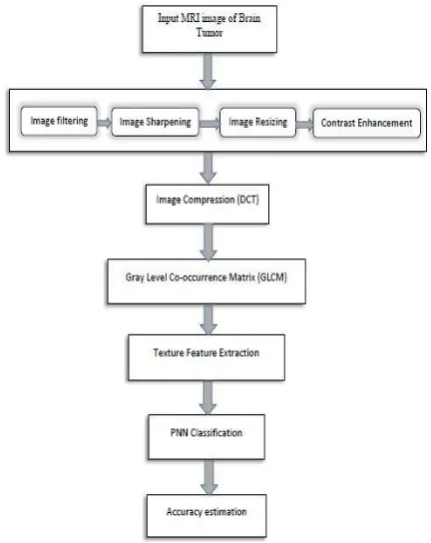
The proposed prototype is designed using GLCM based feature extraction architecture where the MRI image is processed to classify the tumour. Basic classification architecture is shown in Figure 1 below.
Figure 1: GLCM based PNN architecture
III.
PRE-PROCESSINGPre-processing includes image filtering, image resizing, contrast enhancement and image sharpening.
A. Image Filtering
B. Image Resizing
To take different random images as input, the method needs a step to resize the images into a particular shape to avoid further complexity.
C. Image Sharpening
The use of median filter blurs the image which needs a sharpness of image. In this work, the image sharpening is done by the MATLAB default image sharpening function.
D. Contrast Enhancement
For the enhancement of contrast, histogram equalization is used. Histogram equalization is a method in image processing of contrast adjustment using the image's histogram.
IV.
DCTBy removing the redundancy DCT achieves better energy compaction. As the larger number of coefficients gets wiped out, the greater the bits are saved for the same loss.
V.
FEATURE-EXTRACTIONTexture features of a particular image reveals certain properties of that particular image which can be further used to justify the image.
Gray Level Co-occurrence Matrix (GLCM) is used in this work to extract the features of an image. It is a statistical method to examine the texture with the help of the spatial relationship of the pixels. Using the spatial relationship, it calculates the number of times those pixel pairs occurs in that image and put that value in the corresponding position of new matrix, which is the desired GLCM. The Gray Level Co-occurrence Matrix is followed by the Discrete Cosine Transformed Matrix. The extracted features can be used for the robust and accurate classification of the brain tumour.
22 different features were extracted for this following work. Each of these features will reveal certain properties of the spatial distribution. These features values will be used as the input of the probabilistic neural network (PNN) to classify the brain tumour. The three classes of the brain tumour will directly depend on these 22 feature values.
TABLE 1:22 EXTRACTED FEATURES OF AN IMAGE
FEATURES
CONTRAST SUM ENTROPY
DISSIMILARITY SUM VARIANCE
ENERGY DIFFERENCE
VARIANCE
ENTROPY DIFFERENCE
ENTROPY HOMOGENEITY
MATLAB
INFORMATION
MEASURE OF
CORRELATION 1 HOMOGENEITY
PAPER
INFORMATION
MEASURE OF
CORRELATION 2
VARIANCE CLUSTER
PROMINENCE INVERSE
DIFFERENCE NORMALIZED
CLUSTER SHADE
INVERSE DIFFERENCE MOMENT NORMALIZED
AUTOCORRELATION
MAXIMUM PROBABILITY
CORRELATION PAPER
VI.
PNNDESIGNFigure 2: Basic network architecture of PNN [5]
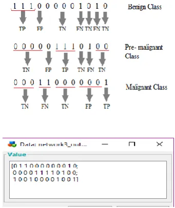
Here, the black vertical bar is our input layer [5]. In this work, for a particular image P, the dimension will be 22 × 1. We will train the network with 30 different images. So here, the weight matrix W will be 30 × 22. The radial basis layer will perform a layer by layer multiplication which will produce |W- P|*b. The rad-bas function will be . Each of these values will be multiplied by the layer weight matrix M. In the competitive layer, C will compare the outputs from three classes and will put 1 to the largest value. The rest will be 0. So the 1 will be our desired class.
VII.
SIMULATION RESULTS AND DISCUSSIONWe extracted features of 30 images after doing the pre-processing and DCT. These feature values were used as the input of our network.
Figure 3: (from left) original Image, contrast enhanced image and DCT image
These 30 images were used to train the network.
Figure 4: Contrast enhanced images of training dataset
In probabilistic neural network, Spread value [4] has great influence on its performance, and probabilistic neural network will generate bad prediction results if it is improperly selected.
Figure 6: Output of network 2 for spread value =5
Figure 7: Output of network 3 for spread value =10
Figure 8: Output of network 4 for spread value =15
A. Accuracy Estimation
Here we used 12 samples to classify the brain tumour between benign, pre malignant and malignant tumour. We used the spread value [4] as the smoothing factor.
TABLE 2:ACCURACY, SPECIFICITY AND SENSITIVITY OF BENIGN TUMOUR
Sprea d value
T P
T N
F P
F N
Accura cy
Specifici ty
Sensiti vity
1 3 6 1 2 75% 85.7% 60%
5 3 6 1 2 75% 85.7% 60%
10 2 7 2 1 75% 77.78% 66.67 % 15 2 8 2 0 83.33% 80% 100% 20 1 8 3 0 75% 72.72% 100% 25 0 8 4 0 66.67% 66.67% 0 30 0 8 4 0 66.67% 66.67% 0
TABLE 3:ACCURACY, SPECIFICITY AND SENSITIVITY OF PRE-MALIGNANT TUMOUR
Spread
value
TP TN FP FN Accuracy Specificity Sensit
ivity
1 3 8 1 0 91.67% 88.89% 100%
5 3 7 1 1 83.33% 87.5% 75%
10 4 7 0 1 91.67% 100% 80%
15 4 7 0 1 91.67% 100% 80%
20 4 7 0 1 91.67% 100% 80%
25 4 6 2 0 83.33% 100% 80%
30 4 5 0 3 75% 100% 57.14
%
In case of pre-malignant tumour, the accuracy was fixed for the spread value of 1, 10 and 15. If we increase the spread value further, the accuracy will decrease.
TABLE 4:ACCURACY, SPECIFICITY AND SENSITIVITY OF MALIGNANT TUMOUR Sprea d value T P T N F P F N Accurac y Specificit y Sensitivit y
1 2 6 2 2 66.67% 75% 50%
5 1 6 3 2 58.33% 66.67% 33.33% 10 2 6 2 2 66.67% 75% 50% 15 3 6 1 2 75% 85.71% 60% 20 3 5 1 3 66.67% 83.33% 50% 25 3 5 1 3 66.67% 83.33% 50% 30 2 5 2 3 58.33% 71.42% 40%
For malignant class, the accuracy decreases from spread value=15.
TABLE 5:ACCURACY, SPECIFICITY AND SENSITIVITY OF OVERALL SYSTEM
From the following table, we can see that if we increase the spread value from 15, the accuracy decreases. So that 15 is the optimal spread value for 30 trained data to achieve best accuracy. In our case, the system was more accurate for pre-malignant type tumour.
VIII.
CONCLUSIONIn this work, PNN has been implemented for classification of MR brain image. PNN is adopted as it has faster training speed and simple structure. Thirty samples of brain MRI were used to train the PNN classifier and tests were run on twelve sets of images to examine classifier accuracy. The developed classifier was examined under different spread values as a smoothing factor. Experimental result indicates that PNN classifier is workable with an accuracy ranged from 83.3% to 72% according to the spread value. Maximum accuracy of 83.33% was achieved by spread value of 15. This work will act as supportive tool for radiologists and will help doctors for fast diagnosis based on which the treatment plan can be decided.
IX.
REFERENCES1. Sonali B. Gaikwad and Madhuri S. Joshi “Brain Tumour Classification using Principal Component Analysis and Probabilistic Neural Network”, International Journal of Computer Applications (0975 – 8887) Volume 120 – No.3, June 2015. 2. GPreethi and Mr.V.Sornagopal.” MRI Image
Classification Using GLCM Texture Features” IEEE GCC Conference and Exibition,Pages: 295-298,2011. 3. Issac H.Bankman, Hand book of Medical Image
processing and Analysis, Academic Press, 2009. 4. Shobana G and Ranjith Balakrishnan “Brain Tumour
5. Othman, Mohd Fauzi and Mohd Ariffanan Mohd Basri. "Probabilisticneural network for brain tumour classification." Intelligent Systems, Modelling and Simulation (ISMS), 2011 Second International Conference on. IEEE, 2011
6. Geordiadis, Et all, “Improving brain tumour characterization on MRI by probabilistic neural networks and non-linear transformation of textural Features”,computer methods and program in biomedicine, vol 89,pp2432,2008.
7. Specht, Donald F. "Probabilistic neural networks for classification, mapping, or associative memory." Neural Networks, 1988., IEEE International Conference on. IEEE, 1988.
8. Noramalina Abdullah, Umi Kalthum Ngah, Shalihatun Azlin Aziz. "Image Classification of Brain MRI Using Support Vector Machine" 978-1-61284-896-9/11/$26.00 ©2011 IEEE.
9. Kailash D.Kharat & Pradyumna P.Kulkarni &M.B.Nagori “Brain Tumour Classification Using Neural Network Based Methods”.
10. Georgiadis. Et all , “ Improving brain tumour characterization on MRI by probabilistic neural networks and non-linear transformation of textural features”,Computer Methods and program in biomedicine, vol 89, pp24-32, 2008.
11. Pauline John “Brain Tumour Classification Using Wavelet And Texture Based Neural Network” International Journal Of Scientific & Engineering Research Volume 3, Issue 10, October-2012 1 ISSN 2229-5518.
12. Varada S.Kolge ,Prof.K.V.Kulhalli, "A novel approach to automated Brain tumour Classification using Probabilistic Neural Network", International Journal ojComputational Engineerin ResearchS (ijcrtonline) Vol. 2 Issue.7.201 2.
![Figure 2: Basic network architecture of PNN [5]](https://thumb-us.123doks.com/thumbv2/123dok_us/1227351.1627537/4.595.50.223.559.616/figure-basic-network-architecture-pnn.webp)