© 2019 by the Serbian Biological Society How to cite this article: Bošković EV, Galović VO, Karaman MA. Spatial 435 distribution of genets in populations of Saprotrophic basidiomycetes, Mycetinis
alliaceus, Marasmius rotula and Gymnopus androsaceus, from Serbian and Montenegrin forests. Arch Biol Sci. 2019;71(3):435-41.
Spatial distribution of genets in populations of saprotrophic basidiomycetes, Mycetinis
alliaceus, Marasmius rotula and Gymnopus androsaceus, from Serbian and Montenegrin
forests
Eleonora V. Bošković1,*, Vladislava O. Galović2 and Maja A. Karaman1
1University of Novi Sad, Faculty of Sciences, Department of Biology and Ecology, Trg Dositeja Obradovića 2, 21000 Novi
Sad, Serbia
2University of Novi Sad, Institute of Lowland Forestry and Environment, Antona Čehova 13, 21000 Novi Sad, Serbia
*Corresponding author: [email protected]
Received: February 18, 2019; Revised: April 4, 2019; Accepted: April 9, 2019; Published online: April 10, 2019
Abstract: Saprotrophic basidiomycetes play a crucial role in leaf-litter decomposition, especially in nitrogen-limited boreal and temperate forests. Populations of this group of fungi have been inadequately investigated. We examined the popula-tions of three different saprotrophic species (Mycetinis alliaceus, Marasmius rotula and Gymnopus androsaceus)in forests in Serbia and Montenegro. To determine the distribution of genets at each of the three investigated sites, molecular analysis was conducted using the inter-simple sequence repeats (ISSR) method. Seven to fifteen genets (genotypes, individuals) were identified on each site and the majority of them were represented by a single sporocarp. The sizes of the genets with two or more sporocarps were estimated to range from 0.3 to 4.0 m. Results obtained in this study suggest that populations of these three species can consist of numerous and relatively small genets.
Keywords: genet size; (GACA)4; (GTG)5; ISSR; litter-exploiting
INTRODUCTION
Fungi play an important role in leaf-litter decomposi-tion as they contribute up to 90% of the total respiradecomposi-tion of soil organisms [1]. Saprotrophic basidiomycetes, along with ectomycorrhizal species [2], are the key organisms responsible for sequestration and release of carbon in the forest floor, especially in nitrogen-limited boreal and temperate forests [3]. In spite of their critical role in these environments, little is known about the size and spatial distribution of genets of saprotrophic basidiomycetes. The mycelial nature and cryptic lifestyle of fungi makes studies of their populations relatively difficult, but the use of molecular markers can help reveal spatiotemporal population dynamics of selected fungal species [4].
The ISSR method was shown to be useful in the identification of genets in local populations of ec-tomycorrhizal species of basidiomycetes [5-8]. This molecular technique uses a single 16-18 bp primer composed of a repeated sequence to amplify
numer-ous inter-microsatellite sequences at multiple loci throughout the genome [9]. Amplified fragments create multilocus and polymorphic band patterns that are often polymorphic between different genets and can be used for their distinction [9]. Thus far, there are no published findings regarding the use of ISSRs
on Mycetinis alliaceus and Gymnopus androsaceus, and
only one report on the use of these molecular markers
in Marasmius rotula [10].
The sizes and distributions of genets in popula-tions of saprotrophic basidiomycetes, especially in the litter-exploiting group, have been rarely investigated [11-16]. In most of these studies the identification of genets was performed by somatic-incompatibility (SI) testing which may not accurately reveal their dis-tribution in all cases [17]. Population studies of two litter-exploiting basidiomycetes, G. androsaceus and M.
rotula [11,12], have found that the majority of genets
to genets of ectomycorrhizal species which are usu-ally between 7 to 30 m [5,17], but can grow up to 600 m in diameter (e.g. a genet of Amanita pyramidifera
[18]). It was suggested that the size and density of some ectomycorrhizal species’ genets varied depend-ing on forest age, with fewer larger genets occurrdepend-ing in older forests, while numerous smaller individuals were detected in younger stands [19,20]. It was sug-gested that ectomycorrhizal fungi forming small genets frequently reproduce sexually and re-establish from spores [21-23], while fungi developing large genets are capable of expanding by mycelia over time [19,24]. The fungal species investigated in this study are common, widespread and usually produce numerous sporocarps in a relatively small area throughout the summer and autumn in temperate broadleaf and mixed forests. All this makes the chosen species good models for population studies of litter-exploiting basidiomycetes.
Mycetinis alliaceusis a litter-decaying species in
Fagus forest and is known from most Fagus-areas in
Europe [25]. Its fruit bodies (sporocarps) are mostly found on decaying stems and twigs of Fagus sylvatica, rarely on the leaves, but it was also recorded on Carpi-nus and coniferous needles [26]. Gymnopus androsaceus
is a common species, found from the lowlands up to alpine habitats in Mediterranean, temperate, boreal and arctic zones. Its fruit bodies are gregarious on lit-ter of coniferous trees. It can be found less frequently on the leaves of broad-leaved trees, dwarf shrubs and herbs, sometimes even growing endophytically on living plants [26]. The fungus spreads in the litter layer by means of black rhizomorphs. G. androsaceus
is an effective degrader of lignin and cellulose, which makes it one of the major decomposers of needle lit-ter [12]. Marasmius rotula is widespread in the boreal and temperate zones of the northern hemisphere. Its sporocarps are usually found in groups on wood (branches, trunks, bark) of broadleaved trees, but they can also be collected from cupules of Fagus, basal parts of grass, and twigs and needles of Pinus [26].
The aim of the present study was to determine the numbers and sizes of the genets of M. alliaceus,
G. androsaceus and M. rotula from three different
forest sites in Serbia and Montenegro. The obtained findings should contribute to a better understanding of the ecology of these three species of saprotrophic basidiomycetes.
MATERIALS AND METHODS
Investigated sites and sporocarp sampling
Sporocarps of all species analyzed in this studywere collected from three sites located on two mountains in the Republic of Serbia, Mt. Stara Planina and Mt. Tara, and on a mountain in the territory of the Repub-lic of Montenegro, Mt. Biogradska Gora. These sites were within protected forest areas in three national parks, with no cutting of trees nor removal of dead wood in the last 25 to 40 years. This makes these sites good representatives of natural, undisturbed forests that are acceptable for investigation of natural fungal populations. Detailed descriptions of the investigated sites, samples of fungi and their substrates are given in Supplementary Table S1.
All present sporocarps within the investigated sites were collected and studied. Between 12 and 23 (10 sporocarps from Mt. Stara Planina and 13 sporocarps from Mt. Tara of M. alliaceus; 15 sporocarps from Mt. Tara of G. androsaceus; 12 sporocarps from Mt. Biogradska Gora of M. rotula) sporocarps from each analyzed species were collected from the three sites. At each site, individual sporocarps were mapped using a GPS device and by measuring the physical distance between them using a tape meter.
Molecular analysis
DreamTaq DNA polymerase (Thermo Fisher Scientific, Massachusetts, USA). Cycling conditions consisted of initial denaturation at 95°C for 2 min, followed by 30 cycles at 95°C for 30 s, at 50°C for 30 s and at 72°C for 1 min for (GTG)5 primer, and 40 cycles at 94°C for 1 min,at 48°C for 1 min and at 72°C for 1 min for (GACA)4 primer, and final extension at 72°C for 10 min. PCR products were visualized on 1.5% agarose gels prepared with 2 μL ethidium bromide (1 mg/ mL), and using a 1 kb DNA Ladder (Thermo Fisher Scientific, Massachusetts). Gels were documented with the BioDocAnalyze System (Analytik Jena AG, Germany). Gel band patterns were analyzed visually. Sporocarps which showed identical band patterns on each gel, that is for both (GTG)5 and (GACA)4 prim-ers, were considered to belong to the same individual (genet) and were grouped accordingly.
RESULTS
DNA fragment analysis
Reproducible banding patterns were obtained with both primers for the majority of the collected sporo-carps (Supplementary Figs. S1-S3). The number and
the size of bands varied between the analyzed species and between the primers: (GTG)5 primer produced 5-14 (750-3500 bp), 5-9 (825-2500 bp) and 8-16 (440-6000 bp) fragments for Mycetinis alliaceus, Marasmius
rotula and Gymnopus androsaceus respectively, while
(GACA)4 primer produced 2-9 (300-4000 bp) frag-ments for M. alliaceus, 2-6 (1000-2000 bp) fragments
for M. rotula and 1-7 (440-2750 bp) fragments for G.
androsaceus. (GACA)4 primer produced fewer
frag-ments than (GTG)5 primer in all analyzed species. Clustering of sporocarps based on the band pat-terns obtained with both primers were congruent, but there were some inconsistencies with a few samples
of M. alliaceus and M. rotula (Table 1). Samples MA4,
MA5, MA6 and MA7 of M. alliaceus were clustered into two genets (genets A and B; Table 1, Fig. 1A) ac-cording to primer (GTG)5; the results of amplification with (GACA)4 primer showed different grouping of samples – MA4 and MA7 had identical band patterns (genet D; Table 1, Fig. 1A) while MA5 and MA6 had unique products of amplification (genets ma5 and ma6; Table 1, Fig. 1A). According to the band patterns obtained with (GACA)4 primer, samples MR3, MR8 and MR6 of M. rotula represent the same genet (genet A; Table 1, Fig. 2), but groupings based on (GTG)5 Table 1. ISSR groups determined by band patterns obtained with two primers (GTG)5 and (GACA)4 for sporocarps of Mycetinis alliaceus, Marasmius rotula and Gymnopus androsaceus collected from the investigated localities: Mt. Stara Planina, Mt. Tara and the National Park (NP) Biogradska Gora. Columns “(GTG)5” and “(GACA)4”show designations of the collected sporocarps. Bolded designations refer to sporocarps which had identical band patterns with both primers. Designations of individual genets are given in the column “genet”. Genets which were designated with capital letters A, B or C are presented with two or more sporocarps, while genets presented with one fruit body have the same designation as the sporocarp but are in lowercase letters
Mycetinis alliaceus Marasmius rotula Gymnopus androsaceus
Mt. Stara planina Mt.Tara NP Biogradska gora Mt. Tara
(GTG)5 (GACA)4 genet (GTG)5 (GACA)4 genet (GTG)5 (GACA)4 genet (GTG)5 (GACA)4 genet
MA1 MA1 ma1 MA11 MA11 ma11 MR1 MR1 mr1 GA1 GA1 ga1
MA2 MA2 ma2 MA12 MA12 ma12 MR2 MR2 mr2 GA3 GA3 ga3
MA3 MA3 ma3 MA13 MA13 ma13
MR3 MR8
MR3 MR8
MR6 A
GA4 GA4 ga4
MA4
MA5 MA4MA7 A or D MA14MA15 MA14MA15 ma14ma15 GA5GA6 GA5GA6 ga5ga6
MA6
MA7 B MA17MA16 MA16MA17 ma16ma17 MR12MR4 MR12MR4 B GA7GA8 GA7GA8 ga7ga8
MA8 MA8 ma8 MA18 MA18 ma18 MR7
MR9 MR10 MR11
MR7 MR9 MR10 MR11
C
GA9 GA9 ga9
MA9
MA10 MA10 C MA19MA20 MA19MA20 ma19ma20 GA10GA11 GA10GA11 ga10ga11
MA5 ma5 MA21 MA21 ma21 GA12 GA12 ga12
MA6 ma6 MA22 MA22 ma22 MR5 MR5 mr5 GA13 GA13 ga13
MA23 MA23 ma23 MR6 mr6 GA14 GA14 ga14
primer products distinguished them into two genets (genets A and mr6; Table 1, Fig. 2).
Genets of Mycetinis alliaceus collected from the localities Mt. Stara Planina and Mt. Tara
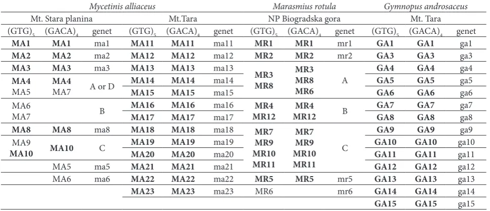
Ten sporocarps of M. alliaceus collected from Mt. Stara Planina were grouped to 9 distinct genets (Table 1). Six genets were represented by a single sporocarp (genets ma1, ma2, ma3, ma5, ma6 and ma8). Sporocarps MA4, MA5 and MA6 were found fruiting on the same twig, while sporocarp MA7 was found in proximity of 20 cm on another branch on the forest floor. In spite of that these, sporocarps were collected from two discrete substrates (twigs), and they probably represent one or two distinct genets (genets A and B; Table 1, Fig. 1A). All 13 sporocarps of M. alliaceus collected from Mt. Tara had unique PCR fingerprints so each of them represents a distinct genet (Fig. 1B).
Genets of Marasmius rotula from the investigated site in the National Park Biogradska Gora
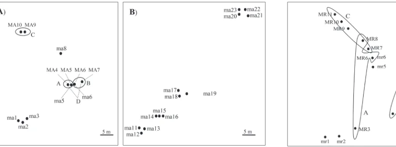
In the locality Mt. Biogradska Gora, 12 sporocarpsof
M. rotula were collected and they were grouped into
7 genets (Table 1). All sporocarps of M. rotula were collected from an area of approximately 4x4 m (Fig. 2). Genet A was comprised of 2 or 3 sporocarps (based on primers (GTG)5 or (GACA)4, respectively) with a diameter of at least 4 m. Genet B was comprised of 2
sporocarps which were 3 m distant from each other. Genet C was represented by 4 sporocarps all found within a 2 m diameter. This genet partially overlapped with genet A.
Genets of Gymnopus androsaceus from the locality Mt. Tara
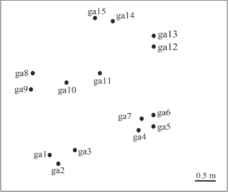
Fifteen sporocarps of G. androsaceus were collected on the investigated site on Mt. Tara from an area of 2x2 m (Fig. 3). Each collected sporocarp was found fruiting single on a pine needle. All sporocarps showed unique PCR fingerprints with both (GTG)5 and (GACA)4 primers (Table 1), so it can be assumed that each of them represented a distinct genet.
DISCUSSION
At site one (on Mt. Stara Planina), 10 collected spo-rocarps of Mycetinis alliaceus were grouped into 9 genets. As these were mostly represented by a single sporocarp, the sizes of these genets could not be esti-mated. Sporocarps MA4, MA5, MA6 and MA7 were found in close proximity (within 15 cm), all fruiting on the same twig except sporocarp MA7 which was found about 20 cm away on another branch on the forest floor. Although there were some inconsisten-cies between fingerprints obtained with (GTG)5 and (GACA)4 primers in this group of samples, these results Fig. 1. Schematic map of the positions of the sporocarps of Mycetinis alliaceus
collected from the localities Mt. Stara Planina (A) and Mt. Tara (B). Encircled dots represent sporocarps which belong to the same genotypes according to the ISSR band patterns. Genets A and B are not drawn to scale, the sporocarps were found in close proximity (ca 20 cm). Designations of sporocarps and genets are explained in Table 1.
show that the genets of M. alliaceus were not limited to discrete substrates (e.g. a dead branch or twig), but can spread on other similar substrates found in relatively close proximity. Although some studies support the “one-log-one-genet” hypothesis [14,16], the results obtained in this study show that the same principle may not apply for smaller wood substrates (branches and twigs), or at least for the genets of M. alliaceus.
The second population of M. alliaceus was found on Mt. Tara, where 13 sporocarps were collected. In the field, all sporocarps were clustered in 4 groups which were several meters apart. Within each group, sporocarps were found in close proximity of 0.3 to 1.5 m. Results of PCR amplification with both (GTG)5 and (GACA)4 primers showed that all sporocarps collected from this site had a unique band pattern, thus repre-senting a unique small genet. For the species Suillus
bovinus and Suillus variegatus, it was determined that
at sites which are subject to major disturbances (e.g. clear-cutting), their genets were small and appeared in high density [19,28,29]. Such activities have not been recently performed on the site, so the result could indicate that we detected a young population, recently developed from basidiospores. It must be emphasized that, although the presence of a sporocarp indicates the presence of the parent mycelium in the soil, the absence of sporocarps does not necessarily mean that mycelia are absent from the soil [5,29,30]. The
prob-ability that there were other genets present in the soil is high, but they were not producing sporocarps at the time when the samples were collected.
Analysis of (GTG)5 and (GACA)4 banding patterns
of Marasmius rotula sporocarps revealed 7 identified
genets within 12 collected sporocarps. The banding patterns obtained by both primers for samples MR3, MR6, and MR8 were not completely congruent, but they were grouped into the same genet. Out of all seven identified genets, four were represented by only one sporocarp, thus it was not possible to determine their approximate diameters, while other three had diameters ranging from 2 to 4 m. These results are in agreement with our previous study [11] where three distinct genets were identified from an area of similar size as in the present study. The approximate sizes of the two genets identified in the study of Bošković et al. [11] were 2 m in diameter and one even appeared to be 15 m in diameter. Results from both studies indicate that even though they colonize distinct substrates, the genets of M. rotula can occupy relatively large areas of the forest floor. It was also observed that the growth of a particular genet was not necessarily prevented by a second genet, since partial overlap occurred in the case with genets A and C. Holmer and Stenlid [12] re-ported a similar phenomenon with genets of Gymnopus
androsaceus (as Marasmius androsaceus). Although
when growing in culture plates, distinct genets of the same species do not overlap, and in field conditions genets can grow in different layers of forest litter [1] and therefore do not come into contact. This enables them to coexist in the same space and produce fruit bodies; however, further investigations are needed to support this claim.
A population of G. androsaceus was investigated on site two located on Mt. Tara. Fifteen sporocarps were collected and they all showed unique ISSR banding patterns obtained by both primers, thus each of them presents a unique genet. Since all fruit bodies were found in close proximity (Fig. 3), all detected genets have relatively small diameters of up to a few dozen centimeters. These results contrast with the findings of Holmer and Stenlid [12] who found several large genets of G. androsaceus that consisted of numerous sporocarps (10-30) and a few smaller genets repre-sented by one or several fruit bodies (2-4) on each Fig. 3. Schematic map of the positions of the sampled Gymnopus
of the four sites investigated in their study. The sites investigated in both studies were similar in size. It is possible that the population examined in this work was young and recently developed from basidiospores and consequently all genets appeared to be relatively small. The other possibility is not only that Holmer and Stenlid [12] investigated inbred populations (on all four sites sporocarps were collected from an area of 96x288 cm), but they also used SI testing that can fail to separate closely-related genets [31,32]. More investigations are needed to obtain more accurate data about G. androsaceus populations.
The results presented in this study suggest that populations of saprotrophic basidiomycetes, M.
al-liaceus, M. rotula and G. androsaceus, can consist of
numerous and relatively small genets when compared to the genets of ectomycorrhizal species [5,6,8]. Also, it was shown that the genets of M. alliaceus and M.
rotula were not restricted to a single discrete substrate
(twig, bark, and branch).
Funding: This study was carried out within projects supported by the Ministry of Education, Science and Technological Develop-ment of the Republic of Serbia, No III43002.
Author contributions: Eleonora Bošković conducted all field and laboratory work and provided the draft manuscript. All authors were involved in planning the research as well as in the processing of the obtained data and analyses of the results. All authors contrib-uted to the discussion of the results and editing of the manuscript.
Conflict of interest disclosure: The authors declare no conflict of interest.
REFERENCES
1. Osono T. Ecology of ligninolytic fungi associated with leaf litter decomposition. Ecol. Res. 2007;22:955-74.
2. Lindahl BD, Tunlid A. Ectomycorrhizal fungi - potential organic matter decomposers, yet not saprotrophs. New Phy-tol. 2015; 205:1443-47.
3. Tlalka M, Bebber D, Darrah PR, Watkinson SC. Mycelial net-works: Nutrient uptake, translocation and role in ecosystems. In: Boddy L, Frankland J, van West P, editors. British Mycologi-cal Society Symposia Series. Academic Press; 2008. p. 43-62. 3. Anderson JB, Kohn LM. Genotyping, gene genealogies and
genomics bring fungal population genetics above ground. Trends Ecol. Evol. 1998;13:444-49.
5. Sawyer NA, Chambers SM, Cairney JWG. Molecular inves-tigation of genet distribution and genetic variation of Corri-narius rotundisporus in eastern Australian sclerophyll forests. New Phytol. 1999;142:561-8.
6. Zhou Z, Miwa M, Hogetsu T. Analysis of genetic structure of a Suillus grevillei population in a Larix kaempferi stand by polymorphism of inter-simple sequence repeat (ISSR). New Phytol. 1999;144:55-63.
7. Zhou Z, Miwa M, Hogetsu T. Genet Distribution of Ectomycor-rhizal Fungus Suillus grevillei Populations in Two Larix kaemp-feri Stands over Two Years. J. Plant Res. 2000;113:365-74. 8. Hirose D, Kikuchi J, Kanzaki N, Futai K. Genet distribution of
sporocarps and ectomycorrhizas of Suillus pictus in a Japanese white pine plantation. New Phytol. 2004;164:527-41 9. Wang, S, Miao, X, Zhao, W, Huang, B, Fan, M, Li, Z, Huang,
Y. Genetic diversity and population structure among strains of the entomopathogenic fungus, Beauveria bassiana, as revealed by inter-simple sequence repeats (ISSR). Mycol. Res. 2005;109:1364-72.
10. Nagaoka T, Ogihara Y. Applicability of Inter-Simple Sequence Repeat Polymorphisms in Wheat for Use as DNA Markers in Comparison to RFLP and RAPD Markers. Theoretical and Applied Genetics. 1997;94:597-602.
11. Bošković E, Karaman M, Galović V. Spatial distribution of genets in population of saprotrophic fungi Marasmius rotula on Mt. Stara planina. Matica Srp J Nat Sci. 2017;133:143-50. 12. Holmer L, Stenlid J. Population structure and mating system
in Marasmius androsaceus Fr. New Phytol. 1991;119:307-14. 13. Kay E, Vilgalys R. Spatial distribution and genetic relation-ships among individuals in a natural population of the oyster mushroom Pleurotus ostreatus. Mycologia. 1992;84:173-82. 14. Huss MJ. Spatial distribution among mycelial individuals of
Lycoperdiforon pyrme occurring on decaying logs. Mycol Res. 1993;97:1119-25.
15. Murphy JF, Miller OK. The population biology of two litter decomposing agarics on a southern Appalachian Mountain. Mycologia. 1993;85:769-76.
16. Keirle MR, Avis PG, Hemmes DE, Mueller GM. Testing the “one-log-one-genet” hypothesis: methodological challenges of population sampling for the Hawaiian wood-decay fungus Rhodocollybia laulaha. Mycologia. 2014;106:896-903 17. Anderson IC, Chambers SM, Cairney JWG. Use of
molecu-lar methods to estimate the size and distribution of mycelial individuals of the ectomycorrhizal basidiomycete Pisolithus tinctorius. Mycol Res. 1998;102:295-300.
18. Sawyer N, Chambers SM, Cairney JWG. Distribution of Ama-nita spp. genotypes under eastern Australian sclerophyll veg-etation. Mycol Res. 2003;107:1157-62.
19. Dahlberg A, Stenlid J. Size, distribution and biomass of genets in populations of Suillus bovinus (L.:Fr.) Roussel revealed by somatic incompatibility. New Phytol. 1994;128:225-34. 20. Dahlberg A, Stenlid J. Spatiotemporal patterns in
ectomycor-rhizal populations. Can J Bot. 1995;73;1222-30.
21. Gryta H, Debaud JC, Effosse A, Gay G, Marmeisse R. Fine-scale structure of populations of the ectomycorrhizal fungus Hebeloma cylindrosporum in coastal sand dune forest ecosys-tems. Mol Ecol. 1997;6:353-64.
22. Gherbi H, Delaruelle C, Selosse MA, Martin F. High genetic diversity in a population of the ectomycorrhizal basidiomy-cete Laccaria amethystina in a 150-year-old beech forest. Mol Ecol. 1999;8:2003-13.
in the ectomycorrhizal fungus Tricholoma scalpturatum: New insights from population genetics and spatial autocorrelation analysis. Mol Ecol. 2008;17:4433-45.
24. Bonello P, Bruns TD, Gardes M. Genetic structure of a natu-ral population of the ectomycorrhizal fungus Suillus pungens. New Phytol. 1998;138:533-42
25. Courtecuisse R, Duhem B. Guide des champignons de France et d’Europe S.A. Lausanne (Switzerland): Delachaux et Niestlé; 1994. 480 p.
26. Antonín V, Noordeloos ME. A Monograph of Marasmius, Collybia, and Related Genera in Europe Part 1: Marasmius, Setulipes, and Marasmiellus. Eching, Germany: IHW-Verlag; 1993. 229 p.
27. Doyle JJ, Doyle JL. A rapid DNA isolation procedure for small quantities of fresh leaf tissue. Phytochem Bull. 1987;19:11-5 28. Dahlberg A, Stenlid J. Population structure and dynamics in
Suillus bovinus as indicated by spatial distribution of fungal clones. New Phytol. 1990;115:487-93.
29. Dahlberg A. Population ecology of Suillus variegatus in old Swedish Scots pine forests. Mycol Res. 1997;101:47-54. 30. Gardes M, Bruns TD. Community structure of
ectomycor-rhizal fungi in a Pinus muricata forest: above- and below- ground views. Can J Bot. 1996;74:1572-83.
31. Jacobson KM, Miller OK, Turner BJ. Randomly amplified polymorphic DNA markers are superior to somatic incom-patibility tests for discriminating genotypes in natural popu-lations of the ectomycorrhizal fungus Suillus granulatus. Proc Natl Acad Sci USA. 1993;90:9159-63.
32. Guillaumin JJ, Anderson JB, Legrand P, Ghahari S, Berthelay S. A comparison of different methods for the identification of genetics of Armillaria spp. New Phytol. 1996;133:333-43.
Supplementary Data
Supplementary Table S1 and Figs. S1-S3.