ENDOTHELIAL DYSFUNCTION IN
ATHEROSCLEROSIS
RAJESH KUMAR KHARBANDA
Centre for Clinical Pharmacology and Toxicology, The
Ray ne Institute, University College London,
5 University Street, London.
THESIS SUBMITTED FOR THE DEGREE OF DOCTOR
OF PHILOSOPHY OF THE UNIVERSITY OF LONDON,
ProQuest Number: U641886
All rights reserved
INFORMATION TO ALL USERS
The quality of this reproduction is dependent upon the quality of the copy submitted.
In the unlikely event that the author did not send a complete manuscript and there are missing pages, these will be noted. Also, if material had to be removed,
a note will indicate the deletion.
uest.
ProQuest U641886
Published by ProQuest LLC(2015). Copyright of the Dissertation is held by the Author.
All rights reserved.
This work is protected against unauthorized copying under Title 17, United States Code. Microform Edition © ProQuest LLC.
ProQuest LLC
789 East Eisenhower Parkway P.O. Box 1346
Abstract
Endothelial dysfunction may serve as a mechanism to explain the vasoconstriction, inflammation, thrombosis and abnormal flow regulation in atherosclerosis, and play a role in the development of atheroma, and pathophysiology of its complications.
This thesis describes experiments to characterise the mechanisms o f endothelial dysfunction that could contribute to the pathogenesis o f human atherosclerosis and its complications, in three main areas:
1. Mechanisms of flow-mediated dilatation
■\
The role of endothelial nitric oxide (NO) in flow-mediated dilatation (FMD) was investigated in healthy volunteers and hypercholesterolaemic patients. FMD to transient-flow stimuli was found to be NO-dependent and reduced in hypercholesterolaemia. In contrast, FMD to sustained-flow stimuli was largely NO- independent and preserved in these patients. These data suggest a second mechanism of FMD exists in humans, which is preserved in certain atherosclerosis risk groups.
2. The effect of acute inflammation on endothelial function
dysfunction in humans and a link between inflammation and acute cardiovascular risk.
3. The effect of ischaemia-reperfusion on endothelial function
Table of Contents
A bstract...2
Table of Contents... 4
List of F igures...6
List of T a b le s...6
Abbreviations...7
Acknowledgements...9
Publications... 10
CHAPTER 1: In troduction ... 11
1.1 Aims and scope of this thesis... 12
1.2 Atherosclerosis and the endothelium... 15
1.3 Role o f endothelial nitric oxide in flow-mediated dilatation in early atherosclerosis... 36
1.4 The effect of acute inflammation on endothelial function... 38
1.5 The effect o f ischaemia-reperfusion on endothelial function... 44
CHAPTER 2: General m ethods...58
2.1 Human forearm m odel... 59
2.2 Methods to change blood flow in conduit vessels... 59
2.3 Model of acute systemic inflammation... 59
2.4 Forearm model o f ischaemia-reperfusion injury... 60
2.5 Assessment of endothelial function in humans in vivo... 61
2.6 Assessment of neutrophil function... 68
2.7 Assessment of systemic inflammation...70
2.8 Drugs and reagents... 71
2.9 Statistical methods... 71
CHAPTER 3: Role of endothelial nitric oxide in flow-mediated dilatation in early atherosclerosis...72
3.1 Background... 73
3.2 Experimental protocols...74
3.3 Results... 80
3.4 Discussion... 90
CH APTER 4: The effect of inflammation on endothelial function of the arterial v a scu latu re...94
4.1 Background... 95
4.2 Experimental protocols... 96
4.3 Results...97
CHAPTER 5: Mechanisms of inflammation induced endothelial dysfunction
... 107
5.1 B ackground...108
5.2 Experimental protocols... 109
5.3 Results... I l l 5.4 D iscussion... 116
CHAPTER 6: Effect of ischaemia-reperfusion on endothelial function and modulation by local ischaemic preconditioning... 120
6.1 B ackground... 121
6.2 Experimental protocols... 122
6.3 Results...125
6.4 D iscussion... 137
CHAPTER 7: Remote preconditioning of the endothelium in humans in vivo and in experimental myocardial infarction... 141
7.1 B ackground... 142
7.2 Experimental protocols... 144
7.3 Results...148
7.4 D iscussion... 152
CHAPTERS: Conclusion...154
8.1 Role o f endothelial nitric oxide in flow-mediated dilatation in early atherosclerosis... 155
8.2 The effect of acute inflammation on endothelial function... 156
8.3 Effect o f ischaemia-reperfusion on endothelial fimction... 158
8.4 Conclusion... 159
List of Figures
Figure 1-1; Endothelial function and atherosclerosis... 35
Figure 1-2: NO in ischaemia-reperfusion injury... 47
Figure 1-3: Mechanisms o f endothelial injury in ischaemia-reperfusion... 49
Figure 1-4: Mechanisms of preconditioning... 53
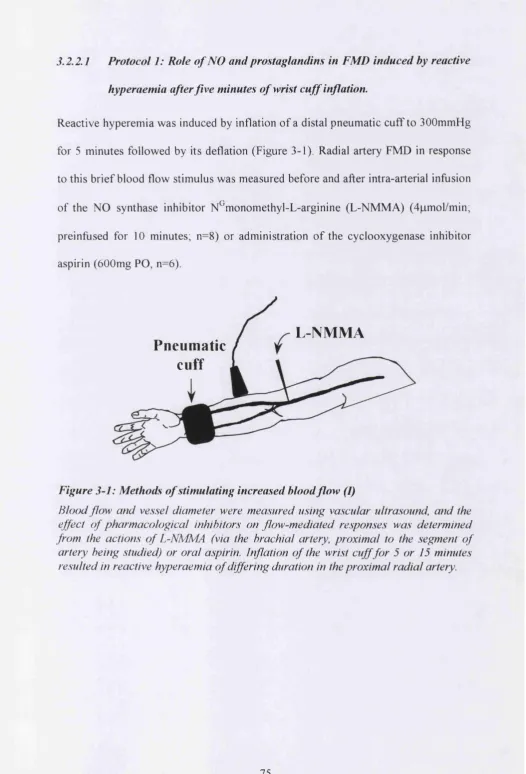
Figure 3-1: Methods o f stimulating increased blood flow (I)...75
Figure 3-2: Methods of stimulating increased blood flow (II)...77
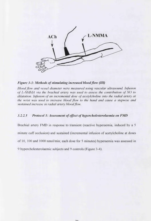
Figure 3-3: Methods of stimulating increased blood flow (III)... 78
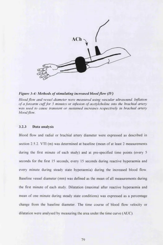
Figure 3-4: Methods of stimulating increased blood flow (IV )... 79
Figure 3-5: Effect of L-NMMA on radial artery F M D ...81
Figure 3-6: Effect of L-NMMA on radial artery FMD (II)... 83
Figure 3-7: FMD of radial artery with hand warming... 85
Figure 3-8: FMD of radial artery with distal infusion of acetylcholine...86
Figure 3-9: Brachial artery FMD in hypercholesterolaemia... 89
Figure 4-1: Forearm blood flow (FBF) responses before and after vaccination 100 Figure 4-2: FMD and GTN responses before and after vaccination... 102
Figure 5-1: Effect of oral aspirin pre-treatment on vaccine associated endothelial dysfunction... 112
Figure 5-2: Effect of oral aspirin pre-treatment on vaccine associated cytokine responses...114
Figure 5-3: Effect of intra-arterial aspirin on FBF response to bradykinin following vaccination...115
Figure 6-1: Effect of ischaemia-reperfusion on resistance vessel endothelial function. ... 127
Figure 6-2: Effect o f ischaemia-reperfusion on resistance vessel smooth muscle function... 128
Figure 6-3: Effect of ischaemia-reperfusion on conduit vessel function...129
Figure 6-4: Radial artery FMD following ischaemia-reperfusion...130
Figure 6-5: Effect of ischaemic preconditioning on the response o f forearm vessels to ischaemia-reperfiision:...132
Figure 6-6: Effect of ischaemia-reperfusion on neutrophil function... 134
Figure 6-7: Effect of ischaemic preconditioning on neutrophil function...136
Figure 7-1: Effect of remote preconditioning on vascular responses to ischaemia-reperfusion in hum ans...149
Figure 7-2: Effect of remote preconditioning on myocardial infarction (animal model)...151
List of Tables
Table 1: Characteristics of subjects studied in Protocol 5 - Assessment of effect of hypercholesterolaemia on FMD...88Abbreviations
ACh acetylcholine
ANOVA analysis of variance
AUC area under curve
BH4 tetrahydrobiopterin
BK bradykinin
BP blood pressure
cGMP 3',5' guanosine monophosphate
COX cyclo-oxygenase
GRP C-reactive protein
EDHF endothelium derived hyperpolarising factor ELISA enzyme-linked immunosorbent assay eNOS endothelial nitric oxide synthase
ET endothelin
FACS flow activated cell sorter
FBF forearm blood flow
FITC fluoroscein isothiocyanate
FMD flow mediated dilatation
GTN glyceryl trinitrate
HDL high-density lipoprotein
ICAM intercellular adhesion molecules
XL interleukin
IPC ischaemic preconditioning
IR ischaemia-reperfusion
Ka t p ATP-sensitive potassium channel
LDL low density lipoprotein
L-NMMA NG-monomethyl-L-arginine
MFI median fluorescence intensity
NADPH reduced nicotinamide adenine dinucleotide phosphate
NO nitric oxide
PAI-1 plasminogen activator inhibitor 1 PDGF platelet-derived growth factor
PGI2 prostacyclin
PNC platelet neutrophil complex
PKC protein kinase C
RH reactive hyperaemia
sGC soluble guanylate cyclase
TGF-b tissue growth factor b
TNF-a tumour necrosis factor alpha
tPA tissue plasminogen activator
TXA2 thromboxane A2
VTI velocity time integral
Acknowledgements
The work described in this thesis was carried out during a clinical PhD studentship from the British Heart Foundation, and would not have been possible without the help and support of many individuals.
Raymond MacAllister and Michael Mullen initiated the work described in this thesis and provided constant supervision and guidance. Raymond MacAllister has always had time, enthusiasm and dedication in training me in the ways o f academic medicine.
Publications
The following publications represent work presented in this thesis:
Hingorani AD, Cross J, K harbanda RK, Bhagat K, Palacios M, Griffin GE, Deanfield J, MacAllister RJ, Vallance P. Acute systemic inflammation impairs endothelium-dependent dilatation in humans. Circulation,2000; 102: 994-999.
K h arbanda R, Mullen MJ, Cross J, Donald AE, Taylor M, Vallance P, Deanfield JE, MacAllister RJ. Mechanisms of flow-mediated dilatation in human conduit arteries in vivo. Circulation Research, 2001; 88: 145-151.
K harbanda RK, Peters M, Walton B, Kattenhom M, Mullen M, Klein N, Vallance P, Deanfield J, MacAllister RJ. Ischemic preconditioning prevents endothelial injury and systemic neutrophil activation during ischemia-reperfusion in humans in vivo. Circulation, 2001; 103: 1624-1630.
K harbanda RK, Walton B, Allen M, Klein N, Hingorani AD, MacAllister RJ, Vallance P. Prevention o f inflammation induced endothelial dysfunction: a novel vasculo-protective action of Aspirin. Circulation, 2002; 105: 2600-2604.
K harbanda RK, Mortensen U.M, White PA, Kristianson S, Schmidt M, Hoschtitzky A, Vogel M. Sorenson K, Redington AN, MacAllister RJ. Transient limb ischemia induces remote ischemic preconditioning in vivo. Circulation, 2002;
CHAPTER 1 :
1.1
Aims and scope of this thesis
This thesis explores three areas of relevance to the further understanding of the contribution o f endothelial dysfunction to the pathogenesis of atherosclerosis: the role of endothelial nitric oxide (NO) in flow-mediated dilatation (FMD) in early atherosclerosis, and the effects of acute inflammation and ischaemia-reperfiision on endothelial function.
Atherosclerosis and its complications are a major cause of morbidity and mortality worldwide. There is a long pre-clinical phase in this disease with the lesions of atherosclerosis present from a young age. The clinical complications of atherosclerosis are diseases o f middle age. The reasons that individuals develop atherosclerosis, and why some present with its clinical complications remain unclear. One major factor involved in the development of atherosclerosis is endothelial dysfunction. Endothelial dysfunction has also been implicated in precipitating ischaemic complications of atherosclerosis, and determining the extent of injury following such complications.
This thesis will present data from human studies that investigate endothelial function in experimental models representing different stages o f atherosclerosis; specifically, the studies described investigate aspects of the role o f the endothelium in the preclinical (presymptomatic) phase, the precipitation of ischaemic complications of atherosclerosis and the determination of tissue damage resulting from ischaemic complications.
Role o f endothelial nitric oxide in flow-mediated dilatation in early atherosclerosis
Reduced NO-bioactivity has been implicated in the pathogenesis of atherosclerosis in many studies, but other endothelial dilator pathways might be involved. Clearly, it is important to define the mechanisms of endothelial dysfunction, as an essential prerequisite to therapeutic intervention. Much o f what is understood about endothelial dysfunction in humans comes from studies of FMD in peripheral conduit vessels, yet uncertainties remain about precisely what is being measured by this technique. The contribution o f NO to flow mediated vasoreactivity o f the vasculature is discussed (section 1.3) as a prelude to its investigation in Chapter 3.
The effect o f acute inflammation on endothelialfunction
The effect o f ischaemia-reperfusion on endothelial function
Nitric oxide (NO) has anti-atherogenic properties and is also involved in the pathophysiology o f the complications o f atherosclerosis. The major clinical syndromes such as stroke and myocardial infarction are characterised by ischaemia, and subsequent reperfusion. The role of the vascular endothelium in these syndromes is understood in animal models, but its role in humans is less well defined. The studies described in Chapter 7 and 8 explore a human model o f ischaemia- reperfiision injury and the effects on endothelial function.
Aim o f this thesis
The aims o f this thesis are to investigate:
1. The role of endothelial nitric oxide in flow-mediated dilatation in early atherosclerosis
1.2
Atherosclerosis and the endothelium
1.2.1 Pathogenesis of atherosclerosis
Cardiovascular and cerebrovascular diseases cause over 15 million deaths every year - one third of the global total (Tunstall-Pedoe et a l, 1994; Reddy, 1998). The incidence of circulatory diseases is increasing in developing countries, partly because lifestyles (and the attendant risk factors) are becoming similar to those common in industrialised countries.
Post-mortem studies have demonstrated that abnormalities of atherosclerosis are present as early as 6 months of age (Stary, 1989; Enos et a l, 1953; McGill, et a l,
1995). Advanced lesions begin to appear from the fourth decade and are the predominant lesion type in middle-aged and older persons. It has been shown in recent intra-vascular coronary ultrasound studies that nearly 17% o f donor hearts from individuals aged less than 20 years o f age have detectable atherosclerosis, and this proportion is nearly 50% in donors over 50 years old (Tuzcu et a l, 2001). Serial studies in heart transplant recipients have confirmed that such atheroma can regress over time, supporting the concept that the process is dynamic in nature (Tsutsui et
a/., 2001).
lesions, which have a dense extracellular lipid core, and Type V lesions, in which intimai lipid deposits are thickened by fibrous tissue.
The deposition o f macrophages in the intima is the earliest abnormality found in arteries (Masuda & Ross, 1990; Katsuda etal., 1992). These cells are filled with lipid in the form o f cholesterol ester and might regulate local lipid levels in the vessel wall. Immunochemistry, Northern blot analysis of messenger ribonucleic acid (mRNA) and in-situ hybridisation have shown the presence of major histocompatibility complex, cluster of differentiation (CD) antigens, and cytokines in macrophages from human specimens, and animal models of early atherosclerosis (de Boer et a l, 1999). As lesions progress, the number of smooth muscle cells also increases (Stary & Malinow, 1982). These cells accumulate lipid but are also the source of the collagen and extracellular matrix, which form the plaque.
Atherosclerotic plaques are dynamic structures and their clinical effects are related to the physical obstruction to blood flow, but they also provide a focus for thrombosis or embolism if they rupture. There is growing evidence that plaques change rapidly, and that non-occlusive plaques may be more likely to fissure and rupture to precipitate acute obstruction to blood flow leading to myocardial infarction or stroke. Studies using intra-vascular coronary ultrasound in patients have highlighted that plaques that chronically occlude the coronary lumen differ from those that cause acute occlusion. These studies have shown that plaques located within the intima and media are more likely to be associated with unstable acute coronary syndromes, and less likely to cause luminal stenosis (Vamava & Davies, 2001). Such plaques also have a greater number of inflammatory cells (Varnava et a l, 2002).
Current theories about the pathogenesis of atherosclerosis begin with Virchow’s
inhibition hypothesis, which proposed that the cellular proliferation seen in the intima was a form o f ‘low-grade inflammation’ or reaction to infiltration o f proteins and lipids from the blood. The von Rokitansky encrustation theory proposed that small thrombi composed of platelets, fibrin, and leukocytes collect over sites of endothelial injury and then organise with smooth muscle proliferation (von Rokitansky, 1852; Virchow, 1856). Ross’s early response-to-injury hypothesis (Ross
become converted to lipid-filled foam cells, perpetuating the inflammatory response and expanding the lesion. The central role for endothelial denudation was revised more recently as it became clear that developing lesions in experimental models have an overlying intact endothelium, and it is now thought that endothelial dysfunction is sufficient to initiate atherosclerosis (Ross, 1999).
1.2.2 The healthy endothelium
The endothelium, a single layer of cells lining the luminal surface of blood vessels, is a direct interface between circulating blood and local tissue and regulates vascular tone, cell adhesiveness, and coagulation, by generating local mediators. These mediators include vasodilators, of which the most important are nitric oxide (NO), prostacyclin (PGI2), and endothelium derived hyperpolarising factor (EDHF). The
endothelium, also generates a number of locally acting vasoconstrictors, including thromboxane (TX) and endothelin (ET).
Nitric oxide
Shear stress is tangential force generated by viscous blood flowing over the endothelial surface. It varies in a pulsatile manner, and increases as blood flow increases. In the arterial circulation changes in shear stress induce endothelial dependent vasodilatation as an early response (Davies, 1995). This is predominantly through increased NO production, as shear can activate eNOS by serine phosphorylation through a calcium-independent mechanism (Fleming & Busse, 2003). ^
The activity o f the eNOS enzyme requires the presence of co-factors such as reduced nicotinamide adenine dinucleotide phosphate (NADPH), flavine adenine dinucleotide (FAD), and tetrahydrobiopterin (BH4). BH4 has been shown to stabilise the dimeric form o f eNOS, to act as an electron donor and as an allosteric regulator of eNOS activity (Cosentino & Luscher, 1999). NO is a gas and diffuses to adjacent smooth muscle where it increases soluble guanylate cyclase (sGC) activity. Under basal conditions sGC is a dimer containing two haem molecules. NO binds to haem, which initiates a conformational change resulting in increased activity o f the enzyme (Ignarro, 1990). The product of sGC is cyclic 3’,5’ guanosine monophosphate (cGMP), which reduces intracellular calcium within the smooth muscle cell, and so causes smooth muscle relaxation (Fiscus, 1988).
1989). Similar studies have been performed in the coronary, pulmonary and systemic vasculature, and have demonstrated the importance of endogenous NO production in the regulation of blood flow in humans in different vascular beds (Quyyumi et a l,
1995b; Stamler a/., 1994; Celermajer a/., 1994).
Prostaglandins
Endothelial cells produce a range of prostaglandin molecules, particularly prostacyclin and thromboxane A]. Prostacyclin is a vasodilator that binds to specific receptors on the target cell to activate adenylate cyclase and increase cyclic AMP (cAMP) levels. In smooth muscle this causes relaxation and inhibits aggregation in platelets; in contrast thromboxane A2 has vasoconstrictor properties and aggregates
platelets (Vallance, 1992). Prostaglandins also contribute to vascular tone in the coronary vasculature (Duffy et a l, 1999).
The precursor for prostaglandins is arachidonic acid, which is released from membrane lipids by phospholipase A. Arachidonic acid is metabolised by cyclo- oxygenase (COX) - also known as prostaglandin H synthase (Davidge, 2001). There are two isoforms o f COX (1 and 2), and these differ by a single amino acid substitution in the active enzyme site. COX-1 is constitutively expressed, whereas COX-2 is induced by inflammatory cytokines and growth factors.
Endothelium-derived hyperpolarising factor (EDHF)
EDHF is a compound that causes hyperpolarisation o f smooth muscle cells and endothelium-dependent vasorelaxation (Feletou M & Vanhoutte PM, 1996). Although NO and prostacyclin also hyperpolarise vascular smooth muscle, the activity of EDHF is not affected by NOS or COX inhibitors in many types of vessel (Feletou & Vanhoutte, 1999; Campbell & Harder, 1999). However, inhibiting calcium activated potassium channels, and inwardly rectifying potassium channels in the smooth muscle cells blocks the hyperpolarisation. The exact identity of EDHF is uncertain, and there may be more than one type of EDHF responsible for hyperpolarisation in different blood vessels. For example, EDHF activity has been ascribed to potassiums ions in the subendothelial space, cytochrome-P450, metabolites of arachidonic acid and myoendothelial gap junctions (Beny & Schaad, 2000, Campbell etal. 1996).
It has not been easy to characterise EDHF because of the toxicity to humans of many of the potassium channel blockers that have been used to block EDHF in animals. However, barium and oubain, which have been proposed as inhibitors of potassium mediated EDHF-like vasodilatation, reduce dilatation to potassium in the human forearm, providing indirect evidence of EDHF-like activity (Dawes et a l, 2002). Cytochrome-P450 inhibitors have also been implicated in potassium mediated vasodilatation in the human forearm (Halcox et a l, 2001).
Endothelins
of cytokines in vitro. Human endothelial cells are the major source of endothelin-1 (ET-1). Endothelins act through two receptor subtypes (ETa and ETb) and are coupled to phospholipases and protein kinase C. Depending upon the concentration and the receptor subtype activated, endothelins can also stimulate NO release (Miyauchi T & Masaki T, 1999). Infusing endothelin into the forearm vasculature reduces blood flow in healthy subjects, and antagonists of endothelin increase forearm blood flow, suggesting basal release (Clarke et a l, 1989; Haynes & Webb,
1994). Endogenous ET-1, acting through the ETa receptor, contributes to
maintenance o f basal coronary artery tone and may have a role in regulating coronary collateral blood flow in humans (Kyriakides et a l, 2000).
Regulation o f circulating cells and cell adhesion
The endothelium also regulates the function of circulating cells by releasing locally active mediators or expressing molecules on the endothelial surface that act as receptors for complementary molecules on the circulating cell surface. In the healthy artery, the leukocytes, platelet and red blood cells do not adhere to endothelium or migrate into the local tissues since the endothelium provides an efficient barrier. Cell-cell adhesion is dependent on the interaction o f three major families o f cell adhesion molecules: the selectins, the integrins and the immunoglobulin superfamily.
rapidly expressed on the cell surface following stimulation but E-selectin is not stored and requires transcription-dependent protein synthesis. Cytokines, bacterial toxins, and oxidants are able to upregulate the expression o f P- and E-selectin on endothelial cells. A major ligand for all three selectins is a specific sialyl-Lewis^ - type glycoprotein on circulating neutrophils and platelets.
Integrins are heterodimeric proteins made up of a and P subunits, pi integrins have a common p subunit (CD29), which is linked to specific a subunits, pi integrins are involved in the adhesion of eosinophils, lymphocytes, monocytes and natural killer cells to activated endothelial cells. The P% integrins consist of a common p subunit (CD 18), which may be linked to one of four a subunits (CD 11 a, CD 11b, C D llc or CD lid ). These are expressed on leukocytes only and are important in regulating monocyte, lympocyte and neutrophil traffic across the endothelium. The ligands for integrins depend upon the a subunit and are in two major groups: the immunoglobulin supergene family and large matrix proteins, such as fibronectin, thrombospondin, fibrinogen and complement components (Diamond & Springer,
1994).
adhesion of leukocytes. VCAM-1 is not normally expressed on endothelial cells, but after stimulation by cytokines there is marked upregulation and enhanced binding of monocytes and lymphocytes. The upregulation can be rapid, through effects on cell stores, or changes in transcription o f adhesion molecules and de-novo protein synthesis. These endothelial adhesion molecules regulate the transmigration of circulating cells such as platelets, leukocytes and monocytes (Simon et a l, 1995).
Locally active mediators influence the interaction between the endothelium and circulating cells. TNF-a, platelet activating factor, and IL-1 (Tsao et a l, 1995) are pro-adhesive substances which upregulate adhesion molecule expression on leukocytes and/or the endothelium, whilst NO is a powerful anti-adhesive molecule, acting both to vasodilate and to reduce expression o f adhesion molecules (Panes & Granger, 1998, Z&hQx et a l, 1995).
Coagulation andfibrinolysis
Thrombosis is central to the progression of atherosclerosis and the process of acute arterial occlusion leading to acute coronary syndromes or stroke. Thrombus formation is a balance between the coagulation and fibrinolytic factors present in blood (Triplett, 2000). Primary haemostasis involves platelet aggregation which leads to the production of thrombin, activation of the coagulation pathway and fibrin formation (Dahlback, 2000). Under normal physiological conditions the endothelium is anticoagulant but endothelial dysfunction may produce a pro-coagulant state.
antithrombin (previously known as antithrombin III), which is a relatively weak inhibitor, but its action is enhanced by heparin-like molecules expressed on the surface of endothelial cells (Sagripanti & Carpi, 2000). The second endothelial anticoagulant pathway is the protein C system. Endothelial cells bind thrombin via thrombomodulin receptors. Once thrombin is bound, the complex activates protein C which breaks down coagulation cofactors and inactivates plasminogen activator inhibitor 1 (PAI-1). Endothelial cells also synthesise tissue factor pathway inhibitor, which inactivates the extrinsic pathway (Sagripanti & Carpi, 2000).
Fibrinolytic pathways digest fibrin and reduce clot formation. The balance between tissue-type plasminogen activator (tPA) and PAI-1 determines blood fibrinolytic activity. tPA converts plasminogen to plasmin and PAI-1, which inhibits the activity of tPA, is the principal determinant of the conversion o f plasminogen to plasmin (Tsikouris et al., 2002). Under basal conditions, the endothelium produces little PAI- 1 and the lytic pathway is most active (Pothula et a l, 2000). NO inhibits platelet aggregation, and the expression o f adhesion molecules such as the fibrinogen receptor (Ilb/IIIa). NO also enhances the activity of the fibrinolytic pathway by stimulating the release of tPA. In animal models, inhalation o f NO prolongs bleeding time, although this effect has not been demonstrated in humans (Krejcy et a l, 1995).
1.2.3 Endothelial dysfunction in atherosclerosis
Endothelial ‘injury’ can disturb normal endothelial vasomotor function, promote the adhesion and transmigration of monocytes and platelets, and favour thrombosis. The following sections will review the abnormalities of the endothelium that have been described in atherosclerosis.
In animal models o f atherosclerosis, there are distinct morphological changes o f the endothelium, including loss o f the normal orientation of endothelial cells in the direction of flow, retraction o f endothelial cells from adjacent cells and rounded appearance o f cells. At the subcellular level there is an increase in cytoplasmic filaments on the abluminal surface (Stary, 1990). Human data are difficult to obtain because there are is a delay in fixation following death and some changes may be induced by fixation itself. Importantly, an intact endothelium is found in early lesions but changes are described in more advanced lesions. Autopsy studies are consistent with the data from experimental models.
Animal studies
Experimental animal models of atherosclerosis (induced by feeding high cholesterol diets) have demonstrated impaired endothelium-dependent relaxation o f conduit vessels vessels at an early stage of atherosclerosis, prior to significant anatomical changes developing (Verbeuren et a l, 1990; Bossaller et a l, 1987; Jayakody et a l,
atherosclerosis’ These functional abnormalities are found in the presence of a
structurally normal endothelium, and are present very early following cholesterol feeding. Even 10 minutes of exposure of blood vessels to a high concentration of purified low density lipoprotein (LDL) can inhibit agonist stimulated endothelium- dependent relaxation (Andrews et a l, 1987). Similar studies have also demonstrated endothelial dysfunction in the resistance vessels in these models o f atherosclerosis (Reddick et a l, 1994; Bonthu et a l, 1997).
Human studies
Human studies have demonstrated reduced endothelium-dependent dilatation in peripheral conduit vessels o f young patients with high cholesterol, diabetes, a family histoiy of heart disease or smokers (Stary, 2000; Celermajer et a l, 1992; Clarkson et a l, 1996a; Clarkson et a l, 1996b) and in the resistance vessels o f patients with high cholesterol and hypertension (Stroes et al, 1997; Panza et a l, 1993). The methods used to study these are described in detail in Chapter 2. Young subjects without manifest atherosclerosis have abnormal endothelium-dependent vasodilator function, which can be detected as early as the first decade of life. In these risk factor groups, clinical interventions that reduce future cardiovascular events, such as lipid lowering therapy and administration o f angiotensin-converting inhibitor, also improve the vasodilator function o f peripheral arteries (Mullen et a l, 1998).
(receptor-mediated dilators) are reduced in arteries with atherosclerosis. Infusion of acetycholine may even cause vasoconstriction in ‘angiographically normal’ arteries in patients with risk factors for atherosclerosis (Ludmer et a l, 1986; Quyyumi et a l,
1995c). These vessels retain the ability to respond to the endothelium-independent vasodilator glyceryl-trinitrate (GTN).
Epicardial coronary vessels also dilate in response to increased flow, but dilatation is reduced in patients with atherosclerosis (Lefroy et a l, 1993; Quyyumi et a l, 1995a; Egashira et a l, 1996). There is a temporal sequence of abnormality, with reduced response to acetylcholine and serotonin initially, followed by substance P and then flow. This suggests that different mechanisms may be affected at different stages of atherosclerosis (Drexler & Zeiher, 1991).
Abnormal endothelium-dependent vasodilatation is an important aspect of the defect identified in experimental models and human studies at all stages of atherosclerosis and its complications. This may result from a deficiency o f NO or other vasodilator mediators, or enhanced vasoconstrictor mechanisms.
Ahnormalities in the L-arginine/NOpathway
As described above, animal models of atherosclerosis exhibit reduced eNOS activity within the vasculature. Moreover, eNOS inhibition accelerates atherosclerosis in animals, an effect that can be reversed by supplementation with arginine (Bouchard et al, 2000; Naruse et al, 1994).
groups) and with established atherosclerosis (Vallance, 1996). In comparisons with healthy controls, a diminished response to L-NMMA has been taken as evidence for reduced NO bioactivity (Calver et al, 1994). Endothelium-dependent dilatation in humans is not wholly attributable to NO (particularly in the resistance vasculature) and so endothelial dysfunction measured by the response to endothelium-dependent agonists does not specifically reflect the NO pathway. NO appears to be involved in the dilator mechanism o f FMD, which is universally reduced in early and established atherosclerosis.
The abnormalities in vasoreactivity may be the result of defects at several points in the process by which NO is generated. These include defects in: (a) the signal transduction mechanisms in the endothelial cell, (b) defects in the mechanism by which NO is produced, (c) or an increase in mechanisms that cause breakdown of NO (Harrison, 1997).
Abnormal signal transduction
Experimental data from porcine models of atherosclerosis show that the endothelium-dependent responses are impaired at an early stage o f atherosclerosis due to an abnormality in the pertussis toxin sensitive Gi protein. As atherosclerosis progresses, the responses to bradykinin and ADP become impaired as a result of abnormality in Gq subunit (Shimokawa et al, 1991). In human vessels Gia expression is affected by ageing, hypertension, and hypercholersterolaemia (Tsutsui et a l,
Reduced production o f NO
Abnormalities of the provision or utilisation o f L-arginine, the substrate for eNOS, may account for reduced NO Circulating L-arginine concentrations are several-fold higher than necassary for eNOS to function, and therefore reduced absolute levels are unlikely to be an important mechanism. However, there are circulating endogenous L-arginine analogues, which act as competitive antagonists and may reduce the availability of L-arginine. These accumulate in experimental models o f atherosclerosis, in patients with hypercholesterolaemia and those with renal failure (Vallance a/. , 1992, Bogere^a/., 1998).
L-arginine is broken down by the enzyme arginase I. This is present on endothelial cells but can also be induced by lipopolysaccharide and interferon, and may deplete levels o f L-arginine (Buga et a l, 1996). In animal models, L-arginine supplementation can retard the progression of atherosclerosis (Cooke et a l, 1992) but such studies have not been performed in humans. Although L-arginine supplementation can restore conduit and resistance vessel endothelium-dependent dilatation in humans with risk factors for atherosclerosis, it has little effect in patients with established coronary atherosclerosis (Walker et a l, 2001). The exact mechanisms responsible for these effects are not known.
increase the risk of developing atherosclerosis, and this may reflect a functional difference in these polymorphisms (Hingorani et a l, 1999).
eNOS dimer is stabilised by BH4, and there are several other mechanisms by which this co-factor can alter enzyme activity. In the presence of a reduction in BH4, eNOS-dependent NO production is reduced and superoxide production is increased. In isolated humans vessels from patients with diabetes, BH4 reduces endothelial superoxide production (Guzic et a l, 2002). BH4 infused directly into the forearm circulation restores receptor mediated endothelium-dependent dilatation in patients with hypercholesterolaemia (Stroes et a l, 1997).
NO is a free radical and reacts with superoxide to produce peroxynitrite. Superoxide is the major oxidant radical produced under physiological conditions as part of normal cellular metabolism and either reacts with free radical species, or is broken down by superoxide-dismutase (SOD) to produce hydrogen peroxide and oxygen. The hydrogen peroxide is then further metabolised to oxygen and water by catalase (Freeman a/., 1995).
The major source of superoxide in the vasculature is NAD(P)H oxidase. NAD(P)H oxidase is expressed in endothelial cells, and smooth muscle cells (Kapadia et a l,
Defect in other endothelial pathways in atherosclerosis
Abnormal endothelium dependent vasodilatation may also result from defects in other vasodilator or vasoconstrictor pathways, and like NO, the actions of these molecules are not restricted to their effects on vascular tone alone.
Thromboxane is a potent vasoconstrictor, platelet-aggregating agent, and activator of adhesion molecule expression on monocytes. In animal models of atherosclerosis, thromboxane levels are increased and prostanoid synthesis is inhibited. Selective thromboxane antagonists inhibit atherosclerosis in these models (Pratico et a l,
2001). In patients with hypercholesterolaemia, or those with established atherosclerosis, aspirin improves impaired endothelium-dependent dilatation (Husain
et a l, 1998; Noon et al, 1998). These data suggest that the activity of vasoconstrictor prostanoids may be increased in human atherosclerosis.
EDHF
Endothelin
In humans, it is not clear if increased endothelin-mediated constriction contributes to reduced endothelium-dependent dilatation in disease states. Although circulating levels of endothelin- 1 are elevated in patients with heart failure, there are no
differences in the response to forearm blood flow following infiision of selective antagonists between patients and control subjects (Love et a l, 2000). In patients with hypertension, there is no clear relationship between circulating endothelin levels and blood pressure. However, the response to endothelin blockade is greater in hypertensive patients compared to controls (Taddei et al, 1999). Similar findings have been reported in obese and diabetic subjects (Mather et al, 2002). There is thus evidence for increased endothelin activity in risk factor groups and an association with impaired endothelium-dependent dilatation.
Endothelial dysfunction predicts clinical manifestations o f atherosclerosis
Several clinical studies have now shown that the endothelial function of the coronary arteries is a better predictor of a major cardiac event (death, myocardial infarction, coronary artery bypass surgery or angioplasty) than measures of atherosclerosis (Schachinger et a l, 2000, Suwaidi et al, 2000, Halcox et al, 2002). However, these studies are small and the event rate is low. In one study, 157 patients without obstructive coronary atherosclerosis were followed up for a mean period of 28 months and coronary endothelial function assessed and response to ACh was an independent predictor of event rate (Suwaidi et al, 2000). In a longer term follow-up study, coronary endothelial function (assessed as response to ACh) was also found to predict cardiovascular event rate, independent of other cardiovascular risk factors and the degree o f atherosclerosis at a mean follow-up of over 7 years (Schachinger et a l, 2 0 0 0).
Since there is a close relationship between coronary and peripheral endothelial function, this has also been investigated as a prognostic marker. In a study of 225 newly diagnosed hypertensive subjects, peripheral artery endothelial function, assessed by response to intra-arterial agonist infusion, provided an independent marker o f future cardiovascular events (Perticone et a l, 2001). Another study investigated brachial artery endothelial function in 187 patients prior to vascular surgery and found impaired FMD predicted cardiovascular events at 30-day follow- up (Mehta et a l, 2 0 0 1). These studies suggest that endothelial function is a predictor
E n d o th e lia l d y sfu n ctio n
*
Precursor lesion Early lesion
Ageing, risk factors
C om plex lesion
throm bosis
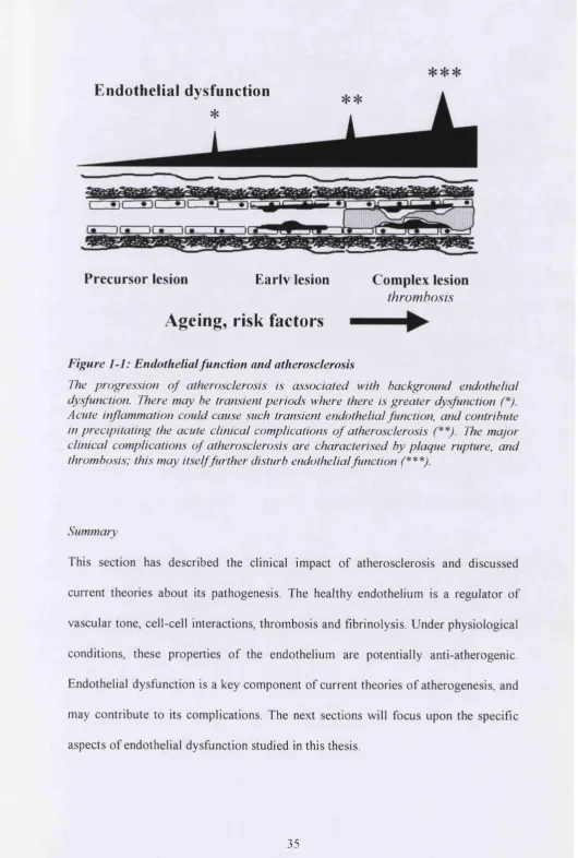
Figure 1-1: Endothelial function and atherosclerosis
The progression o f atherosclerosis is associated with background endothelial dysfunction. There may he transient periods where there is greater dysfunction (*). Acute inflammation could cause such transient endothelial function, and contribute in precipitating the acute clinical complications o f atherosclerosis The major clinical complications o f atherosclerosis are characterised by plaque rupture, and thrombosis; this may itself further disturb endothelial function (***),
Summary
1.3
Role of endothelial nitric oxide in flow-mediated dilatation in
early atherosclerosis
Conduit arteries dilate in response to an increase in blood flow (Nabel et a l, 1990; Anderson & Mark, 1989). This physiological response is dependent on the presence of an intact endothelium (Holtz et a l, 1984; Smiesko et a l, 1985), and forms the basis of the measurement of FMD in vivo as an assessment of endothelial function. Abnormalities o f FMD have been demonstrated in patients with clinical coronary artery disease and in younger subjects with risk factors for atherosclerosis (section
1.2).
Endothelial cells are sensitive to shear stress and respond by synthesising factors that regulate vascular smooth muscle tone (Davies, 1995) Endothelium-derived vasodilators that have been identified include NO and prostacyclin. In humans, dilatation o f conduit arteries in response to reactive hyperaemia (RH) is reduced by inhibitors of NO synthesis, suggesting an important role for NO in FMD (Joannides
et a l, 1995). However, a number of studies have suggested that, under different physiological conditions, FMD occurs by mechanisms that are independent of NO production. In animals and human coronary arteries, FMD in response to a sustained flow increase, induced by distal infusion o f adenosine, appears resistant to the effects of inhibition of NO synthesis (Canty & Schwartz, 1994; Shiode e ta l, 1996).
responses in humans has important implications for understanding the regulation of vascular tone and interpreting the results o f endothelial function tests that use flow as a stimulus.
1.4
The effect of acute inflammation on endothelial function
Inflammation and atherosclerosis
There is considerable evidence to support the concept that inflammation is important in the pathogenesis o f atherosclerosis. The response-to-injury hypothesis emphasises the importance o f the endothelium in the process of atherosclerosis. ‘Injury’ alters the phenotype of the endothelium to promote cell adhesion, migration of inflammatory cells and promote thrombosis. The expression of adhesion molecules on endothelial cells and inflammatory cells is increased initially and promotes monocyte and T-cell migration across the endothelium. These cells release a range o f cytokines, hydrolytic enzymes and growth factors, which lead to smooth muscle proliferation and focal necrosis. In this model, inflammation initiates and propagates the process of atherosclerosis.
Animal studies
Although it is clear that local inflammation occurs within atherosclerosis, the role of systemic inflammation and infection as a driving process is less clear. Two recent studies have addressed this question. In one model Apo-E knockout mice infected with herpes virus demonstrated accelerated atherosclerosis, and a rabbit model using a high-cholesterol diet to induce atherosclerosis, showed markedly enhanced atherosclerosis in animals with a superimposed systemic inflammatory response initiated with endotoxin (Alber et a l, 2000; Lehr et a l, 2001). Studies in animal models have also implicated infection as a cause of endothelial dysfunction, prior to the morphological changes of atheroma (Hatsukami et a l, 2000)
Human studies
Epidemiological and observational data have suggested that, in individuals in the long preclinical phase o f atherosclerosis, there are elevations in circulating inflammatory markers. Cohort studies show that circulating levels o f C-reactive protein (GRP), interleukin- 6 (IL-6) and intracellular adhesion molecule-1, serum
amyloid A, E-selectin, P-selectin, TNF-a, fibrinogen, von Willebrand factor, PAI-1, or evidence o f prior infection with common pathogens are associated with atherosclerosis and that these are independent predictors of future atherosclerotic complications (Ridker, 1994; Ridker, 1998; Ridker, 2000d; Ridker et a l, 2000c; Ridker et a l, 2000b; de Lemos et a l, 2000; Eikelboom et a l, 1999; Kiechl et al,
Endothelial function and inflammation
Cross-sectional studies have demonstrated that serum levels of GRP show inverse correlation with forearm blood flow responses to acetylcholine in a cohort with established coronary artery atherosclerosis (Fichtlscherer et a l, 2000). Furthermore, this relationship was independent of classical risk factors and endothelial function improved as CRP levels reduced with treatment. Although it remains unclear whether this is an effect of CRP itself, or reflects some other inflammatory process, these findings provide evidence that inflammation is associated with endothelial dysfunction, which may provide the setting for the development of atherosclerosis or its complications.
Acute coronary syndromes
The incidence o f acute coronary events is increased following inflammatory processes, including respiratory tract infections and surgery and elevated inflammatory markers in otherwise healthy individuals and patient groups are predictive of future acute vascular events (Milazzo et al, 1999; Rossi et a l, 2002;
et a l, 1995).
Studies in patients with coronary artery disease support the importance of inflammation in the transition to unstable coronary syndromes. Patients presenting with unstable angina have elevated levels of inflammatory cytokines on admission (CRP, IL-6, SAA) and the levels of these cytokines correlate with risk developing o f
suggesting that it was not ischaemia per se that was responsible for the elevation in cytokines (Liuzzo e ta l, 1996)
More direct evidence for coronary plaque inflammation is available from atherectomy studies. In one such study, specimens from 110 patients were studied by immunohistochemistry for macrophages, CD-3 positive lymphocytes and smooth muscle cells. Patients who developed unstable angina pectoris in the follow-up period had a greater number of macrophages and T-lymphocytes than those who did not (Meuwissen et a l, 2001). Plaques from patients with unstable angina also have greater inflammatory cell infiltration and activation (Piek et a l, 2000).
An elegant study by Rothwell et al tested the hypothesis that irregular plaques should occur in multiple vascular beds in some individuals more frequently than would be expected by chance, suggesting a systemic cause of instability. These investigators studied carotid angiograms and concluded that individuals with ‘irregular plaques’ were more likely to have irregular plaque on the contralateral carotid, previous myocardial infarction and a non-stroke vascular death on follow-up. Whilst such clustering of unstable plaques suggests a systemic cause (Rothwell et a l, 2000), no markers of inflammation were measured in this study.
These clinical studies suggest that a systemic process may occur which shifts the balance from stable atherosclerosis to an ‘unstable’ picture, with greater probability of plaque rupture and superimposed thrombus formation. Associated with these changes, is abnormal tissue perfusion due to vasoconstriction.
coronary plaque, causing spasm, thrombosis, and an acute coronary syndrome, (b) acute inflammation might also have direct pro-coagulant effects, which, in the presence of endothelial dysfunction could contribute to acute thrombotic complications, including myocardial infarction, (c) inflammation may directly influence the nature of the immune cell activity within a plaque, and initiate plaque rupture and thrombosis.
The fibrous cap is made up of smooth muscle cells and collagen. The collagen gives the cap strength, and T lymphocytes within the plaque can signal smooth muscle cells to reduce collagen production, and stimulate macrophages to increase production of collagen degradation enzymes such as matrix metalloproteinases (Gidron et a l, 2002). Circulating cytokines might have similar effects on the cellular components o f plaques (Libby et a l, 1995). Another process that may be important in causing weakness in the fibrous cap is smooth muscle cell death. Plaques isolated from patients with unstable angina show a reduced number o f smooth muscle cells and a greater proportion o f cell death by apoptosis in these plaques compared to those with stable angina (Bauriedel et al, 1999). Enzymes that regulate or stimulate apoptosis (e.g. caspases) are expressed to a high degree in atherosclerotic plaques in animal models and, in isolated cell systems, incubating smooth muscle cells with leukocytes or monocytes causes smooth muscle cell death by apoptosis (Boyle et al,
If the hypothesis that acute systemic inflammation is an important process in the transition from stable to unstable atherosclerosis is true and the endothelium is an important component of this then it would be expected that inflammation would cause endothelial dysfunction. This concept has been studied directly in humans in vivo, using the dorsal hand vein as a model system. TNF-a, IL-ip, IL- 6 and
endotoxin cause venous endothelial dysfunction, and aspirin and hydrocortisone protect the endothelium in this model (Bhagat et al., 1999). However, there is no direct evidence that acute systemic inflammation causes endothelial dysfunction of the arterial vasculature in vivo.
1.5
The effect of ischaemia-reperfusion on endothelial function
Therapies, such as thrombolysis or acute angioplasty, re-establish blood flow to ischaemic tissues after acute arterial occlusion in unstable angina, acute myocardial infarction, and stroke. These treatments are essential to prevent ischaemic cell death but the process of reperfiision allows infiltration of inflammatory cells into the tissue and this may contribute to further tissue injury. Tissue injury following ischaemia- reperfusion is a composite of damage caused by ischaemia and the additional damage caused by reperfusion.
1.5.1 Ischaemic injury
Reduced oxygen supply to tissues during ischaemia prevents mitochondrial respiration, depleting cells o f intracellular adenosine triphosphate (ATP) with subsequent build up o f anaerobic metabolites, increased adenosine monophosphate (AMP) and adenosine. ATP dependent membrane ion pump activity is reduced, leading to increased intracellular calcium and the leakage of intracellular ions and enzymes to the extracellular space.
free radical scavengers. Although neutrophils and tissue phagocytes produce large amounts of ROS, the endothelium is also another important source.
During ischaemia, there is not only loss o f antioxidant molecules from the cytoplasm but the calcium-sensitive second messenger systems that regulate these enzymes become disrupted. ROS levels therefore increase and cause failure of cell membrane integrity, damage to intracellular proteins, disruption o f cytoskeletal elements, cell swelling, and ultimately cell death via necrosis or apoptosis (Pohlman & Harlan,
2000).
1.5.2 Reperfusion injury
Cell death during reperfiision occurs in part because of persistent effects of ischaemia but also secondary to reperfiision injury. This is a complex inflammatory reaction in tissue after a period of ischaemia, where the recruitment of inflammatory mediators, cytokines and the cellular components o f the defence mechanism themselves cause and amplify tissue injury.
There is activation of the complement system during reperfiision and products such as C3a and C5a are potent chemotactic agents. The C5-9 membrane attack complex can cause direct cell damage (Ward, 1971). Administration of monoclonal antibodies against C5a or soluble C5a scavengers reduces the extent of myocardial infarction in animal models (Amsterdam et a l, 1995; Buerke et a l, 1998). Other chemokines that have been implicated in the process of reperfiision injury are platelet activating factor (PAF), TNTa, IL-1, IL-6, Interleukin- 8 (IL-8) and the anti-inflammatory Interleukin-
Animal models also indicate an important role for neutrophils in reperfiision injury. Neutrophil-depletion, selective blockade of neutrophil adhesion molecules, or neutrophil adhesion molecule knockout, protects against reperfusion injury in animal models (Litt et a l, 1989; Zhao et a l, 1997). In tissues, neutrophils undergo a respiratory burst, releasing phagocytic contents including myeloperoxidase and superoxide, which contribute to tissue injury (Hansen, 1995; Jordan et a l, 1999).
Platelets are potentially important in several aspects of reperfusion injury. There is evidence o f platelet activation and adhesion to blood vessels during reperfusion, which might exacerbate tissue injury by causing microvascular thrombosis and spasm (Lefer et a l, 1998; Lefer, 2000). Furthermore, the mediators that are released are able to activate other inflammatory cells such as neutrophils and monocytes, and they may adhere to such cells to promote their transmigration through the vessel. Animal models show that platelet depletion or synthetic peptides which block P- selectin interactions reduce ischaemia-reperfusion injury (Seko et a l, 1996).
1.5.3 Role of the endothelium in ischaemia-reperfusion injury
molecules (Hearse et a i, 1993; Quillen et a i, 1990). These changes might amplify endothelial and tissue injury by promoting neutrophil and platelet activation and adhesion. In addition to the increased infiltration of activated inflammatory cells, endothelial dysfunction of resistance vessels causes abnormal tissue perfusion, which may further exacerbate tissue injury.
NO in ischaemia-reperfusion
HOCL
Preconditioning
^ ONOO permeability
N O
CAMs
endothelium Arg NO
vasodilatation
Figure 1-2: NO in ischaemia-reperfusion injury
A reduced vasodilator capacity occurs, even in the presence o f structurally normal endothelial cells. This mainly reflects reduced NO activity, although other regulators of vascular tone have not been systematically studied. The reduction in NO may be due to reduced substrate or cofactor provision (L-arginine or BH4 reduction), or may reflect increased breakdown by ROS, as has been shown in animal models (Lefer, 1995; Tiefenbacher et a l, 1996; Dhalla et a l, 2000). Reduced NO activity initiates potentially deleterious processes leading to tissue injury (Lefer & Lefer, 1996).
In addition to the direct effects of ischaemia on endothelial cells, the presence of inflammatory cytokines (such as TNF-a and IL-1) can directly disrupt endothelial cell function and promote expression o f adhesion molecules (such as P-selectin, ICAM- 1 and VCAM-1). Adhesion of activated inflammatory cells to the
^ ^ T N F a
p . Vpregulatior, o fINSULT inflammatory cells adhesion molecules
and chemokines
I
Neutrophil adhesion
ENDOTHELIAL INJURY AND DYSFUNCTION
Figure 1-3: Mechanisms o f endothelial injury in ischaemia-reperfusion
Ischaemia may cause direct damage, hut the major mechanisms are thought to he due to the direct actions o f inflammatory cytokines on the endothelium, and the effects o f inflammatory cells (predominantly neutrophils).
1.5.4 Evidence for ischaemia-reperfusion injury in humans
Although restoring flow in the conduit artery is an essential component of therapy in stroke and myocardial infarction, recent studies have examined the possibility that this alone may not be sufficient to restore perfusion of tissues, and that subsequent impaired tissue perfusion may reflect microvascular endothelial dysfunction (Michaels et a i, 2 0 0 0; Mukherjee & Moliterno, 2 0 0 0; Roe et a i, 2001).
It is impossible to define whether this dysfunction is the result of infarction or causal. Several indirect indices have been used to assess microvascular function in the context of acute myocardial infarction, including myocardial contrast echocardiography. Echolucent contrast is injected, either directly into the infarct related coronary artery or intravenously, to assess perfiision at the microvascular level and this is regarded as an index of microvascular function. These studies have shown that approximately 40% of patients have abnormal microvascular function following successful recanalisation of the epicardial conduit artery (Ito & Iwakura,
1998; Czitrom a/., 1999).
The clinical relevance of such findings has been shown in studies using surrogate markers o f tissue level reperfusion, such as ST segment resolution and magnetic resonance perfusion studies, in patients following thrombolysis for acute infarction. These have consistently shown reduced improvement in left ventricular function and increased mortality in the patients with impaired microvascular function (Feldman et a l, 2 0 0 0).
Several interventional trials have used adjuvant therapy at reperfusion to modulate the processes of reperfusion injury. Drugs such as vasodilators, oxygen free radical scavengers or agents that modulate inflammatory cell function have been studied in animal models and a small number of human studies.
burst capacity (Cronstein et a l, 1985; Cronstein et a l, 1986; Burkey & Webster, 1993; Wollner et a l, 1993; Bullough et a l, 1995; Zhao et a l, 1996). Although small- scale trials o f corticosteroids have shown benefit in reducing the extent of inflammation following bypass surgery or size o f myocardial infarction, this has not been translated into clinical benefit (Hafezi-Moghadam et al, 2002). Large-scale human trials of complement activation inhibitors and adhesion molecule antibodies in acute myocardial infarction do not show a reduction in the extent of myocardial infarction (Faxon et a l, 2002) Human studies using agents to block myocardial sodium/hydrogen exchange channels have also failed to show overall clinical benefit, although certain subgroups may gain benefit (Theroux et a l, 2000). Many of these studies are difficult to interpret because the patient populations studied are heterogeneous and often highly selected. Furthermore, the outcomes that are examined may be subject to confounding, including prior extent of coronary disease and perfusion of the myocardium by collaterals. However, to date no agent has been shown to significantly modulate reperfusion injury in a large-scale human trial.
There are no human in vivo models to define the effects of acute ischaemia- reperfusion on endothelial function directly. Such studies are difficult in the coronary or cerebral circulations but the ability to define a model in humans would allow the biology o f this process to be unravelled, with the potential to investigate therapeutic options. The studies described in Chapter 6 characterise a human model of
1.5.5 Ischaemic preconditioning
One innate protective mechanism, which promotes cell survival and preserves function to various pathological stresses, is called preconditioning. Exposure o f the cell to sublethal injury causes the recruitment of endogenous mechanisms that protect the cell from further insults. At present, the defence mechanisms that are upregulated are poorly understood. However, cells become resistant to a wide variety of physiological insults when preconditioned. Numerous physiological stresses can induce this protective preconditioned state (Domenech & Macho, 1998).
Ischaemic preconditioning utilises sub-lethal ischaemia to induce protection from subsequent lethal ischaemia. In animal models o f coronary occlusion this can reduce infarct size by up to 75% (Murry et a l, 1986). In addition to a direct effect on ischaemic tissues, ischaemic preconditioning of the endothelium and immune cells may also occur preventing endothelial injury and neutrophil activation and indirectly reducing tissue injury (Hiasa et a l, 2001).
These triggers act on cell surface receptors to initiate a cascade of second messengers, including activation of phospholipases C and D, generation of diacylglycerol and stimulation o f protein kinase C (PKC). PKC regulates many cellular processes that could be involved in preconditioning, including cell metabolism, ion transport and gene expression. In some species, PKC-induced preconditioning is blocked by inhibitors of protein tyrosine kinases (e.g. genistein), suggesting that tyrosine kinases are distal in the preconditioning pathway to PKC. A third group of kinases, the mitogen activated protein kinases (MAPK) appear to act distal to the tyrosine kinase cascade (Cohen et a i, 2000).
brac^kinin adenosine
oxygen radicals
PLC
PLD
PKC
TK
MAPK
K.-tp channel mitochondrion
Figure 1-4: Mechanisms o f preconditioning
The end effectors of preconditioning remain uncertain but could include ATP- sensitive potassium channels ( Ka t p) . A consensus is emerging that the mitochondrial
Ka t p channels are involved in preconditioning; activation o f these channels by
diazoxide mimics and inhibition by glibenclamide or 5-hydroxydecanoate blocks ischaemic preconditioning. It is not clear how activation of mitochondrial Ka t p
channels might cause preconditioning; and some data suggests that Ka t p channels
might principally be involved in the early triggering rather than late effector phase of preconditioning (Pain et a l, 2000).
The mitochondrial permeability transition pore (MPTP) is non-specific channel on the inner mitochondrial membrane. Opening of this pore leads to cell death through either apoptosis or necrosis. This channel remains closed during ischaemia, but opens during the first few minutes of reperfusion. High mitochondrial calcium, ATP depletion, acidosis and oxidative stress provide the stimulus for this channel to open. Blocking this channel has been shown to reduce ischaemia-reperfusion injury in experimental models. It has recently been shown that activation of the Ka t p channel
inhibits opening o f the MPTP, and so this may act as final effector mechanism in preconditioning (Hausenloy et al, 2002)
(Sheth et a l, 1997). However, the predominant receptor on coronary endothelial cells is the A2A subtype, which mediate protection against anoxia-reperfiision injury (Zhou
e ta l, 1996).
Bradykinin acts upon membrane receptors to stimulate preconditioning. In an isolated guinea-pig heart model, ischaemic preconditioning is abolished by blocking bradykinin B2 receptors (Sheth et a l, 1997). In an in-vivo rat heart model, blockade of B 1 receptors, but not B2 receptors, was involved in vascular preconditioning (Bouchard et a l, 1998). It remains unclear whether the endogenous B2 receptor or the inducible B1 receptor is responsible for endothelial preconditioning, and it is likely that this depends upon the species and model studied.
The second messenger systems that are responsible for ischaemic preconditioning have not been fully characterised in endothelial cells. The Ka t p channels are thought
to be distal effectors in the process of preconditioning. In the rat coronary circulation,
the Ka t p channel blockers such as glibenclamide attenuate endothelial ischaemic
preconditioning (Bouchard & Lamontagne, 1996). Ka t p channels on the plasma
membrane in the myocardium and the vasculature differ, and there is increasing evidence that the mitochondrial Ka t p channel may be more important then those on
the plasma membrane.