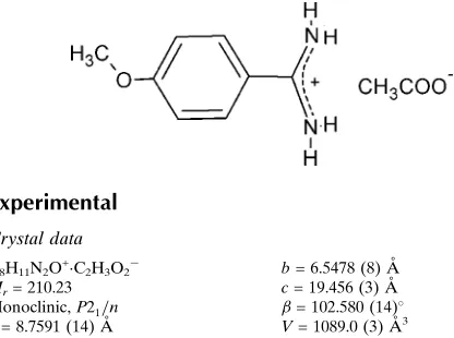
4-Methoxybenzamidinium acetate
Simona Irrera and Gustavo Portalone*
Chemistry Department, "Sapienza" University of Rome, P.le A. Moro, 5, I-00185 Rome, Italy
Correspondence e-mail: [email protected]
Received 29 October 2012; accepted 30 October 2012
Key indicators: single-crystal X-ray study;T= 298 K; mean(C–C) = 0.003 A˚; Rfactor = 0.056;wRfactor = 0.141; data-to-parameter ratio = 15.2.
The title compound, C8H11N2O +
CH3CO2
, was synthesized by a reaction between 4-methoxybenzamidine (4-amidino-anisole) and acetic acid. In the cation, the amidinium group forms a dihedral angle of 11.65 (17) with the mean plane of
the benzene ring. The ionic components are associated in the crystalvia N—H+ O hydrogen bonds, resulting in a one-dimensional structure consisting of dimers and catemers and orientated approximately along thecaxis.
Related literature
For the biological and pharmacological relevance of benza-midine, see: Powers & Harper (1999). For structural analysis of proton-transfer adducts containing molecules of biological interest, see: Portalone, (2011a); Portalone & Irrera (2011). For the supramolecular association in proton-transfer adducts containing benzamidinium cations, see; Portalone (2010, 2011b, 2012); Irrera & Portalone (2012a,b); Irreraet al.(2012). For hydrogen-bond motifs, see Bernstein et al. (1995). For standard bond lengths, see: Allenet al.(1987).
Experimental
Crystal data
C8H11N2O+C2H3O2
Mr= 210.23 Monoclinic,P21=n a= 8.7591 (14) A˚
b= 6.5478 (8) A˚
c= 19.456 (3) A˚ = 102.580 (14)
V= 1089.0 (3) A˚3
MoKradiation = 0.10 mm1
0.210.180.15 mm
Data collection
Oxford Diffraction Xcalibur S CCD diffractometer
Absorption correction: multi-scan (CrysAlis PRO; Agilent, 2011)
Tmin= 0.980,Tmax= 0.986
14433 measured reflections 2365 independent reflections 1834 reflections withI> 2(I)
Rint= 0.040
Refinement
R[F2> 2(F2)] = 0.056 wR(F2) = 0.141 S= 1.08 2365 reflections 156 parameters
H atoms treated by a mixture of independent and constrained refinement
max= 0.21 e A˚
3
min=0.15 e A˚
[image:1.610.51.259.539.694.2]3
Table 1
Hydrogen-bond geometry (A˚ ,).
D—H A D—H H A D A D—H A
N1—H1A O1 0.91 (3) 1.94 (3) 2.847 (2) 175 (2) N1—H1B O1i
0.91 (2) 1.98 (2) 2.832 (2) 155 (2) N2—H2A O2 0.94 (3) 1.83 (3) 2.776 (2) 176 (2) N2—H2B O2ii
0.88 (2) 1.95 (2) 2.817 (2) 168 (2)
Symmetry codes: (i)x;y;zþ1; (ii)x1 2;yþ
1 2;zþ
1 2.
Data collection: CrysAlis PRO(Agilent, 2011); cell refinement:
CrysAlis PRO; data reduction: CrysAlis PRO; program(s) used to solve structure:SIR97 (Altomareet al., 1999); program(s) used to refine structure:SHELXL97(Sheldrick, 2008); molecular graphics:
ORTEP-3(Farrugia, 1997); software used to prepare material for publication:WinGX(Farrugia, 1999).
Supplementary data and figures for this paper are available from the IUCr electronic archives (Reference: TK5166).
References
Agilent (2011).CrysAlis PRO. Agilent Technologies, Yarnton, England. Allen, F. H., Kennard, O., Watson, D. G., Brammer, L., Orpen, A. G. & Taylor,
R. (1987).J. Chem. Soc. Perkin Trans. 2, pp. S1–19.
Altomare, A., Burla, M. C., Camalli, M., Cascarano, G. L., Giacovazzo, C., Guagliardi, A., Moliterni, A. G. G., Polidori, G. & Spagna, R. (1999).J. Appl. Cryst.32, 115–119.
Bernstein, J., Davis, R. E., Shimoni, L. & Chang, N.-L. (1995).Angew. Chem. Int. Ed. Engl.34, 1555–1573.
Farrugia, L. J. (1997).J. Appl. Cryst.30, 565. Farrugia, L. J. (1999).J. Appl. Cryst.32, 837–838.
Irrera, S., Ortaggi, G. & Portalone, G. (2012).Acta Cryst.C68, o447–o451. Irrera, S. & Portalone, G. (2012a).Acta Cryst.E68, o3083.
Irrera, S. & Portalone, G. (2012b).Acta Cryst.E68, o3244. Portalone, G. (2010).Acta Cryst.C66, o295–o301. Portalone, G. (2011a).Chem. Centr. J.5, 51. Portalone, G. (2011b).Acta Cryst.E67, o3394–o3395. Portalone, G. (2012).Acta Cryst.E68, o268–o269. Portalone, G. & Irrera, S. (2011).J. Mol. Struct.991, 92–96.
Powers, J. C. & Harper, J. W. (1999).Proteinase inhibitors, edited by A. J. Barrett & G. Salvesen, pp. 55–152. Elsevier: Amsterdam.
Sheldrick, G. M. (2008).Acta Cryst.A64, 112–122.
Structure Reports
Online
supporting information
Acta Cryst. (2012). E68, o3277 [doi:10.1107/S1600536812044911]
4-Methoxybenzamidinium acetate
Simona Irrera and Gustavo Portalone
S1. Comment
The present work is part of a general structural analysis of proton-transfer adducts containing molecules of biological
interest (Portalone, 2011a; Portalone & Irrera, 2011). In this context, benzamidine derivatives, which have shown strong
biological and pharmacological activity (Powers & Harper, 1999), are being used in our group as bricks for
supramolecular construction (Portalone, 2010, 2011b, 2012). Indeed, the bidentate hydrogen-bonding interaction between
the amidinium and the carboxylate functional groups can be a powerful organizing force in solution and in the solid-state.
We report here the single-crystal structure of the title molecular salt, 4-methoxybenzamidinium acetate, (I), which was
obtained by a reaction between 4-methoxybenzamidine (4-amidinoanisole) and acetic acid.
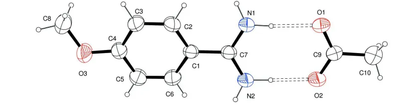
The asymmetric unit of (I) comprises one non-planar 4-methoxybenzamidinium cation and one acetate anion (Fig. 1).
In the cation the amidinium group forms dihedral angle of 11.65 (17)° with the mean plane of the phenyl ring, which
agrees with the values observed in protonated benzamidinium ions (14.4 (1) - 32.7 (1)°, Portalone, 2010, 2012; Irrera et
al., 2012). The lack of planarity in all these systems is obviously caused by steric hindrances between the H atoms of the
aromatic ring and the amidine moiety. This conformation is rather common in benzamidinium-containing small-molecule
crystal structures, with the only exception of benzamidinium diliturate, where the benzamidinium cation is planar
(Portalone, 2010). The pattern of bond lengths and bond angles of the 4-methoxybenzamidinium cation agrees with that
reported in previous structural investigations (Irrera et al., 2012; Portalone, 2010, 2012; Irrera & Portalone, 2012a,
2012b). In particular the amidinium group, true to one's expectations, features C—N bonds within experimental error
[1.312 (2) and 1.306 (2) Å], evidencing the delocalization of the π electrons and double-bond character.
In the acetate moiety the C—O bond lengths indicate delocalization of the negative charge on both O atoms, since the C
—O bond lengths [1.250 (2) and 1.249 (2) Å] are intermediate between single Csp2—O and double Csp2═O, and
correlate well with values for carboxylate anions [1.247 - 1.262 Å, Allen et al., 1987].
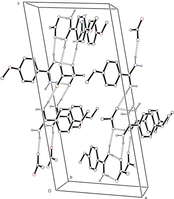
Analysis of the crystal packing of (I), (Fig. 2), shows that each amidinium unit is bound to three acetate anions by four
distinct N—H+···O- strong intermolecular hydrogen bonds (N+···O- = 2.776 (2) - 2.847 (2) Å, Table 1) into a
one-dimensional structure. The ion pairs of the asymmetric unit are joined by two N+—H···O- (±) hydrogen bonds to form
ionic dimers with graph-set motif R2
2(8) (Bernstein et al., 1995). These subunits are then joined as catemers into linear
chains approximately along the crystallographic c axis through the remaining N+—H···O- hydrogen bonds to adjacent
anti-parallel dimers.
S2. Experimental
4-Methoxybenzamidine (0.1 mmol, Fluka at 96% purity) was dissolved without further purification in 8 ml of a 20%
solution of acetic acid and heated under reflux for 3 h. After cooling the solution to an ambient temperature, colourless
All H atoms were identified in difference Fourier maps, but for refinement all C-bound H atoms were placed in calculated
positions, with C—H = 0.93 Å (phenyl) and 0.89 - 1.01 Å (methyl), and refined as riding on their carrier atoms. The Uiso
values were kept equal to 1.2Ueq(C, phenyl). and to 1.5Ueq(C, methyl). Positional and displacement parameters of H
[image:3.610.93.496.152.262.2]atoms of the amidinium group were refined, giving N—H distances in the range 0.88 (2) - 0.94 (3) Å.
Figure 1
The asymmetric unit of (I), showing the atom-labelling scheme. Displacements ellipsoids are at the 50% probability level.
The asymmetric unit was selected so that the two ions are linked by N+.—H···O- hydrogen bonds. H atoms are shown as
Figure 2
Crystal packing diagram for (I), viewed approximately down b. Displacements ellipsoids are at the 50% probability level.
H atoms are shown as small spheres of arbitrary radii. For the sake of clarity, H atoms not involved in hydrogen bonding
have been omitted. Hydrogen bonding is indicated by dashed lines.
4-Methoxybenzamidinium acetate
Crystal data
C8H11N2O+·C2H3O2− Mr = 210.23 Monoclinic, P21/n
Hall symbol: -P 2yn a = 8.7591 (14) Å b = 6.5478 (8) Å c = 19.456 (3) Å β = 102.580 (14)° V = 1089.0 (3) Å3 Z = 4
F(000) = 448 Dx = 1.282 Mg m−3
Mo Kα radiation, λ = 0.71073 Å Cell parameters from 3220 reflections θ = 2.8–28.9°
µ = 0.10 mm−1 T = 298 K
Oxford Diffraction Xcalibur S CCD diffractometer
Radiation source: Enhance (Mo) X-ray Source Graphite monochromator
Detector resolution: 16.0696 pixels mm-1 ω and φ scans
Absorption correction: multi-scan (CrysAlis PRO; Agilent, 2011) Tmin = 0.980, Tmax = 0.986
14433 measured reflections 2365 independent reflections 1834 reflections with I > 2σ(I) Rint = 0.040
θmax = 27.0°, θmin = 3.3° h = −11→11
k = −8→8 l = −24→24
Refinement
Refinement on F2
Least-squares matrix: full R[F2 > 2σ(F2)] = 0.056 wR(F2) = 0.141 S = 1.08 2365 reflections 156 parameters 0 restraints
Primary atom site location: structure-invariant direct methods
Secondary atom site location: difference Fourier map
Hydrogen site location: inferred from neighbouring sites
H atoms treated by a mixture of independent and constrained refinement
w = 1/[σ2(F
o2) + (0.0561P)2 + 0.3698P]
where P = (Fo2 + 2Fc2)/3
(Δ/σ)max < 0.001
Δρmax = 0.21 e Å−3
Δρmin = −0.15 e Å−3
Special details
Geometry. All s.u.'s (except the s.u. in the dihedral angle between two l.s. planes) are estimated using the full covariance matrix. The cell s.u.'s are taken into account individually in the estimation of s.u.'s in distances, angles and torsion angles; correlations between s.u.'s in cell parameters are only used when they are defined by crystal symmetry. An approximate (isotropic) treatment of cell s.u.'s is used for estimating s.u.'s involving l.s. planes.
Refinement. Refinement of F2 against ALL reflections. The weighted R-factor wR and goodness of fit S are based on F2,
conventional R-factors R are based on F, with F set to zero for negative F2. The threshold expression of F2 > 2σ(F2) is
used only for calculating R-factors(gt) etc. and is not relevant to the choice of reflections for refinement. R-factors based on F2 are statistically about twice as large as those based on F, and R- factors based on ALL data will be even larger.
Fractional atomic coordinates and isotropic or equivalent isotropic displacement parameters (Å2)
x y z Uiso*/Ueq
O3 0.2977 (2) 1.0070 (2) 0.35136 (8) 0.0624 (5)
N1 −0.0153 (2) 0.1780 (3) 0.42774 (8) 0.0453 (4)
H1A −0.074 (3) 0.064 (4) 0.4296 (12) 0.063 (7)*
H1B 0.054 (3) 0.217 (4) 0.4677 (13) 0.063 (7)*
N2 −0.1170 (2) 0.2005 (3) 0.31128 (9) 0.0468 (4)
H2A −0.175 (3) 0.081 (4) 0.3149 (12) 0.070 (7)*
H2B −0.133 (3) 0.261 (3) 0.2696 (13) 0.055 (6)*
C1 0.0643 (2) 0.4636 (3) 0.36478 (9) 0.0343 (4)
C2 0.1370 (2) 0.5636 (3) 0.42564 (10) 0.0449 (5)
H2 0.1318 0.5077 0.4690 0.054*
C3 0.2173 (2) 0.7446 (3) 0.42381 (10) 0.0472 (5)
H3 0.2659 0.8088 0.4655 0.057*
C4 0.2248 (2) 0.8294 (3) 0.35969 (10) 0.0429 (5)
C5 0.1539 (3) 0.7298 (4) 0.29855 (10) 0.0584 (6)
C6 0.0756 (3) 0.5502 (3) 0.30090 (10) 0.0510 (5)
H6 0.0293 0.4849 0.2591 0.061*
C7 −0.0247 (2) 0.2737 (3) 0.36775 (9) 0.0356 (4)
C8 0.3954 (3) 1.0997 (4) 0.41088 (13) 0.0624 (6)
H8A 0.3323 (10) 1.130 (2) 0.4474 (7) 0.094*
H8B 0.4395 (17) 1.231 (2) 0.3962 (3) 0.094*
H8C 0.4838 (17) 1.0040 (17) 0.4315 (6) 0.094*
O1 −0.20216 (18) −0.1693 (2) 0.44118 (7) 0.0548 (4)
O2 −0.29092 (18) −0.1444 (2) 0.32662 (7) 0.0550 (4)
C9 −0.2818 (2) −0.2339 (3) 0.38404 (9) 0.0390 (4)
C10 −0.3743 (3) −0.4258 (3) 0.38417 (12) 0.0606 (6)
H10A −0.3120 (10) −0.5337 (17) 0.3841 (9) 0.091*
H10B −0.4519 (17) −0.4292 (13) 0.3458 (7) 0.091*
H10C −0.4143 (17) −0.4291 (12) 0.4226 (7) 0.091*
Atomic displacement parameters (Å2)
U11 U22 U33 U12 U13 U23
O3 0.0827 (11) 0.0586 (9) 0.0449 (9) −0.0302 (8) 0.0113 (8) 0.0040 (7) N1 0.0564 (11) 0.0443 (10) 0.0298 (8) −0.0120 (8) −0.0020 (7) 0.0052 (7) N2 0.0613 (11) 0.0439 (10) 0.0293 (9) −0.0095 (8) −0.0034 (8) 0.0040 (7) C1 0.0389 (10) 0.0354 (9) 0.0271 (9) 0.0018 (7) 0.0039 (7) 0.0009 (7) C2 0.0616 (13) 0.0460 (11) 0.0263 (9) −0.0063 (9) 0.0076 (8) 0.0021 (8) C3 0.0616 (13) 0.0474 (11) 0.0302 (10) −0.0119 (9) 0.0049 (9) −0.0056 (8) C4 0.0485 (11) 0.0423 (10) 0.0379 (10) −0.0043 (8) 0.0092 (8) 0.0012 (8) C5 0.0798 (16) 0.0647 (14) 0.0292 (10) −0.0221 (12) 0.0083 (10) 0.0075 (9) C6 0.0686 (14) 0.0545 (12) 0.0270 (10) −0.0158 (10) 0.0037 (9) −0.0019 (8) C7 0.0411 (10) 0.0358 (9) 0.0279 (9) 0.0021 (7) 0.0030 (7) 0.0007 (7) C8 0.0673 (15) 0.0587 (14) 0.0587 (14) −0.0234 (11) 0.0086 (12) −0.0064 (11) O1 0.0657 (10) 0.0627 (9) 0.0293 (7) −0.0192 (7) −0.0045 (6) 0.0027 (6) O2 0.0742 (10) 0.0551 (9) 0.0281 (7) −0.0154 (7) −0.0053 (7) 0.0019 (6) C9 0.0376 (10) 0.0436 (10) 0.0330 (10) −0.0009 (8) 0.0014 (8) −0.0003 (8) C10 0.0608 (14) 0.0558 (13) 0.0603 (15) −0.0123 (11) 0.0023 (12) 0.0026 (11)
Geometric parameters (Å, º)
O3—C4 1.353 (2) C3—H3 0.9300
O3—C8 1.418 (3) C4—C5 1.380 (3)
N1—C7 1.312 (2) C5—C6 1.367 (3)
N1—H1A 0.91 (3) C5—H5 0.9300
N1—H1B 0.91 (2) C6—H6 0.9300
N2—C7 1.306 (2) C8—H8A 1.0093
N2—H2A 0.94 (3) C8—H8B 1.0093
N2—H2B 0.88 (2) C8—H8C 1.0093
C1—C2 1.381 (2) O1—C9 1.250 (2)
C1—C6 1.389 (3) O2—C9 1.249 (2)
C1—C7 1.475 (2) C9—C10 1.495 (3)
C3—C4 1.380 (3) C10—H10C 0.8930
C4—O3—C8 119.11 (16) C5—C6—C1 121.01 (18)
C7—N1—H1A 120.0 (14) C5—C6—H6 119.5
C7—N1—H1B 121.9 (15) C1—C6—H6 119.5
H1A—N1—H1B 118 (2) N2—C7—N1 118.69 (18)
C7—N2—H2A 119.1 (14) N2—C7—C1 120.84 (16)
C7—N2—H2B 123.6 (14) N1—C7—C1 120.45 (16)
H2A—N2—H2B 117 (2) O3—C8—H8A 109.5
C2—C1—C6 117.64 (17) O3—C8—H8B 109.5
C2—C1—C7 120.99 (16) H8A—C8—H8B 109.5
C6—C1—C7 121.36 (16) O3—C8—H8C 109.5
C1—C2—C3 121.77 (17) H8A—C8—H8C 109.5
C1—C2—H2 119.1 H8B—C8—H8C 109.5
C3—C2—H2 119.1 O2—C9—O1 123.45 (18)
C4—C3—C2 119.52 (17) O2—C9—C10 117.82 (16)
C4—C3—H3 120.2 O1—C9—C10 118.71 (17)
C2—C3—H3 120.2 C9—C10—H10A 109.5
O3—C4—C5 116.00 (17) C9—C10—H10B 109.5
O3—C4—C3 124.78 (17) H10A—C10—H10B 109.5
C5—C4—C3 119.22 (18) C9—C10—H10C 109.5
C6—C5—C4 120.83 (18) H10A—C10—H10C 109.5
C6—C5—H5 119.6 H10B—C10—H10C 109.5
C4—C5—H5 119.6
C6—C1—C2—C3 0.6 (3) C3—C4—C5—C6 0.6 (4)
C7—C1—C2—C3 −178.24 (18) C4—C5—C6—C1 0.5 (4)
C1—C2—C3—C4 0.4 (3) C2—C1—C6—C5 −1.1 (3)
C8—O3—C4—C5 −169.3 (2) C7—C1—C6—C5 177.8 (2)
C8—O3—C4—C3 10.9 (3) C2—C1—C7—N2 167.00 (19)
C2—C3—C4—O3 178.89 (19) C6—C1—C7—N2 −11.9 (3)
C2—C3—C4—C5 −1.0 (3) C2—C1—C7—N1 −11.3 (3)
O3—C4—C5—C6 −179.3 (2) C6—C1—C7—N1 169.9 (2)
Hydrogen-bond geometry (Å, º)
D—H···A D—H H···A D···A D—H···A
N1—H1A···O1 0.91 (3) 1.94 (3) 2.847 (2) 175 (2)
N1—H1B···O1i 0.91 (2) 1.98 (2) 2.832 (2) 155 (2)
N2—H2A···O2 0.94 (3) 1.83 (3) 2.776 (2) 176 (2)
N2—H2B···O2ii 0.88 (2) 1.95 (2) 2.817 (2) 168 (2)